71
폐실질내 다발성 낭종으로 발현한 기관지기원낭
성균관대학교 의과대학 내과학교실, 삼성서울병원 호흡기내과, 영상의학과1, 병리과2, 흉부외과3 최경아, 고원중, 이경수1, 한정호2, 김관민3
Multicystic Pulmonary Parenchymal Lesions in a Young Adult with Hemoptysis
Kyoung A Choi, M.D., Won-Jung Koh, M.D., Kyung Soo Lee, M.D.1, Joungho Han, M.D.2, Kwhanmien Kim, M.D.3
Division of Pulmonary and Critical Care Medicine, Department of Medicine, Radiology1, Pathology2, and Thoracic Surgery3, Samsung Medical Center, Sungkyunkwan University School of Medicine, Seoul, Korea
Bronchogenic cysts are commonly located in the mediastinum or lung parenchyma, and arise from the abnormal budding of the primitive tracheobronchial tube. Cough and pain are the most common symptoms. Bronchogenic cysts appear as spherical or oval masses with smooth outlines and are usually unilocular and noncalcified. We report a young adult with a bronchogenic cyst presenting as multicystic pulmonary parenchymal lesions. This case is very unusual because a multicystic intrapulmonary bronchogenic cyst is very rare in adults. (Tuberc Respir Dis 2007; 62: 71-73) Key words: Bronchogenic cyst, Adult, Hemoptysis.
Address for correspondence: Won Jung Koh, M.D.
Division of Pulmonary and Critical Care Medicine, Department of Medicine, Samsung Medical Center, Sungkyunkwan University School of Medicine, 50 Iron-dong, Gngnam-gu, Seoul 135-710, Korea.
Telephone: (822) 3410-3429 Fax: (822) 3410-3849 E-mail: [email protected]
Received: Nov. 29. 2006 Accepted: Dec. 21. 2006
증 례
환 자: 남자, 26세 주 소: 객혈
현병력: 환자는 7개월 전 건강검진에서 시행한 흉 부엑스레이검사에서 좌측 폐에 6 cm 크기의 낭종 (cyst)이 우연히 발견되었고, 경과관찰 중 2주전부터 간헐적으로 소량의 객혈이 발생하여 입원하였다.
흡연력: 5갑년의 흡연력
과거력 및 가족력: 특이 사항 없음
진찰 소견: 내원 당시 혈압 110/64 mmHg, 맥박수 80회/분, 호흡수 18회/분, 체온 36.3℃이었다. 전신상 태는 급성병색을 보이지는 않았으며 결막 및 공막에 는 특이소견 보이지 않았다. 흉부 청진상 심음 및 폐 음은 정상이었고 복부나 사지 등의 진찰 소견도 정상 이었다.
검사실 소견: 말초 혈액검사상 백혈구 6000/mm3,
혈색소 16.4 g/dL, 혈소판 230,000/mm3이었고, 생화학 검사 소견상 총단백 7.7 g/dL, 알부민 4.9 g/dL, AST/ALT 17/7 U/L, 총 빌리루빈 0.4 mg/dL, 소변검 사 및 심전도 검사결과는 정상이었고, 폐기능 검사상 FEV1/FVC, FEV1 모두 정상 소견이었다.
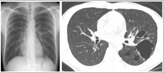
방사선 소견: 흉부 엑스레이 검사에서 좌측 폐에 6 cm 크기의 낭종이 관찰되었고 이외에 양측 폐실질 및 종격동에는 특이 소견이 없었다(Figure 1A). 흉부 전 산화 단층촬영에서 좌상엽의 후분절과 좌하엽의 상분 절을 포함하는 폐실질내 경계가 명확한 다발성 낭종 이 관찰되었다. 낭종 내에 액체 저류 등은 없었으며, 종격동이나 폐문 림프절 확대, 흉막액 삼출 등의 이상 소견은 없었다(Figure 1B).
수술 및 병리조직학적 소견: 상기 검사 결과를 토 대로 개흉 좌하엽 절제술 및 좌상엽의 낭종절제술을 시행하였다. 수술 소견상 흉막의 심한 유착이나 흉막 액 삼출 등의 소견은 없었고, 좌상엽의 낭종은 낭종 내 괴사성 액체가 소량 고여있었고 이를 흡인하여 배 양 검사를 시행하였으나 동정된 균은 없었다. 병리조 직학적 소견상 육안적으로 좌하엽의 상분절내에 내부 에 매끄러운 얇은 막을 지닌 소엽상(lobulated) 낭종 이 있었고, 주위로 여러 개의 소낭종이 관찰되었다.
주위 폐실질은 정상 조직이었다(Figure 2A). 현미경 적 소견상 낭종 내면이 가중층 섬모 원주세포(ciliated
KA Choi et al: Intrapulmonary bronchogenic cyst
72
Figure 2. (A) Left lower lobectomy specimen shows a large lung cyst without communication with bronchus. (B) Photomicrography (original magnification, × 400; H & E staining) shows a cyst wall lined by respiratory epithelium.
Figure 1. (A) Posteroanterior chest radiograph shows 6 cm sized thin-walled air cyst in the left lung. (B) Computed tomography (CT) image shows multicystic intraparenchymal lesions in the superior segment of the left lower lobe and the posterior segment of the left upper lobe.
pseudostratified columnar epithelium)로 되어있었고, 국소적으로 염증과 육아 조직을 동반한 병변이 관찰 되어 폐실질내 기관지기원낭(intrapulmonary bron- chogenic cyst)으로 진단되었다(Figure 2B).
치료 및 경과: 환자는 수술 후 특별한 합병증 없이 퇴원하여 외래에서 경과관찰 중이다.
고 찰
기관지기원낭은 비교적 드문 선천성 질환으로 태생 기의 기관지나 기관지 분지의 이상에 의해 발생한다.
이는 원시전망(foregut)에서 발생하는 기관아(tracheal
bud)의 분화 이상에 의한 것으로 주로 소아에서 발견 되나 성인에서도 드물게 발견된다. 기관지기원낭은 주로 종격동에서 흔하게 관찰되고 폐실질에서 발생하 는 경우는 15-20%로 드물다1. 드물게 피부나 피하 조 직, 목, 심낭, 횡격막, 복강, 척수강 내에서도 발견될 수 있다. 임상 증상은 기관지 교통 여부에 따라 다른 것으로 알려져 있으며 가장 흔한 증상으로 기침, 호흡 곤란, 흉통, 발열, 객혈이 발생하는데2, 이는 기관지 압 박으로 인한 물리적 요인에 의한 증상이거나 이차적 인 세균감염으로 인해 주로 증상이 나타나게 된다.
방사선학적으로는 흉부 엑스레이 검사와 전산화 단 층촬영이 진단을 위해 가장 많이 사용되는데, 폐실질
Tuberculosis and Respiratory Diseases Vol. 62. No.1, Jan. 2007
73 내 낭종의 경우 보통 경계가 분명하고 석회화가 없는
원형 또는 난원형의 종괴로 나타나고, 종격동내 기관 지 낭종은 중간 종격동 혹은 후종격동의 용골(carina) 직하부 혹은 우측 기관지벽을 따라 원형이나 타원형 의 단일 낭종으로 나타나는 것이 전형적이며, 다발성 낭종으로 나타나는 경우는 매우 드물다3-5. 폐실질내 기관지기원낭에서는 공기액체층이 관찰되기도 한다6. 전산화 단층촬영에서는 보통 균일한 밀도의 Houns- field Number(0~20)를 보이는 낭종으로 관찰되고, 공 기액체층으로 나타날 수도 있다. 이는 낭종이 기관지 와 연결되면서 염증을 동반한 경우가 많으며, 폐농양 등과의 감별이 필요하다. 또한 대부분의 기관지기원 낭이 경계가 분명한 것으로 알려져 있으나, 국내 문헌 에서도 보고되었듯이 불분명한 경계와 연조직 음영을 가지고 있어 악성 종양으로 오인되었던 증례도 있었 다7. 따라서 확진은 수술을 통한 조직학적 진단으로 가능하다. 수술이 아닌 경기관지 또는 경피적 낭종 흡 인 생검의 경우 수술을 대체할 수 있는 가능성을 제기 하나 낭종 파열 등의 위험이 높아 아직까지는 받아들 여지지 못하고 있다8. 수술로 완전절제를 한 경우 예 후가 좋으나 불완전절제가 된 경우는 재발률이 높고 합병증 발생도 증가하는 것으로 보고된다2. 또한 무증 상 환자에 비해 증상 발생 후에 수술을 하는 경우 수 술에 따른 합병증 발생이 증가하는 것으로 보고되어 무증상 환자에서도 수술적 완전절제를 시행하는 것이 추천된다2,6,9.
본 증례는 성인에서 다발성 낭종으로 관찰된 폐실 질내 기관지기원낭의 예로 이러한 예는 소아에서 드 물게 보고되나10, 성인에서의 다발성 낭종의 형태로 발견되는 경우는 매우 드물다.
요 약
기관지기원낭은 대체로 방사선학적으로 종격동이
나 폐실질내에 얇고 균일한 벽을 지닌 경계가 분명한 타원형의 종괴나 낭포로 발견되며 증상 유무에 관계 없이 완전 절제를 하는 것이 추천된다. 본 증례는 젊 은 성인에서 객혈을 동반한 폐실질내 기관지기원낭으 로 수술하였던 예로 비전형적으로 다발성 낭종의 형 태로 발견되었기에 문헌 고찰과 함께 보고하는 바이다.
참 고 문 헌
1. Suen HC, Mathisen DJ, Grillo HC, LeBlanc J, McLoud TC, Moncure AC, et al. Surgical management and radiological characteristics of bronchogenic cysts.
Ann Thorac Surg 1993;55:476-81.
2. Sarper A, Ayten A, Golbasi I, Demircan A, Isin E.
Bronchogenic cyst. Tex Heart Inst J 2003;30:105-8.
3. Nakata H, Nakayama C, Kimoto T, Nakayama T, Tsukamoto Y, Nobe T, et al. Computed tomography of mediastinal bronchogenic cysts. J Comput Assist Tomogr 1982;6:733-8.
4. Naidich D, Zerhouni E, Siegelman S. Computed tomography and magnetic resonance of the thorax.
New York: Raven press 1991. p. 120-3.
5. Yoon YC, Lee KS, Kim TS, Kim J, Shim YM, Han J.
Intrapulmonary bronchogenic cyst: CT and pathologic findings in five adult patients. AJR Am J Roentgenol 2002;179:167-70.
6. Aktogu S, Yuncu G, Halilcolar H, Ermete S, Buduneli T. Bronchogenic cysts: clinicopathological presentation and treatment. Eur Respir J 1996;9:2017-21.
7. Kim YW, Lee SH, Hong SC, Lee HH, Park SJ, Lee GJ, et al. A case report of a bronchogenic cyst miscon- ceived to lung cancer. Tuberc Respir Dis 2003;55:
526-30.
8. St-Georges R, Deslauriers J, Duranceau A, Vaillan- court R, Deschamps C, Beauchamp G, et al. Clinical spectrum of bronchogenic cysts of the mediastinum and lung in the adult. Ann Thorac Surg 1991;52:6-13.
9. Patel SR, Meeker DP, Biscotti CV, Kirby TJ, Rice TW.
Presentation and management of bronchogenic cysts in the adult. Chest 1994;106:79-85.
10. Ramenofsky ML, Leape LL, McCauley RG. Broncho- genic cyst. J Pediatr Surg 1979;14:219-24.