Ⅰ. 서 론
다형성 선종은 대타액선과 소타액선에서 기원하는 가장 흔한 양성 상피성 종양으로1), 상피세포, 근상피세포 및 간엽조직 성분 을 포함하는 매우 다양한 양상의 조직소견을 보인다. 염색체 분 석결과, 다형성 선종은 약 70%에서 염색체 구조의 이상소견을 보이며2), 이러한 염색체 이상의 50% 이상이 8번 염색체와 관련된 다고 보고되어 있다3). 염색체 이상의 가장 흔한 유형은 t(3;8)(p21;q12) 상반 전위(reciprocal translocation)이고2-4), 그 밖에 t(5;8)(p13;q12) 전위 등이 보고되어 있다4). 인체 종양발생과 관련
된 염색체 전위는 이상 융합단백을 생성하거나 전사영역 교환 (promoter swapping)에 따른 비정상적 전사를 통해 종양발생에 관여한다. 다형성 선종 발생과 관련된 전위는 사람 염색체 8q12 부위에 존재하는 PLAG1 유전자와 염색체 3p21 부위의β-catenin 유전자(CTNNB1)나 transcription elongation factor SII 유전자, 염색 체 5p13 부위의 leukemia inhibitory factor receptor(LIFR) 유전자의 전사영역(promoter)이 서로 교환되어 PLAG1이 비정상적으로 발 현되는 기전에 주로 의존한다2,5,6).
PLAG1은 7개의 C2H2 zinc finger를 갖는 아미노기 말단과 serine 기가 풍부한 카복시 말단을 갖는 단백질로서 전사조절인자일 것 으로 추정된다7). 앞서 기술한 바와 같은 전위 기전에 의해 성인 의 타액선에서는 비활성 상태를 유지하는 PLAG1의 발현이 증가 되면, PLAG1의 표적유전자가 비정상적으로 발현되어 타액선 종 양이 야기될 것으로 여겨진다.
다형성 선종은 타액선에서 발생되는 구강악안면영역의 비교 적 흔한 종양이며, 유전자 융합이 그 주요 발생기전으로 알려져 있음에도 불구하고, 유전자 융합 이후의 진행과정은 거의 알려
타액선 다형성 선종에서의 PLAG1과 CTNNB1 유전자 융합
김재진∙김은석∙고승오*∙김효분*∙조남표*
충남대학교 병원 구강악안면외과, 전북대학교 치과대학 구강생체과학연구소*
Abstract (J. Kor. Oral Maxillofac. Surg. 2003;29:206-211)
조 남 표
전주시 덕진구 덕진동 664-14
전북대학교 치과대학 구강생체과학연구소 Nam-Pyo Cho
Dept. of Oral Bioscience, School of Dentistry, Chonbuk National University 664-14 Dukjindong, Dukjingu, Chonju, Korea
Tel. 82-63-270-4026 Fax. 82-63-270-4004 E-mail : [email protected]
FUSION OF THE PLAG1 AND CTNNB1 GENES IN PLEOMORPHIC ADENOMA OF THE SALIVARY GLANDS
Jae-Jin Kim, Eun-Seok Kim, Seung-O Ko*, Hyo-Bun Kim*, Nam-Pyo Cho*
Department of Oral and Maxillofacial Surgery, Chungnam National University Hospital, Institute of Oral Bioscience, School of Dentistry, Chonbuk National University*
The pleomorphic adenoma is the most common neoplasm involving both the major and minor salivary glands. It is a benign, slow- growing tumor, but local recurrences can occur. The pleomorphic adenoma gene 1 (PLAG1), which is a novel zinc finger gene, is fre- quently activated by reciprocal chromosomal translocations involving 8q12 in a subset of salivary gland pleomorphic adenomas. This experimental study was preformed to observe the translocation patterns between PLAG1 gene and the three translocation partner genes. We also have analyzed the presence of PLAG1 transcripts by RT-PCR.
CTNNB1/PLAG1 gene fusion was observed in three of nine pleomorphic adnomas. However, LIFR/PLAG1 and SII/PLAG1 gene fusions were not detectable. All of three gene fusions was not detectable in one Warthin’s tumor and three inflammatory salivary gland tissues. PLAG1 transcripts were expressed in all inflammatory salivary gland tissues and tumors except for three pleomorphic adeno- mas. Of particular one pleomorphic adenoma showing CTNNB1/PLAG1 gene fusion did not express PLAG1 transcipt.
Our data indicate that gene fusion involving PLAG1 is a frequent event in pleomorphic adenoma, but correlation between gene fusion involving PLAG1 and PLAG1 transcription is not definite.
Key words: Pleomorphic adenoma, PLAG1, Chromosomal translocation, CTNNB1
※ 이 연구는 한국과학재단 목적기초연구사업(R01-2001-00180)의 지원에 의하여 이루어진 것임.
져 있지 않다. 특히, 국내에서는 다형성 선종의 발생과 관련된 유 전적 변화에 관한 연구가 행해진 바가 없으므로, 본 연구에서는 사람의 다형성 선종을 대상으로 유전자 융합의 존재여부 및 유 전자 융합과 PLAG1 mRNA 발현과의 연관성을 알아보고자 한다.
Ⅱ. 재료 및 방법 1. 연구대상
본 연구에서는 2000년부터 2001년까지 충남대학교병원 구강외 과 및 이비인후과에서 채취된 다형성 선종 9예와 타액선 양성 종 양인 Warthin’s tumor 1예 및 염증성 타액선 조직 3예를 이용하였 다. 양성 종양의 성별 분포는 남성 4명, 여성 6명이고, 발생부위 는 이하선 9예, 악하선 1예였다. 염증성 타액선 조직의 성별 분포 는 모두 남성이었으며, 채취부위는 악하선 이었다. 획득된 조직 의 일부는 파라핀 포매 후 H&E염색을 시행하였으며, 나머지 조 직은 획득 후 즉시 영하 193℃의 액화질소에 액침한 후, 영하 70
℃의 냉동고에 보관하여 실험에 이용하였다.
2. 동결조직으로부터 total RNA의 분리
본 연구에 이용된 total RNA는 guanidine/phenol 방법에 기초하 여 one-step으로 total RNA를 분리할 수 있는 QIAGEN RNeasy Mini Kit(QIAGEN, Hilden, Germany)를 이용하여 추출하였다. 추출된 total RNA는 Gene-Quant pro RNA/DNA Calculator(Amersham Pharmacia Biotech, Cambrige, UK)를 사용하여 정량한 후, formaldehyde가 포함된 gel에서 전기영동하고, ethidium bro- mide(EtBr)로 염색하여 그 농도와 상태를 확인하고 영하 70℃에 서 보관하였다.
3. total RNA로부터 cDNA의 합성
1㎍의 total RNA에 random Hexamer(200μM, Boehringer Mannheim, Mannheim, Germany) 1 ㎕를 첨가하고 고압 멸균된 3 차 증류수로 최종 용량을 7.5 ㎕가 되게 조정한 후 70℃에서 10분 간 변성시켰다. 준비된 시료에 RNase inhibitor를 포함하는 반응액 을 첨가하여 42℃에서 2분간 incubation한 후, reverse transcrip- tase(Superscript II RT, 200U/㎕, Invitrogen, Rockville, MD) 1 ㎕를 첨 가하여 42℃에서 50분간, 70℃에서 15분간 반응시켜 cDNA를 합 성한 후, 영하 80℃에 보관하였다.
4. primer의 선택 및 제작
cDNA 합성 유무를 확인하기 위해 사용된β-actin 유전자의 RT- PCR primer는 OLIGO(Molecular Biology Insights, Cascade, CO) pro- gram을 이용하여 선택하였으며, CTNNB1/PLAG1, LIFR/PLAG1 및 SII/PLAG1 유전자 전위를 확인하기 위한 RT-PCR primer는 Voz 등 이 보고한 염기서열을 참조하였다4).
5. β-actin에 대한 RT-PCR
cDNA의 합성 유무를 확인하기 위하여 정상 조직 세포와 병소 세포에서 항상 발현되는 housekeeping gene인β-actin gene의 RT- PCR를 실시하였다. PCR은 60℃를 annealing temperature로 35 cycle 반복한 후, 1.5% agarose gel 에서 전기영동하여 결과를 확인 하였다.
6. 정상 및 융합 transcripts에 대한 RT-PCR
염증성 타액선 조직 및 다형성 선종 내에 유전자 융합이 존재 하는지를 확인하기 위하여, PLAG1 유전자 영역에서는 reverse primer를, 그 주된 표적유전자들로 알려진 CTNNB1, SII 및 LIFR 유 전자들에서는 forward primer를 각각 선택하여 RT-PCR을 시행하 였다. 정상 transcripts의 에 대한 RT-PCR은 유전자 융합에 의해 발 현이 증가된다고 보고된 PLAG1을 대상으로 하였다.
7. PCR 산물의 염기 서열 확인
RT-PCR 산물이 원하는 유전자임을 확인하기 위하여 염기 서열 을 확인하였다. PCR산물 5 ㎕에 exonuclease I(10U/㎕) 1 ㎕와 shrimp alkaline phosphatase(2U/㎕) 1 ㎕를 첨가한 후 37℃ 에서 15 분 동안 반응시키고, 80℃ 에서 15분 동안 방치하여 효소를 불활 성시켰다. 처리된 PCR산물을 dye terminator cycle sequencing ready reaction kit를 이용하여 ABI prism 377 Genetic Analyzer로 (Perkin Elmer, Foster City, CA, any) 분석하였다.
Ⅲ. 결 과 1. β-actin RT-PCR
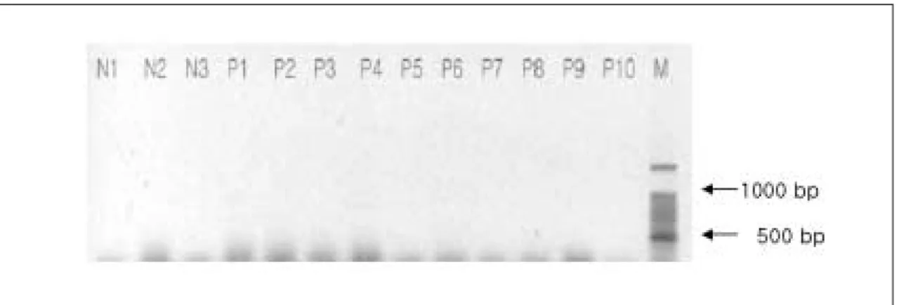
10예의 타액선 종양과 3예의 염증성 타액선 조직 모두 β-actin RT-PCR에서 증폭되었다(Fig. 1).
2. PLAG1과 관련된 유전자 융합의 검색
타액선 다형성 선종에서 흔히 보여지며, 그 주요 발생기전으로 작용한다고 알려진 CTNNB1/PLAG1, LIFR/PLAG1 및 SII/PLAG1의 3 가지 유전자 융합을 RT-PCR로 확인한 결과는 다음과 같다.
A. CTNNB1/PLAG1유전자 융합
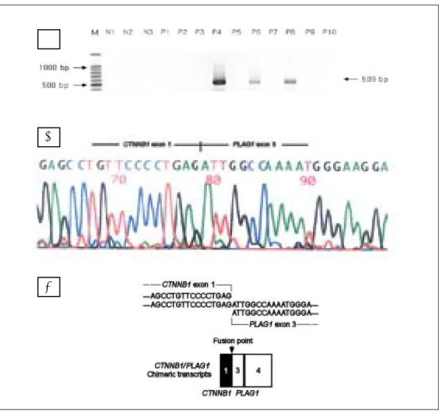
CTNNB1/PLAG1 유전자 융합은 9예의 다형성 선종 중 3예에서 증폭을 보였으며, 기타 염증성 타액선 조직 및 Warthin’s tumor에 서는 증폭이 보여지지 않았다(Fig. 2A, 2B, 2C).
B. LIFR/PLAG1및 SII/PLAG1 유전자 융합
LIFR/PLAG1및 SII/PLAG1 유전자 융합은 타액선 종양 및 염증성 타액선 병소 모두에서 보여지지 않았다(Fig. 3, 4).
Fig. 1. Identification of the existence of cDNA by β-actin RT-PCR. An expected 155 bp product was obtained in all the samples. M, 100 bp ladder molecular marker; N1-3, inflammatory salivary gland tissue; P1-10 except for P9, pleomorphic adenoma; P9, Warthin’s tumor.
Fig. 2. RT-PCR analysis and sequencing analysis of reciprocal fusion transcript involving CTNNB1 and PLAG1. A. An expected 509 bp product was obtained in P4, P6 and P8. M, molecular marker; N1-3, inflammatory salivary gland tissue; P1-10 except for P9, pleomorphic adenoma; P9, Warthin’s tumor. B.
Nucleotide sequence data. All fusion transcripts gave rise to a product of 509 bp consisting of exons 1 of CTNNB1 fused to exons 3-4 of PLAG1. C. Schematic representation of the nucleotide sequences flanking the fusion point of the chimeric transcript. Fusion transcripts junctioned between exon 1 of CTNNB1 and exon 3 of PLAG1. M, 100 bp ladder molecular marker.
A
B
C
3. PLAG1 발현의 검색
PLAG1 mRNA는 실험에 사용된 3예의 모든 염증성 타액선 조직 들과 3예의 다형성 선종에서 발현되었다(Fig. 5). PLAG1 transcript
가 발현된 3예의 다형성 선종 중 2예는 CTNNB1/PLAG1 유전자 융 합이 존재하였으며, 1예는 유전자 융합이 존재하지 않는 선종이 었다.
Fig. 3. RT-PCR analysis of reciprocal fusion transcript involving LIFR and PLAG1. An expected 474 or 579 bp product was not obtained in all of the samples. M, molecular marker; N1-3, inflammatory salivary gland tissue; P1-10 except for P9, pleomorphic adenoma; P9, Warthin’s tumor.
Fig. 4. RT-PCR analysis of reciprocal fusion transcript involving LIFR and PLAG1. An expected 557 or 662 bp product was not obtained in all of the samples. M, molecular marker; N1-3, inflammatory salivary gland tissue; P1-10 except for P9, pleomorphic adenoma; P9, Warthin’s tumor.
Fig. 5. Identification of the existence of cDNA by PLAG1 RT-PCR. An expected 523 bp product was obtained in N1-3, P3, 4, 6, 10. M, molecular marker; N1-3, inflammatory salivary gland tissue; P1-10 except for P9, pleomorphic adenoma; P9, Warthin’s tumor.
Ⅳ. 고 찰
다형성 선종은 타액선에서 가장 흔히 발생하는 양성 종양으로, 상피조직, 근상피조직 및 간엽조직 등의 다양한 조직소견을 보 인다. 주로 30�50세의 젊은 성인에서 호발하며, 모든 타액선에 서 발생할 수 있으나 대부분 이하선에서 나타나며8), 악하선 및 소화관 상부의 소타액선에서 일부 발생된다9). 다형성 선종은 양 성종양임에도 불구하고 5�30% 정도의 비교적 높은 재발율을 보이나, 정상조직까지 완전히 절제한 경우는 5%이하로 줄일 수 있으며. 일부 다형성 선종은 악성 종양으로 전환되기도 한다9).
다형성 선종은 염색체 전위가 흔히 보여져서 염색체 8q12 부위 와 관련된 염색체 전위를 가지는 군이 39%, 염색체 12q13-15 부 위와 관련된 염색체 전위를 가지는 군이 8%, 염색체 8q12 또는 12q13-15 부위와 관련이 없는 염색체 이상을 가지는 군이 23%를 차지한다고 보고되어 있다10-12). 최근에는 8q12 부위에서의 유전 적 변화가 PLAG1과 관련되며, PLAG1 promoter가 활발히 전사되 는 다른 유전자들의 promoter가 교환되는 promoter swapping 기 전에 의한다고 알려져 있다13). 이러한 promoter swapping에 의해 결국 PLAG1의 발현은 증가되고, 상반 전위(reciprocal transloca- tion)된 다른 유전자의 발현은 감소되는 효과를 가져온다13). PLAG1유전자와 흔히 상반 전위되는 유전자들로는 염색체 3p21 부위에 존재하는 β-catenin의 유전자인 CTNNB1, 5p13 부위의 LIFR 유전자 및 3p21.3-22 부위의 transcription elongation factor SⅡ 유전 자 등이 알려져 있다2,5,6).
β-catenin은 세포간 부착과 WG/WNT signaling pathway에 있어 중요한 기능을 수행하는 단백질로서, 종양 발생과도 관련이 있 는 것으로 알려져 있다5). PLAG1과 CTNNB1의 전위는 양 유전자 의 5′noncoding region에서 일어나며 coding sequence를 보존하 면서 regulatory control element를 교환함으로써, PLAG1은 과발현 되고 CTNNB1의 발현은 감소된다13). LIFR유전자 및 transcription elongation factor SⅡ 유전자와의 전위도 CTNNB1과 동일한 기전 에 의해 PLAG1의 과발현을 야기한다2,6).
다형성 선종에서의 염색체 전위에 의해 흔히 그 발현이 증가되 는 PLAG1단백은 7개의 C2H2 zinc finger를 갖는 아미노기 말단과 serine기가 풍부한 카복시 말단을 갖는 구조를 지녀서 전사조절 인자로서 작용할 것으로 여겨진다4,7). 즉, 발현이 증가된 PLAG1은 그 표적유전자의 전사에 이상을 초래하여 종양을 발생시킬 것으 로 여겨진다. Kas 등은 PLAG1이 태반, 태아조직 등의 발생조직이 나, 8q12 염색체 전위를 보이는 양성 타액선 종양에서만 발견된 다고 보고하였으나13), Queimado 등은 양성 타액선 종양, 악성 타 액선 종양, 정상 타액선 조직에서 발견될 수 있다고 보고한 바 있
다14,15). Astrom 등은 염색체 8q12 부위의 전위가 없는 정상적인 핵
형을 갖는 17개의 다형성 선종과 PLAG1 염색체 부위와는 관련이 없는 12q13-15 염색체 부위에 전위를 갖는 10개의 다형성 선종에 대한 PLAG1 발현을 검색한 결과, 정상 핵형을 갖는 다형성 선종 에서는 76%(13/17), 염색체 12q13-15 전위를 갖는 다형성 선종에 서는 50%(5/10)에서 PLAG1 발현이 관찰되어 다형성 선종에서의 PLAG1발현이 핵형과 관계없다고 보고한 바 있다6).
PLAG1에 의해 전사가 조절되는 표적유전자에 대해서는 현재 까지 거의 알려져 있지 않으나, 최근 Voz 등은 IGF-II를 그 표적유 전자로 처음 제시한 바 있다4). IGF-Ⅱ는 태아 발생과 종양발생에 있어 중요한 역할을 수행하는 펩타이드계 성장인자로, PLAG1이 과발현된 타액선 종양에서는 과발현되나, PLAG1 과발현이 없는 다형성 선종과 정상 타액선에서는 발견되지 않는다고 보고하고
있다16-19). 그러나, Kerr 등은 포유동물의 정상 타액선에서도 IGF-
Ⅱ가 합성, 분비된다고 보고한 바 있다20).
이상의 문헌고찰에서 다형성 선종이 여러 유형의 염색체 전위 와 흔히 관련됨은 명확하나, 염색체 전위에 의한 PLAG1 발현의 증가 및 PLAG1에 의한 IGF -Ⅱ의 전사조절 등은 다소 다른 결과 가 보고되어 있고4), 그 연구 또한 매우 제한적으로 행해진 바 있 다. 본 연구는 사람의 타액선 조직 및 종양을 실험에 이용함으로 써 시료 수가 다소 적었으나, PLAG1과 관련된 3가지 주된 유전자 융합 중 CTNNB1/PLAG1 유형이 9예의 다형성 선종 중 3예만 보여 졌으며 기타 유전자 융합은 보여지지 않았다. 또한 염증성 타액 선 조직 및 Warthin’s tumor에서는 모든 유전자 융합이 보여지지 않았다.
PLAG1 mRNA는 실험에 사용된 3개의 모든 염증성 타액선 조직 들과 P2, P4, P6의 다형성 선종에서 발현되었다. 본 실험결과는 PLAG1의 발현이 유전자 융합과 절대적 상관관계가 없으며, 타액 선 종양 외의 염증성 타액선 조직에서도 발현될 수 있음을 보여 서 Kas 등의 실험결과와 달리2,13), Queimado 등이 주장과 일치되 는 소견이었다15). 또한 PLAG1과 관련된 유전자 융합이 보여진 다 형성 선종 1예에서는 PLAG1이 검출되지 않음으로써 유전자 융 합이 PLAG1 발현을 항상 증가시키지 않을 수 있음을 암시하였 다.
이상의 실험 결과는 다형성 선종에서 PLAG1과 관련된 염색체 전위가 비교적 흔하나, 이러한 전위가 PLAG1의 발현증가와 절대 적 상관관계를 지니지는 않는 것으로 여겨지며, 염색체 전위 이 외의 PLAG1 발현조절 기전이나 PLAG1의 전사 표적단백 등을 확 인하는 연구가 추가적으로 행해져야 할 것으로 여겨진다.
Ⅴ. 결 론
사람의 타액선에서 발생된 다형성 선종 9예, Warthin’s tumor 1 예 및 염증성 타액선 조직 3예를 대상으로 PLAG1과 관련된 유전 자 융합 및 PLAG1 mRNA 존재여부를 분석한 결과는 다음과 같 다.
1. 9예의 다형성 선종 중 3예에서 CTNNB1/PLAG1 유전자 융합이 보여졌으며, 다른 종류의 유전자 융합은 보여지지 않았다.
2. 1예의 Warthin’s tumor와 3예의 염증성 타액선 조직에서는 모 든 종류의 유전자 융합이 보여지지 않았다.
3. PLAG1발현은 유전자 융합이 보여지는 다형성 선종 외의 염 증성 타액선 조직에서도 보여졌으며, 특히 유전자 융합이 존 재한 다형성 1예에서는 PLAG1이 발현되지 않았다.
이상의 결과를 종합하면, 다형성 선종에서는 PLAG1과 관련된 유전자 융합이 비교적 흔히 존재하나, 유전자 융합이 PLAG1
mRNA 발현증가와 절대적 상관관계를 지니지는 않았다.
참고문헌
1. Ellis GL, Auclair PL, Gnepp DR. Surgical pathology of the salivary glands. 1st ed. Philadelphia, W. B. Saunders Co. 1991.
2. Voz ML, Astrom AK, Kas K, Mark J, Stenman G, Van de Ven WJ:
The recurrent translocation t(5;8)(p13;q12) in pleomorphic adeno- mas results in upregulation of PLAG1 gene expression under control of the LIFR promoter. Oncogene 1998;16:1409-1416.
3. Kas K, Roijer E, Voz M, Meyen E, Stenman G, Van de Ven WJ: A 2- Mb YAC contig and physical map covering the chromosome 8q12 breakpoint cluster region in pleomorphic adenomas of the salivary glands. Genomics 1997;43:349-358.
4. Voz ML, Agten NS, Van de Ven W, Kas K: PLAG1, the main translo- cation target in pleomorphic adenoma of the salivary glands, is a positive regulator of IGF-Ⅱ. Cancer Res 2000;60:106-113.
5. Peifer M: β-catenin as oncogene: the smoking gun. Science 1997;275(5307):1752-3.
6. Astrom AK, Voz ML, Kas K, Roijer E, Wedell B, Mandahl N, et al.:
Conserved mechanism of PLAG1 activation in salivary gland tumors with and without chromosome 8q12 abnormalities : identification of SⅡ as a new fusion partner gene. Cancer Res 1999;59:918-923.
7. Kas K, Voz ML, Hensen K, Meyen E, Van de Ven WJ:
Transcriptional activation capacity of the novel PLAG family of zinc finger proteins. J Biol Chem 1998;273:23026-23032.
8. Neville BW, Damm DD, Allen CM, Bouquot JE. Oral & Maxillofical Pathology. 1st ed. Philadelphia, W. B. Saunders Co. 1995.
9. Peterson LJ. Principles of Oral & Maxillofacial Surgery. 1st ed.
Philadelphia, J. B. Lippincott Co, 1992.
10. Sandros J, Stenman G, Mark J: Cytogenetic and molecular observa- tions in human and experimental salivary gland tumors. Cancer Genet Cytogenet 1990;44:153-167.
11. Bullerdiek J, Wobst G, Meyer-Bolte K, Chilla R, Haubrich J, Thode B, et al.: Cytogenetic subtyping of 220 salivary gland pleomorphic adenomas: correlation to occurrence, histological subtype, and in vitro cellular behavior. Cancer Genet Cytogenet 1993;65:27-31.
12. Mark J, Dahlenfors R, Wedell B: Impact of the in vitro technique used on the cytogenetic patterns in pleomorphic adenoma. Cancer Genet Cytogenet 1997;95:9-15.
13. Kas K, Voz ML, Roijer E, Astrom AK, Meyen E, Stenman G, et al.:
Promoter swapping between the genes for a novel zinc finger pro- tein and β-catenin in pleomorphic adenomas with t(3;8)(p21;q12) translocation. Nat Genet 1997;15:170-174.
14. Schoenmakers EF, Wanschura S, Mols R, Bullerdiek J, Van den Berghe H, Van de Ven WJ: Recurrent rearrangements in the high mobility group protein gene, HMGI-C, in benign mesenchymal tumors. Nat Genet 1995;10:436-444.
15. Queimado L, Lopes C, Du F, Martins C, Bowcock AM, Soares J, et al.: Pleomorphic adenoma gene 1 is expressed in cultured benign and malignant salivary gland tumor cells. Lab Invest 1999;79:583- 589.
16. Toretsky JA, Helman LJ: Involvement of IGF-Ⅱ in human cancer. J Endocrinol 1996;149:367-372.
17. El-Badry OM, Romanus JA, Helman LJ, Cooper MJ, Rechler MM, Israel MA: Autonomous growth of a human neuroblastoma cell line is mediated by insulin-like growth factor Ⅱ. J Clin Invest 1989;84:829-839.
18. Daughaday WH: The possible autocrine/paracrine and endocrine roles of insulin-like factors of human tumors. Endocrinol 1990;127:1- 4.
19. Gearing DP, Thut CJ, VandeBos T, Gimpel SD, Delaney PB, King J, et al.: Leukemia inhibitory factor receptor is structurally related to the IL-6 signal transducer, gp130. EMBO J 1991;10:2839-2848.
20. Kerr M, Lee A, Wang PL, Purushotham KR, Chegini N, Yamamoto H, et al.: Detection of insulin and insulin-like growth factors Ⅰ and
Ⅱ in saliva and potential synthesis in the salivary glands of mice.
Biochem Pharmacol 1995;49(10):1521-1531.