67 http://dx.doi.org/10.4196/kjpp.2014.18.1.67
ABBREVIATIONS: MIRI, myocardial ischemia reperfusion injury;
IPostC, ischemic postconditioning; JNK, c-Jun NH2-terminal kinase;
MAPK, mitogen-activated protein kinase; p38-MAPK, p38 mitogen- activated protein kinases; TUNEL, terminal deoxynucleotidyl trans- ferase dUTP nick end labeling; IR, ischemia reperfusion; LAD, left anterior descending.
Received November 14, 2013, Revised December 21, 2013, Accepted January 6, 2014
Corresponding to: Weidong Zhou, Department of Cardiology, The People’s Hospital of Suining, Suining 221200, XuZhou, Jiangsu, China. (Tel) 86-516-83262106, (Fax) 86-516-83262251, (E-mail) [email protected]
*These authors contributed equally to this work.
This is an Open Access article distributed under the terms of the Creative Commons Attribution Non-Commercial License (http://
creativecommons.org/licenses/by-nc/3.0) which permits unrestricted non-commercial use, distribution, and reproduction in any medium, provided the original work is properly cited.
Naloxone Postconditioning Alleviates Rat Myocardial Ischemia Reperfusion Injury by Inhibiting JNK Activity
Anzhou Xia
1,*, Zhi Xue
1,*, Wei Wang
1,*, Tan Zhang
1, Tiantian Wei
1, Xingzhi Sha
2, Yixun Ding
2, and Weidong Zhou
21
Department of Pharmacology, Xuzhou Medical College, Xuzhou 221004, Jiangsu Province,
2Department of Cardiology, The People’s Hospital of Suining, Suining 221200, Xuzhou, Jiangsu Province, China
To investigate the alteration of c-Jun N-terminal kinase (JNK) activity after myocardial ischemia reperfusion injury (MIRI) and further explore the effect of naloxone postconditioning on MIRI. Forty male Sprague Dawley rats were randomly divided into five groups: sham operation (sham, n=8);
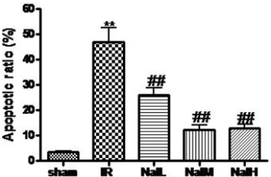
ischemia reperfusion (IR, n=8); IR+naloxone 0.5 mg/kg (Nal L, n=8); IR+naloxone 1.0 mg/kg (Nal M, n=8); IR+naloxone 2.0 mg/kg (Nal H, n=8). Pathological changes of myocardial tissue were visualized by HE staining. The expression of p-JNK, and the apoptosis of cardiomyocytes were investigated with W estern blotting and the TUNEL assay, respectively. Irregular arrangement and aberrant structure of myocardial fibers, cardiomyocytes with granular or vacuolar degeneration, and inflammatory cells infiltrating the myocardial interstitial regions characterized MIRI in the IR group. Signs of myocardial injury and inflammatory infiltration were less prominent in the Nal-treated groups. The expression of p-JNK in the sham group and in all Nal-treated groups was significantly lower than that in the IR group (p< 0.01). The apoptosis index of cardiomyocytes in the IR group was significantly higher than in the sham group (p< 0.01). The apoptosis indices of cardiomyocytes in all Nal-treated groups were significantly reduced to 55.4%, 26.2%, and 27.6%, respectively, of the IR group (p< 0.01). This study revealed that Naloxone postconditioning before reperfusion inhibits p-JNK expression and decreases cell apoptosis, thus alleviating MIRI.
Key Words: Cell apoptosis, c-Jun N-terminal kinases (JNK), Ischemia reperfusion, Naloxone
INTRODUCTION
Among human diseases cardiovascular disease is a con- dition associated with the highest mortality rates, ischemic heart disease being the most prevalent diagnose [1]. Coro- nary occlusion may cause myocardial necrosis in the region of blood supply and aberrant heart function. The mortally affected cardiac muscle can be rescued by reopening infarct arteries and recovering the blood supply in ischemic tissue.
Among the established methods are thrombolytic therapy, coronary angioplasty, and artery bypass grafting. After- wards the cardiac function can be improved, however, re- perfusion can trigger harmful pathophysiological responses and promote cell apoptosis. These side effects are known
as myocardial ischemia reperfusion injury (MIRI).
To alleviate or remove reperfusion injury is a challenge to the clinician. Ischemic postconditioning (IPostC) was con- firmed to protect heart and brain [2-4], but has been found difficult to conduct clinically. As an exogenous intervention, pharmacological postconditioning presented similar endog- enous protective mechanism as IPostC. Pharmacological postconditioning has demonstrated many advantages, in- cluding predictability, controllability, safety, and conve- nient operation, suggesting that it can be used to clinically prevent ischemia reperfusion injury [5-7]. Naloxone is an opioid receptor antagonist and postconditioning with nalox- one can alleviate focal cerebral ischemia reperfusion injury in rats [8] and ischemic acute kidney injury in the mouse [9]. Our previous study indicated that naloxone can relieve MIRI [10]. Herewith we present further research results that may add to the elucidation of the underlying me- chanisms. We used three different dosage of naloxone (0.5, 1.0, 2.0 mg/kg) to investigate the relationship between dos- age and response.
The c-Jun NH2-terminal kinase (JNK) signaling is an im-
portant element in mitogen-activated protein kinase (MAPK) signaling and has been shown to be involved in multiple physiological and pathological progressions. It has not reported yet whether naloxone postconditioning alle- viates MIRI by affecting JNK signaling. In this study, the effect of naloxone postconditioning on JNK expression and activity after MIRI was investigated using a rat MIRI model. The findings on the protective mechanism can pro- vide the experimental foundation for myocardial protection under ischemia reperfusion.
METHODS Animals
The adult male Sprague Dawley (SD) rats ranging from 230 g to 270 g were provided by the experimental animal center of Xuzhou Medical College (Jiangsu, China; SYXK [Su] 2002-0038).
Reagents and apparatus
Naloxone hydrochloride (lot number: 0801171) was pur- chased from Beijing Hua Su Pharmaceutical Co. Ltd. A one-step terminal deoxynucleotidyl transferase dUTP nick end labeling (TUNEL) kit was obtained from Beyotime Institute of Biotechnology (Jiangsu, China). The primary antibodies specific for JNK and p-JNK were purchased from Cell Signal (USA). A refrigerated centrifuge (model 5804R) and a PowerPac
TMHC vertical electrophoresis apparatus were obtained from Eppendorf (Germany) and Bio-Rad (USA), respectively. A LMS-2B dual-trace physiological re- corder, an ALC-V8 artificial animal respirator, and a BX51 fluorescence microscope were purchased from Chengdu Instrument Factory (Sichuan, China), Shanghai Alcott Biotech Co. Ltd (Shanghai, China), and Olympus (Japan), respectively.
Test groups and treatments
Forty rats were randomly divided into five groups as fol- lows: 1) sham operation (sham, n=8); 2) ischemia re- perfusion (IR, n=8); 3) IR plus low-dose naloxone (Nal L, 0.5 mg/kg; n=8); 4) IR plus medium-dose naloxone (Nal M, 1.0 mg/kg; n=8); 5) IR plus high-dose naloxone (Nal H, 2.0 mg/kg; n=8). Rats in the sham group and in the IR group received equal volumes of physiological saline/normal sal- ine while rats in the Nal groups received naloxone by a tail intravenous injection 10 min before reperfusion.
Establishment of myocardial ischemia reperfusion model
Rats were anesthetized with an intraperitoneal injection of 10% chloral hydrate at a dose of 3.5 ml/kg and fixed at supine position. The electrocardiogram (ECG) was observed using an LMS-2B dual-trace physiological recorder. The skin was slit at the center of the neck and the muscle was bluntly separated. The trachea was exposed and cut and the animal respirator was connected to the trachea. Respi- ratory support was given during the whole experiment. The skin was cut along the midclavicular line for 2 cm and the third rib was snipped. The pleura and pericardium were opened and the thymus was separated. The left anterior
descending (LAD) of the coronary artery within the cardiac muscle guided by the vena coronaria sinistra was found be- tween the auricula sinistra and the pulmonary conus. A 5-0 type atraumatic suture needle was passed by the my- ocardial surface, which was 2 cm below the root of the auric- ula sinistra, and the needle was withdrawn at the lateral pulmonary conus. By ligating the LAD 30 min, ischemia was produced followed by reperfusion of the myocardium for 2 h. The significant fall of ST segment of the ECG was selected as the reperfusion criterion. Rats in the sham group underwent the same surgical procedures except that the suture placed under the LAD was not tied.
Sample preparation
After the reperfusion rats were sacrificed by exsanguinat- ing at the abdominal aorta and the left ventricular tissue was obtained. Half of the myocardial tissue was fixed using 10% formaldehyde solution and the paraffin-embedded sec- tions were prepared. Another half of the myocardial tissue was washed with cold saline and stored at -80
oC.
Protein extraction and Western blot analysis
The samples were ground with RIPA buffer and the mix- ture was centrifuged at 10,000 g for 15 min at 4
oC. The supernatants were stored at -20
oC and the protein concen- tration was determined by Lowry assay. Totally, 100 μg of proteins were separated using 12% sodium dodecyl sul- fate polyacrylamide gel electrophoresis (SDS-PAGE). The proteins were transferred to nitrocellulose membranes.
After blocking with 5% BSA in Tris-buffered saline buffer, the membranes were then incubated overnight at 4
oC with primary antibodies. After five times washing for 3 min with washing buffer the membranes were incubated with secon- dary antibodies at a ratio of 1:1000 at room temperature for 2 h. After another five times washing for 3 min with washing buffer the membranes were shown by NBT/BCIP.
The protein bands were scanned and quantified using Image J software. The relative expression of p-JNK was obtained by comparing the optical densities between target protein and β-actin.
Microscopy of myocardium
The paraformaldehyde-fixed myocardial tissues were em- bedded in paraffin, sliced into 4 μm thick sections, and stained with hematoxylin and eosin (HE). Immunostaining was performed on paraffin sections using a microwave- based antigen retrieval technique. The morphological changes of myocardial tissue were observed using a microscope.
Detection of apoptotic cardiomyocytes
Deoxynucleotidyl transferase-mediated dUTP nick end labeling (TUNEL) assay was performed on sections of par- affin-embedded tissue samples using an ApopTag Plus Peroxidase in situ Apoptosis Detection Kit (Chemicon, Temecula, CA). In summary, after digestion (5 mg/ml work- ing strength Proteinase K, 15 min at room temperature) and quenching (3% H
2O
2in methanol, 30 min) steps, equili- bration buffer was applied directly to the sections for 5 min, and working strength TdT enzyme (at a concentration of 1:5 in reaction buffer) was then applied for 1 h at 37
oC.
Positive nuclei were visualized by applying peroxidase-con-
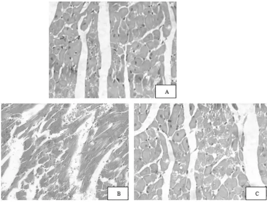
Fig. 1. Pathological and morpho- logical effects of naloxone postcon- ditioning on MIRI rats (HE staining,
×400). (A) Sham group, (B) IR group, (C) Nal M group. Sham group, sham- operated control group; IR, 30 min ischemia prior to 2 h reperfusion;
Nal M, medium-dose naloxone group (1.0 mg/kg).
Group Expression of p-JNK
Sham 0.10±0.01
IR 0.40±0.01**
Nal L 0.18±0.03
##Nal M 0.14±0.02
##Nal H 0.15±0.01
##**Compared with sham group, p<0.01;
##Compared with IR group, p<0.01.
Table 1. Effect of naloxone postconditioning on p-JNK expres- sion of MIRI rats (mean±SD, n=8)
jugated anti-digoxigenin antibody for 30 min, followed by a 0.05% solution of 3,3'-diaminobenzidine tetrahydrochlo- ride for 4 min. For negative controls, TdTenzyme was omitted. Slides were counterstained with methylene blue.
For each myocardial, the number of apoptotic cardiomyo- cytes in 10 non-overlapping renal cortical fields was count- ed under ×400 magnification. The number of TUNEL-pos- itive nuclei was averaged for each field. Myocardial apopto- sis was represented as apoptosis index (AI) calculated as follows: AI=number of TUNEL-positive myocytes/total num- ber of myocytes.
Statistical analysis
Data were presented as mean±standard deviation (SD) and analyzed using Statistical Product and Service Solu- tions (SPSS) version 13.0. Inter-group comparison was done by one-way analysis of variance (one-way ANOVA). Corre- lation analysis was performed using curve estimation.
Differences were considered significant statistically when p<0.05.
RESULTS
Pathological and morphological effects of naloxone postconditioning on MIRI rats
The myocardial structure of rats in the sham group was arranged regularly. The cells presented with normal size, clear boundaries and even coloration (Fig. 1A). The struc- ture of myocardial fibers in the IR group presented aber- rantly and in wavy arrangement. The cells were stained unevenly, some cardiomyocytes showed granular or vacuo- lar degeneration (Fig. 1B). The cardiomyocytes in Nal-treat- ed groups, especially in the high- and medium-dose groups,
presented with mild edema. Few cells showed granular de- generation (Fig. 1C).
Effect of naloxone postconditioning on JNK and p- JNK protein expression in myocardial tissue of MIRI rats
JNK and p-JNK presented as two bands in each treat- ment group. They located at 46 kDa (p-JNK
1) and 54 kDa (p-JNK
2), respectively. The sham group showed weak p-JNK expression. p-JNK expression in the IR group was significantly higher than in the sham group (p<0.01). The p-JNK expression in the Nal L, Nal M, and Nal H groups significantly decreased to 47.6%, 34.5%, and 36.9% of the expression in the IR group, respectively (p<0.01). No sig- nificant difference was observed among these three nalox- one-treated groups (p>0.05; Table 1 and Fig. 2).
Effect of naloxone on myocardial apoptosis in MIRI
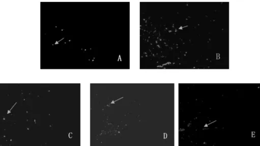
More apoptotic nuclei were found in myocardial tissue
Fig. 3. Effect of naloxone postcon- ditioning on cell apoptosis of MIRI rats (×200). (A) Sham group, (B) IR group, (C) Nal L group, (D) Nal M group, (E) Nal H group. Sham group, sham-operated control; IR, 30 min ischemia prior to 2 h reperfusion;
Nal L, Low-dose naloxone group (0.5 mg/kg); Nal M, Medium-dose nalo- xone group (1.0 mg/kg); Nal H, High- dose naloxone group (2.0 mg/kg).
Fig. 2. Effect of naloxone postconditioning on p-JNK protein expression of MIRI rats. **Compared with sham group: p<0.01;
##