접수번호: 2008-100
Korean Journal of Ophthalmology 2009;23:198-203 ISSN : 1011-8942 DOI : 10.3341/kjo.2009.23.3.198
Reunion of the Rabbit Superior Oblique Tendon After Weakening Procedures
Dae Wook Kang, MD, Ji Hye Oh, MD, Bo Young Chun, MD, Jung Yoon Kwon, MD, PhD
Department of Ophthalmology Kyungpook National University, School of Medicine, Daegu, Korea
Purpose: To investigate the degree of reunion in rabbit eyes of the superior oblique tendon after several surgical weakening procedures.
Methods: A total of 32 rabbits (64 eyes) were used in this study. The rabbits were randomly assigned to four groups, eight rabbits (16 eyes) in the tenotomy group, eight rabbits (16 eyes) in the tenectomy group, eight rabbits (16 eyes) in the disinsertion group and eight rabbits (16 eyes) in the recession group. The degree of reunion or reattachment of the superior oblique tendon on the globe were examined on four eyes in each group at postoperative weeks two, four, six and eight.
Results: At eight weeks, the newly created insertion site remained at the same site in all eyes in the recession group, and the distal end of the superior oblique tendon was reattached at the medial border of the superior rectus muscle in all four eyes in the tenotomy and disinsertion groups, and in three of four eyes in the tenectomy group.
Conclusions: From this experimental study, it was speculated that superior oblique recession is more effective than other superior oblique weakening procedures. This result could be helpful in the prediction of time of recurrence for superior oblique overaction after superior oblique weakening procedures.
Korean J Ophthalmol 2009;23:198-203 ⓒ 2009 by the Korean Ophthalmological Society.
Key Words: Rabbits, Reunion, Superior oblique overaction, Weakening procedures of the superior oblique
Received: October 10, 2008 Accepted: August 5, 2009
Reprint requests to Jung Yoon Kwon, MD. Department of Ophthalmology, Kyungpook National University Hospital, #50 Samdeok-dong 2-ga, Jung- gu, Daegu 700-721, Korea. Tel: 82-53-420-5812, FAX: 82-53-426-6552, E-mail: [email protected]
* This paper was presented in part at the 91th Annual Meeting of the Korean Ophthalmological Society, April, 2004, Pusan, Korea.
Various kinds of superior oblique (SO) weakening procedures such as tenotomy, tenectomy and recession are performed for surgical correction of superior oblique overaction.
1-7In performing superior oblique tenotomy or tenectomy, there is no risk of complication such as scleral perforation; however, recurrence of superior overaction can be seen during follow-up due to reconnection of the cut tendon ends after surgery.
3,6,8,9Recession of the SO muscle is a more controlled and reliable procedure due to graded recession according to superior oblique overaction. However, a major disadvantage of this procedure is the changing of the SO muscle function of depression into one of elevation after creation of a new insertion point located along the equator rather than the anterior portion of the globe.
10-15Recently, a procedure for weakening the SO muscle by leng- thening of the tendon with a silicone band has been developed.
16-18The authors performed four methods for weakening the SO muscle of rabbits and analyzed the degrees of reunion of the SO muscle according to each surgical method.
Materials and Methods
A total of 32 white, New Zealand rabbits (64 eyes) were used in this study.
The subjects were randomly assigned to four groups depen- ding on the type of SO weakening procedure used: eight rabbits (16 eyes) in the tenotomy group, eight rabbits (16 eyes) in the tenectomy group, eight rabbits (16 eyes) in the disinsertion group and eight rabbits (16 eyes) in the recession group.
General anesthesia was attained with intramuscularly ad- ministered ketamine HCL (25 mg/kg) and xylazine HCL (10 mg/kg). During each operation, a conjunctival incision was made radial to the corneoscleral limbus along the medial side of the superior rectus (SR) insertion. Then, separation of the SR and SO was carefully performed, and the SO tendon was exposed near the nasal border of the SR by a muscle hook under direct visualization.
In the disinsertion group, disinsertion of the SO was per-
formed, and careful dissection of the tendon capsule, and sepa-
ration of the SR from the SO was done. In the tenotomy group,
the SO tendon was exposed on a muscle hook under direct
visualization near the nasal border of the SR. After assuring
that the entire tendon was hooked, it was then transected at 2
mm nasal to the nasal border of the SR insertion. In the tenec-
tomy group, the SO was transected at about 4 mm from the nasal
side of the nasal border of the SR insertion. In the recession
group, the SO tendon was disinserted, passed under the SR,
and a 6-0 vicryl suture was secured in the tendon. The tendon
DW Kang, et al. REUNION OF SO TENDON AFTER WEAKENING PROCEDURES
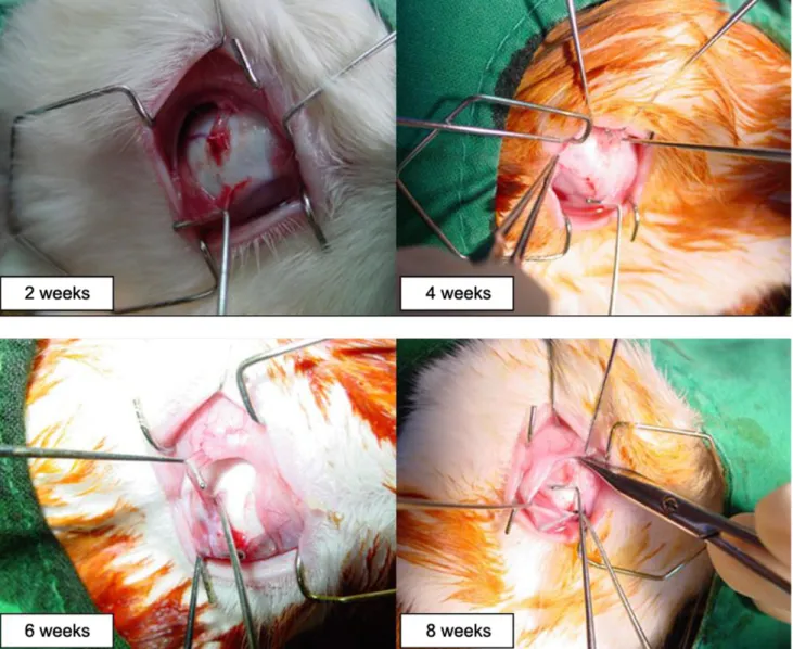
Fig. 1. In the recession group, there was no change in the recessed SO tendon position in all 16 eyes through postoperative week eight.
was then sutured to the sclera at a point located 4 mm nasal to the nasal border of the SR insertion. The conjunctiva was closed using interrupted sutures.
The status of reunion or reattachment of the superior oblique tendon on the globe was examined in four eyes in each group at postoperative weeks two, four, six and eight.
On examination, reopening of the sutured conjunctiva and exploration of the SO was done. Careful dissection around the SR muscle and exposure of the new insertion of the SO were performed. The degree of reunion or anterior displacement was recorded.
Results
In the recession group, there was no change in position of the recessed SO tendon, and all 16 eyes were well positioned through postoperative week eight (Fig. 1).
In the tenotomy group, there was no observable distal end of the tenotomized SO tendon at postoperative week two for
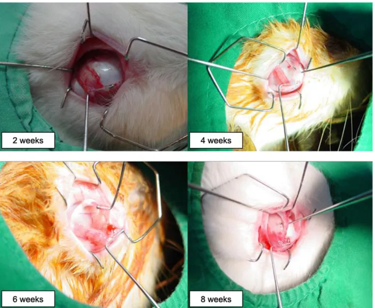
all four eyes. At four weeks, the proximal end of the tenotomized SO tendon was repositioned anteriorly and partially reattached to the sclera and the nasal border of the SR in three eyes. At six weeks, the previously tenotomized end of SO tendon was newly inserted into the nasal border of the SR in three eyes and reattached into the sclera posterior to the previously tenotomized area in one eye. At eight weeks, there was reattachment of the tenotomized ends of the SO tendon into the nasal border of the SR in all four eyes (Fig. 2).
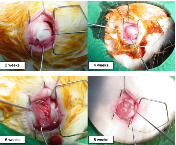
In the tenectomy group, there was no observable distal end of the tenectomized SO tendon at postoperative week two for all four eyes. At four weeks, the tenectomized end of the SO tendon was loosely reattached in a fan-shape to the nasal side of the SR with connective tissue in two 2 eyes. At six weeks, there was reattachment of the tenectomized SO tendon into the nasal border of the SR in two eyes. At eight weeks, there was reattachment of the tenectomized SO tendon with connective tissue along the nasal border of the SR in three eyes (Fig. 3).
In the disinsertion group, there was no identifiable end of the
Korean J Ophthalmol Vol.23, No.3, 2009
Fig. 2. In the tenotomy group, there were no visible cut ends of the SO tendons at postoperative week two in all four eyes (left upper). At four weeks, the proximal end of the SO tendon was repositioned anteriorly and partially reattached to the sclera in three eyes (right upper). At six weeks, there was reattachment of the proximal end of the SO tendon into the nasal border of the SR in four eyes (left lower). At eight weeks, there was reattachment of the proximal end of the SO tendon into the nasal border of the SR in all four eyes (right lower).
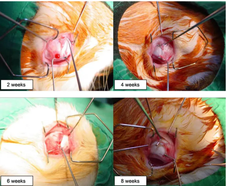
disinserted SO tendon in three eyes; however, loose reattachment of the disinserted SO tendon insertion with connective tissue along with the nasal border of the SR was observed at posto- perative week two in one eye. At four weeks, there was loose reattachment of the disinserted SO tendon to the SR 4 mm posterior to the nasal insertion of the SR in two eyes, loose reattachment of the disinserted SO tendon in the nasal insertion of the SR in one eye, and there was no observable end of the disinserted SO tendon in one eye. At six weeks, there was reattachment of the disinserted SO tendon into the nasal insertion of the SR in four eyes. At eight weeks, the previously disinserted SO tendons of all four eyes were reattached to the SR 2-4 mm posterior to the nasal insertion of the SR (Fig. 4)(Table 1).
D iscussion
Superior oblique tendon weakening procedures have been performed to treat SO overaction and Brown syndrome. In 1946, Berke
19performed SO tenotomy with variable results and several postoperative complications. Probably, this was due to blind hooking of the SO muscle through a superonasal, conjunctival incision. In 1970, Parks and Helveston
20began to perform SO weakening procedures under direct visualization of the SO insertion, and SO tendon recession was first introduced by Ciancia and Diaz.
21In 1974, Prieto-Diaz
22presented a mo- dification of SO recession, which consisted of reinsertion of the tendon behind the equator of the globe in order to avoid frequent complications like a limitation of depression in ab- duction and an over-convergence in extreme downgazing.
Surgeries for SO overaction are relatively less common than
are those for esotropia or exotropia. Superior oblique tendon
weakening procedures are technically difficult due to the location
DW Kang, et al. REUNION OF SO TENDON AFTER WEAKENING PROCEDURES
Fig. 3. In the tenectomy group, there were no visible cut ends of the SO tendons at postoperative week two in all four eyes (left upper). At four weeks, the proximal end of the SO tendon was loosely reattached in a fan-shape to the nasal border of the SR in two eyes (right upper). At six weeks, there was reattachment of the SO tendon into the nasal border of the SR in two eyes (left lower). At eight weeks, there was reattachment of the SO tendon along the nasal border of the SR in three eyes (right lower).
of the superior oblique insertion quite posterior to the globe.
Excessive tissue damage could occur in the attempt to expose and allow direct visualization of the SO, which frequently leads to postoperative adhesions, lid edema, temporary ptosis and severe hemorrhage.
4,10,12,14To prevent these possible compli- cations, it is necessary to avoid blindly hooking the SO muscle and taking extreme caution in handling ocular tissue during direct visualization of the SO insertion.
At present, tenotomy, tenectomy and recession of the SO muscle are the most common procedures for diminishing the action of the SO muscle. Efforts to improve SO weakening surgery have led to the development of such procedures as Z-lengthening, split tendon lengthening, and disinsertion.
1-7However, these methods are not often used because their surgical results are no better than those of tenotomy or tenectomy.
8,10-14,23One major drawback of SO tenotomy and tenectomy is that
they are “all or none” procedures.7 If a large separation occurs
in the cut ends of the tendon, it can lead to SO paresis with in-
cyclotorsion and hypotropia.
11-13It is reported that overcorrection
of the SO tendon by tenotomy or tenectomy is up to 30-85%.
24There is also undercorrection of these procedures due to incom-
plete separation of the SO tendon during surgery or reunion of
the cut ends of the tendon after surgery.
3,6-9,23,25In patients
with bifoveal fusion, intractable cyclovertical diplopia can occur,
and the treatment of this diplopia is very complicated.
6-10,23,26The reason for these complications may be that there is a
lack of control over the amount of separation of the cut ends
of the tendon according to the degree of SO overaction.
7In 1991,
Wright
16introduced a new technique for SO weakening by
elongating the tendon with a segment of a 240 retinal silicone
band. He reported that the use of a silicone expander demon-
strated better results due to the controlled elongation of SO
Korean J Ophthalmol Vol.23, No.3, 2009
Fig. 4. In the disinsertion group, there was loose reattachment of the disinserted SO tendon along the nasal border of the SR at postoperative week two in one eye (left upper). At four weeks, there was loose reattachment of the disinserted SO tendon 4 mm posterior to the nasal insertion of the SR in three eyes (right upper). A six weeks, there was reattachment of the disinserted SO tendon into the nasal insertion of the SR in four eyes (left lower). At eight weeks, the disinserted SO tendons were reattached to the SR 2-4 mm posterior to the nasal insertion of the SR in all four eyes (right lower).
Table 1. The status of reattachment of the superior oblique tendon after weakening procedures
Weakening procedure
Time weeks Two Four
weeks Six weeks Eight weeks
Recession 0/4
*0/4 0/4 0/4
Tenotomy 0/4 3/4 4/4 4/4
Tenectomy 0/4 2/4 2/4 3/4
Disinsertion 1/4 3/4 4/4 4/4
*