Glaucoma is the leading cause of irreversible blind- ness and the second most common cause of blindness after cataract [1]. The main characteristic of glaucoma is a specific optic neuropathy with reduced number of retinal ganglion cells accompanied by characteristic progressive constriction of the visual field [2,3]. Although increased intraocular pressure (IOP) has been considered as a major risk factor for glaucoma [4], there are other concomitant factors affecting the eye such as glutamate excitotoxicity [5], reduced ocular blood flow [6-9], vascular dysregulation [10,11], and reactive oxygen species-related oxidative dam-
age that have been suggested as important etiologic factors in glaucoma pathogenesis [12,13]. The trabecular mesh- work, optic nerve, and retinal ganglion cells are known as the major targets of oxidative damage in the pathological development of glaucoma [14-21].
Why is oxidative stress so important in ocular disease?
Eyes are continually exposed to light in order to collect data from the outer environment, and thus reactive oxygen species are being produced by ionizing or non-ionizing ra- diation when the eyes are open [22]. Reactive oxygen spe- cies are also produced by aerobic metabolism. Even when the eye is in darkness, the retina consumes more oxygen than when it is in light [23]. Ischemia and reperfusion in- jury caused by ocular vascular dysregulation also induce oxidative damage [24,25]. Against this vulnerability to oxi- dative damage, the eye has its own protective mechanism of high levels of both ascorbic acid and glutathione (GSH) [26-32]. Other protective systems including enzymatic Original Article
Circulating Total Glutathione in Normal Tension Glaucoma Patients:
Comparison with Normal Control Subjects
Myoung Hee Park, Jungil Moon
Department of Ophthalmology, Yeouido St. Mary’s Hospital, The Catholic University of Korea School of Medicine, Seoul, Korea
© 2012 The Korean Ophthalmological Society
This is an Open Access article distributed under the terms of the Creative Commons Attribution Non-Commercial License (http://creativecommons.org/licenses /by-nc/3.0/) which permits unrestricted non-commercial use, distribution, and reproduction in any medium, provided the original work is properly cited.
Received: March 5, 2010 Accepted: March 24, 2011
Corresponding Author: Jungil Moon, MD, PhD. Department of Ophthal- mology, Yeouido St. Mary’s Hospital, #10 63(yuksam)-ro, Yeongdeung- po-gu, Seoul 150-713, Korea. Tel: 82-2-3779-1245, Fax: 82-2-3775-1245, E-mail: [email protected]
Purpose: Oxidative stress plays a critical role in the pathogenesis of glaucoma. Glutathione is a major antioxi- dant molecule present in intracellular or extracellular space. Herein, we aimed to examine circulating glutathi- one level in normal tension glaucoma (NTG), which comprises the largest proportion of glaucoma disease in the Korean population.
Methods: Nineteen NTG patients (NTG group) and 30 age- and gender-matched normal control subjects (control group) were included. Antecubital venous puncture was performed between 8 and 10 o’clock in the morning to obtain a 4 mL venous blood sample. Total glutathione level was measured by the spectrophotometric method at 412 nm. Correlation of total glutathione level with mean deviation and pattern standard deviation from the Humphrey visual field test was analyzed in the NTG group.
Results: Total glutathione level in circulating blood was 524.02 ± 231.09 nmol and 586.06 ± 156.08 nmol in the NTG group and the control group, respectively. The difference between these values was not statistically significant (p = 0.121, F = 2.212). Age had no significant effect on circulating total glutathione level in either the NTG group (p = 0.171, r = -0.328) or the control group (p = 0.380, r = -0.166). In the NTG group, circulating total glutathione level had no significant relationship with mean deviation (p = 0.226, F = 1.636) and pattern stan- dard deviation (p = 0.200, F = 1.766) after correcting for age and gender.
Conclusions: In NTG patients, circulating total glutathione levels were not different compared to those of normal subjects.
Key Words: Glutathione, Low tension glaucoma, Oxidative damage
(superoxide dismutase, GSH peroxidase, and catalase) and non-enzymatic antioxidants (cysteines, tocopherols, and retinols) are present in the eye [19]. Under normal physi- ologic conditions, there is a state of equilibrium between the endogenous production of free radicals and their neu- tralization by antioxidant defense mechanisms. If these protective systems fail, damage to the ocular tissues can occur, and various diseases can develop such as cataract [33], age-related macular degeneration [34], and glaucoma [35].
GSH is a major non-enzymatic antioxidant present in both intracellular and extracellular spaces [36,37]. The GSH redox system protects ocular tissues from the damage induced by low H2O2 concentrations through direct and indirect mechanisms [38,39]. A considerable amount of evidence suggests GSH depletion as an important risk fac- tor for glaucoma. In the aqueous humor of primary open angle glaucoma (POAG) patients, activity of antioxidant enzymes, including glutathione peroxidase, is increased and total antioxidant potential is decreased, suggesting that chronic oxidative damage occurs in outflow pathways [40].
An increase in serum autoantibody against glutathione- S-transferase has been reported in glaucoma patients [41].
Genetic polymorphisms have been detected for GSH trans- ferase isoenzyme, and the GSTM1-null genotype has been found to be significantly common in patients with primary open-angle glaucoma compared to controls [42]. Patients with POAG exhibit low levels of circulating glutathione [43]. In an in vivo study, a mouse retina with systemic GSH reduction showed increased retinal neuronal apoptosis [44]
and altered expressions of bcl-2 family mRNA and heat shock protein mRNA [45], suggesting subsequent retinal neuronal protective mechanisms may be changed by sys- temic GSH depletion.
Normal tension glaucoma (NTG) is the most common type of glaucoma in Korea and Japan [46-48]. This is unique in comparison with Western populations. The prev- alence of NTG is about 3.5-fold higher than that of POAG with increased IOP in Korea (The Namil Study, the Kore- an Glaucoma Society) [48]. However, the pathophysiologic mechanism of NTG is little known. Vascular dysregula- tion in the optic nerve head and some influence of high normal intraocular pressure are known as mild risk factors [10,11,49]. Although IOP is still the major target of treat- ment for NTG in current practice [50], attention is shifting to other modalities such as neuroprotection, improvement of ocular blood flow, and antioxidant supplementation [51].
Moreover, some patients with POAG are also facing these new forms of therapy due to the progression of optic neu- ropathy despite well-controlled IOP [52] due to the multi- factorial etiology of glaucoma.
Therefore, information about the status of systemic GSH level in NTG will help to elucidate the role of oxidative stress in the disease progression of NTG. The purpose of
this study was to determine the status of circulating total GSH levels in patients with normal tension glaucoma and to compare levels with those of people without glaucoma.
Materials and Methods
Study sample
Patients with NTG attending the glaucoma clinic of the Catholic Eye Center at St. Mary’s Hospital (Seoul, Korea) between June 2008 and October 2008 were considered for inclusion in this prospective study. Ethical approval was obtained from the committee of St. Mary’s Hospital insti- tutional review board, and written informed consent was received from all subjects before entry into the study.
Patients were diagnosed as having NTG if IOP measure- ments were less than 22 mmHg by Goldmann applanation tonometry, characteristic glaucomatous cupping of the op- tic disc was observed on funduscopic examination, normal open anterior chamber angles was seen on gonioscopy, and repeatable visual field defects consistent with the diagnosis of glaucoma were observed in the results obtained with program 24-2 of the Humphrey field analyzer (Carl Zeiss Meditec, Dublin, CA, USA). The study’s ocular exclusion criteria included any IOP measurement greater than 21 mmHg, narrow iridocorneal angles, evidence of secondary open-angle glaucoma, history of previous intraocular sur- gery, and other nonglaucomatous ocular diseases including cataract, diabetic retinopathy (DR) and age-related macu- lar degeneration (ARMD). The control group was recruited from a group of age- and gender-matched volunteers and was composed of subjects who had never had glaucoma or other ocular diseases such as cataract, DR, or ARMD.
Other exclusion criteria for both groups were smoking and a history of any chronic systemic disease with pre- sumed low GSH level, including autoimmune diseases, alcoholic liver disease, cancer, and diabetes mellitus. After this selection, the experimental group was narrowed to 19 patients with NTG, and that control group contained 30 subjects.
Blood sampling
Subjects were instructed to fast from midnight to 8 a.m. on the morning of the test. All blood samples were obtained by a qualified registered nurse in the morning, between 8 and 10 a.m. Four milliliters of blood were col- lected in EDTA-treated tubes (to prevent oxidation) by venipuncture to the antecubital vein. One thousand micro- liters of blood was then transferred into a centrifuge tube for initial processing. Red blood cells (RBC) were sepa- rated from plasma by centrifugation at 600 ×g for 10 min- utes. The pellet RBCs were then transferred into another microcentrifuge tube and washed twice with 3 volumes of
phosphate buffered saline (pH = 7.4). A 200 microliter ali- quot of RBC pellets was placed in another microcentrifuge tube, to which 200 microliters of 5% 5-sulfosalicylic acid (SSA) were added and then incubated for 10 minutes at 4˚C. After centrifugation at 10,000 ×g for 10 minutes, 100 microliters of the supernatant were immediately cooled at -70˚C. All of these procedures were performed within 2 hours from the time of blood collection. The next steps of the GSH analysis were performed within 10 days.
Glutathione analysis
Total GSH levels were assessed by the 5, 5’-dithiobis-(2- nitrobenzoic acid) recycling procedure, as described in a previous study [52]. The glutathione assay kit from Sigma (Glutathione assay kit, product code CS0260; St Louis, MO, USA) was used.
A standard curve from 3.125 to 50 nanomoles in double- fold increments using a GSH standard solution was pre- pared for each 96-well plate. The standards contained the same final concentrations of SSA as used for the samples.
To each well of a 96-well plate, 150 microliters of working buffer and 10 microliters of standard or sample were added in duplicate, and the plate was incubated at room tempera- ture for 5 minutes. Finally, 50 microliters of 0.16 mg/mL nicotinamide adenine dinucleotide phosphate (NADPH) was added, and the plate was read at 412 nm using a 96- well plate reader. A standard curve was then generated by linear regression.
Statistical analysis
The statistical analysis was performed on a computer equipped with SPSS ver. 12 (SPSS Inc., Chicago, IL, USA).
Data are expressed as the mean ± standard deviation. Dif- ferences between groups at baseline for age, mean devia- tion (MD) and pattern standard deviation (PSD) were cal- culated using Student’s t-test. Difference between groups at baseline for gender was calculated using the χ2-test (Fisher’s exact test). Differences between the two study groups in blood GSH level were computed by analysis of covariance using age as a cofactor. Differences between males and females in blood GSH level were computed by
Student’s t-test for each study group. A correlation was performed to determine the association of age and blood GSH level. A multiple regression was performed to test the influences of age, gender, and blood GSH level on MD and PSD from the 24-2 test pattern of the Humphrey field ana- lyzer. Results with p = 0.05 were considered statistically significant.
The minimum sample size needed was calculated from the formula [(u + v)(σ12 + σ22) / (μ1 - μ2)2] with 90% power and a 5% level of significance. The sample size needed was determined to be 18 for each group [(1.28 + 1.96)2(1002 + 1002) / (300 - 200)2] using the reported data [44].
Results
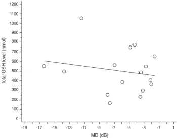
Nineteen patients with NTG and 30 normal control sub- jects were included in this study. The demographics of the study groups are described in Table 1. There was no signif- icant difference in age (p = 0.056) or gender (p = 0.337, χ2
= 0.921) distribution between the NTG group and control group. There were significant differences in MD (p = 0.000) and PSD (p = 0.000) between the two groups.
Total glutathione level in circulating blood was 524.02 ±
Table 1. Patient demographics (mean ± SD)
Control NTG p-value
No. of patients 30 19
Age (yr) 52.70 ± 6.70 56.26 ± 8.48 0.056
Sex (male : female) 20 : 10 10 : 9 0.377
MD -0.75 ± 1.24 -5.66 ± 4.32 0.000
PSD 1.58 ± 0.94 5.06 ± 4.03 0.000
Statistical analysis was performed with Student’s t-test (age, MD, and PSD) and Fisher’s exact test (sex).
NTG = normal tension glaucoma; MD = mean deviation; PSD = pattern standard deviation.
Total GSH level (nmol)
1100 1000 900 800 700 600 500 400 300 200 100
Control NTG
Fig. 1. Median and interquartile range values of total circulating glutathione (GSH) level in the control group and normal tension glaucoma (NTG) group. Total GSH levels were not statistically different between the two groups (p = 0.121, F = 2.212).
231.09 nmol and 586.06 ± 156.08 nmol in the NTG group and the control group, respectively. Circulating total GSH levels were not statistically different between the NTG group and control group (p = 0.121, F = 2.212) (Fig. 1). In the NTG group, the mean glutathione level was 589.41
± 239.64 nmol in males and 451.35 ± 210.39 nmol in fe- males. In the control group, the mean glutathione level was 618.98 ±208.16 nmol in males and 569.61 ± 125.70 nmol in females. There was no statistically significant differ- ence in total GSH level between males and females in the NTG group (p = 0.424) (Fig. 2) or in the control group (p
= 0.202) (Fig. 3). There was no significant assotciation be-
tween age and total GSH level in the NTG group (p = 0.171, r = -0.328) (Fig. 4) or in the normal group (p = 0.380, r = -0.166) (Fig. 5). In the NTG group, after correcting for age and gender influences on systemic total GSH level, there was no significant association between GSH level and vi- sual field parameter (MD: p = 0.226, R2 = 0.260; PSD: p = 0.200, R2 = 0.275) (Figs. 6 and 7).
Discussion
Glutathione is normally present in the cornea, lens, tra- becular meshwork, ciliary body, and retina. Changes of
Total GSH level (nmol)
Age (yr)
40 45 50 55 60 65 70
1100 1000 900 800 700 600 500 400 300 200
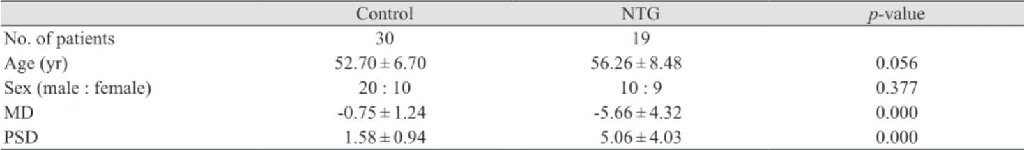
Fig. 5. Correlation between total glutathione (GSH) level and age in the control group. There was no significant association be- tween total GSH level and age (r = -0.166, p = 0.380).
Total GSH level (nmol)
Control male Control female 1100
1000 900 800 700 600 500 400 300 200
Total GSH level (nmol)
1100 1000 900 800 700 600 500 400 300 200 100
NTG male NTG female
Fig. 2. Median and interquartilic range values of total glutathione (GSH) level in the normal tension glaucoma (NTG) group; com- parison between male and female patients. There was no signifi- cant difference between males and females (p = 0.424) in total circulating GSH level.
Total GSH level (nmol)
1100 1000 900 800 700 600 500 400 300 200 100
40 45 50 55 60 65 70
Age (yr)
Fig. 4. Correlation between total glutathione (GSH) level and age in year in the normal tension glaucoma group. There was no sig- nificant association between total GSH level and age (r = -0.328, p = 0.171).
Fig. 3. Median and interquartilic range values of total glutathione (GSH) level in the control group; comparison between male and female patients. There was no significant difference between males and females (p = 0.202) in total circulating GSH level.
glutathione and related enzyme activity in the lens, cornea, retina, and other eye tissues occur with aging, cataract, diabetes, irradiation, and administration of some drugs.
Many ocular diseases including keratoconus, cataract, glaucoma, diabetic retinopathy, and age-related macular degeneration may occur by oxidative damage induced by an abnormal glutathione redox state.
In terms of glaucoma, the major interest lies on two specific structures, the trabecular meshwork and retina, because the trabecular meshwork is a rate-limiting struc- ture of aqueous outflow pathway controlling intraocular pressure, and the retina is one of the target structures of glaucomatous optic neuropathy.
Studies on the calf trabecular meshwork (TM) have shown the presence of glutathione, glutathione reductase, and glucose-6-phosphate dehydrogenase, indicating that the TM is well supplied with the capacity to generate and use NADPH to maintain GSH. It has been supposed that GSH is able to protect the TM against H2O2-induced oxi- dative damage, which would decrease the aqueous humor outflow [19]. Altered GSH and GSH activities have also been reported in the trabecular meshwork and aqueous humor of patients with glaucoma [40,41,53-56]. All of these results imply that reduced activity of GSH in the TM may influence aqueous outflow facility and thus elevate IOP.
Additional studies of the role of glutathione in glau- coma pathogenesis are related to studies of the trabecular meshwork. However, the retina is a major target organ in glaucoma pathogenesis, and glutathione also exists in the retina. Therefore, abnormal glutathione function within the retina may play an important role in glaucoma.
Although the function of glutathione in the retina is not
fully understood, there is some evidence that it may have a role in neuroprotection under harmful conditions such as ischemia and oxidative damage. In the retina, glutathione is predominately localized to Muller cells and horizontal cells. According to a study by Schutte and Werner [57], us- ing immunocytochemical methods for glutathione, post- mortem ischemic injury longer than 10 minutes resulted in strong labeling of neurons, particularly of retinal ganglion cells, whereas Muller cells were essentially devoid of im- munoreactivity. This means that glutathione molecules were transferred from Muller cells to retinal ganglion cells to protect ganglion cells from ischemic injury. In a study by Carter-Dawson et al. [58], Muller cells in glaucomatous monkey retinas showed significantly greater immunoreac- tivity for glutathione than control retinas, increasing with the duration of elevated intraocular pressure.
According to our previous study, a 15% reduction of to- tal retinal GSH level by intraperitoneal injection of buthio- nine sulfoximine caused retinal neuronal apoptosis in mice [44]. To reach this level of GSH in the retina, circulating total GSH might need to be lowered even further. In the same mouse, total GSH level in the heart was reduced to 60% of that of the control mouse (unpublished data). We hypothesized that systemic total glutathione level might be changed in NTG patients in whom there may be ad- ditional factors other than increased IOP for glaucomatous damage. If there is little influence of IOP on glaucomatous optic neuropathy in NTG pathogenesis, and if oxidative damage is one of the major risk factors for NTG, the anti- oxidant capacity of GSH might have been changed in NTG patients. However, as the results of this study show, there was no difference in circulating GSH level between the
Total GSH level (nmol)
PSD (dB) 1200
1100 1000 900 800 700 600 500 400 300 200 100 0
0 1 2 3 4 5 6 7 8 9 10 11 12 13 14 15
Fig. 7. Correlation between total glutathione (GSH) level and pattern standard deviation (PSD) in the normal tension glaucoma group. After correcting for age and gender influence, there was no significant association between GSH level and PSD (p = 0.200, R2 = 0.275).
Total GSH level (nmol)
MD (dB) 1200
1100 1000 900 800 700 600 500 400 300 200 100 0
-19 -17 -15 -13 -11 -9 -7 -5 -3 -1 1
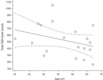
Fig. 6. Correlation between total glutathione (GSH) level and mean deviation (MD) in the normal tension glaucoma group.
After correcting for age and gender influence, there was no sig- nificant association between GSH level and MD (p = 0.226, R2 = 0.260).
NTG group and control group. This difference in results between human and animal subjects is possibly due to the difference in background factors such as genetic homoge- neity and diet.
Systemic glutathione level can change within a day and is affected by diet and medications. We obtained blood samples at a fixed time (8 a.m.) after fasting, as in the pre- vious report [43], in order to reduce the sampling bias.
Gherghel et al. [43] demonstrated that patients with POAG exhibit low levels of circulating glutathione, sug- gesting a general compromise of the antioxidative defense.
In Gherghel’s study, total GSH level in the POAG group was about 75% (225.27 ± 83.03) of that of the control group (332.23 ± 98.50). In addition to the IOP range, there is one more thing we should consider about the difference in results between Gherghel’s study and ours. In contrast to Caucasians, Korean and Japanese people have a higher prevalence of NTG than POAG with a 3.5-fold elevation in pressure. In Choe and Hong [46]’s study of Koreans, the estimated prevalence of POAG in the population over 20 years of age was 2.04%, and the prevalence of NTG was 1.71% (83.8% of POAG). In Iwase et al. [47]’s study of Japa- nese individuals, the estimated prevalence of POAG in the population older than 40 years of age was 3.9%, and the prevalence of cases of POAG with IOP levels of 21 mmHg or less was 3.6% (92.3% of POAG). Korean and Japanese people might have different risk factors compared to Cau- casian populations. Different racial characteristics may lead to different results. These issues illustrate the impor- tance of further studies of POAG with elevated IOP in Ko- rea because of the absence of data on systemic glutathione levels in Korean POAG patients.
Although there were no statistically significant differ- ences in total GSH level between the two groups in this study, the range of total GSH level in the NTG group showed a wider distribution compared to that of the con- trol group. The proportions of patients with very low total GSH level and with very high total GSH level were larger in the NTG group. Considering the smaller sample size of the NTG group, this characteristic might be more meaningful. From this point of view, we can also hypoth- esize that the homeostasis of systemic glutathione-related antioxidant capacity may be inadequately controlled in the NTG patient. Both a low level and a high level of total GSH may be related to insufficient anti-oxidant activity against reactive oxygen species.
It is known that blood GSH level is influenced by gen- der, with men demonstrating higher levels than women.
Our results found that mean total GSH level was slightly higher in men then in women (NTG group, 589.41 ± 239.64 nmol in males and 451.35 ± 210.39 nmol in females; control group, 618.98 ± 208.16 nmol in males and 569.61 ± 125.70 nmol in females), but there were no significant differences in blood GSH level between men and women in either the
NTG group (p = 0.424) or the control group (p = 0.202).
This might be due to either the small sample size or racial characteristics. There are few data on normal circulating GSH levels in the Korean population [59,60]. Further popu- lation-based studies are needed to fully explain the results of this study.
Age is known as one of the influencing factors on sys- temic GSH level. It has been reported that about half of healthy elderly people show low blood GSH level. A nega- tive correlation between age and blood GSH level has also been reported. Our results demonstrated a slightly decreas- ing trend of GSH level as age increases but found no sig- nificant correlation between age and total circulating GSH level in either the NTG group or control group. This might be also due to the small sample size.
We also expected that if the systemic total GSH level is related to the range of glaucomatous optic neuropathy, then the extent of visual field damage in NTG patients might be proportional to the decrement of total GSH level. But the results of this study also showed no significant relation between the MD and PSD of the visual field test and total GSH level. We think this might be due to the finding of no significant difference in total GSH level. Since there was no difference of total GSH level between the NTG group and normal subject group, the extent of visual function may not have been influenced by GSH level. Another pos- sibility is that there might be a change in circulating GSH only in patients with fast progression of optic nerve dam- age. If we had had a greater number of patients, we could have divided patients into two groups with progressing and non-progressing optic nerve damage and compared them.
However, because normal tension glaucoma is a slowly progressing disease in most patients, this would require enormous time and effort to recruit a sufficient number of volunteers with progressive disease.
A major concern of this study is that the study results came from only one measurement of serum glutathione level. Therefore, these results might not be able to represent the long-term status of patients. However, because there are few reports about the relationship between normal tension glaucoma and glutathione, this study represents a meaningful step toward understanding the pathophysiol- ogy of normal tension glaucoma.
Conflict of Interest
No potential conflict of interest relevant to this article was reported.
Acknowledgements
This study was supported in part by a research grant from the Investigator-Initiated Studies Program of Merck
& Co., Inc. The opinions expressed in this paper are those
of the authors and do not necessarily represent those of Merck & Co., Inc.
References
1. Quigley HA, Broman AT. The number of people with glaucoma worldwide in 2010 and 2020. Br J Ophthalmol 2006;90:262-7.
2. Liesegang TJ. Glaucoma: changing concepts and future directions. Mayo Clin Proc 1996;71:689-94.
3. Quigley HA, Green WR. The histology of human glau- coma cupping and optic nerve damage: clinicopathologic correlation in 21 eyes. Ophthalmology 1979;86:1803-30.
4. Krakau CE. Intraocular pressure elevation-cause or effect in chronic glaucoma? Ophthalmologica 1981;182:141-7.
5. Shen F, Chen B, Danias J, et al. Glutamate-induced glu- tamine synthetase expression in retinal Muller cells after short-term ocular hypertension in the rat. Invest Ophthal- mol Vis Sci 2004;45:3107-12.
6. Chung HS, Harris A, Evans DW, et al. Vascular aspects in the pathophysiology of glaucomatous optic neuropathy.
Surv Ophthalmol 1999;43 Suppl 1:S43-50.
7. Flammer J, Orgul S, Costa VP, et al. The impact of ocular blood flow in glaucoma. Prog Retin Eye Res 2002;21:359- 8. Butt Z, O’Brien C, McKillop G, et al. Color Doppler imag-93.
ing in untreated high- and normal-pressure open-angle glaucoma. Invest Ophthalmol Vis Sci 1997;38:690-6.
9. Findl O, Rainer G, Dallinger S, et al. Assessment of optic disk blood flow in patients with open-angle glaucoma. Am J Ophthalmol 2000;130:589-96.
10. Flammer J, Haefliger IO, Orgul S, Resink T. Vascular dys- regulation: a principal risk factor for glaucomatous dam- age? J Glaucoma 1999;8:212-9.
11. Flammer J, Mozaffarieh M. What is the present patho- genetic concept of glaucomatous optic neuropathy? Surv Ophthalmol 2007;52 Suppl 2:S162-73.
12. Moreno MC, Campanelli J, Sande P, et al. Retinal oxidative stress induced by high intraocular pressure. Free Radic Biol Med 2004;37:803-12.
13. Galassi F, Renieri G, Sodi A, et al. Nitric oxide proxies and ocular perfusion pressure in primary open angle glaucoma.
Br J Ophthalmol 2004;88:757-60.
14. Alvarado J, Murphy C, Polansky J, Juster R. Age-related changes in trabecular meshwork cellularity. Invest Oph- thalmol Vis Sci 1981;21:714-27.
15. Alvarado J, Murphy C, Juster R. Trabecular meshwork cel- lularity in primary open-angle glaucoma and nonglauco- matous normals. Ophthalmology 1984;91:564-79.
16. Zhou L, Li Y, Yue BY. Oxidative stress affects cytoskel- etal structure and cell-matrix interactions in cells from an ocular tissue: the trabecular meshwork. J Cell Physiol 1999;180:182-9.
17. Sacca SC, Pascotto A, Camicione P, et al. Oxidative DNA damage in the human trabecular meshwork: clinical corre- lation in patients with primary open-angle glaucoma. Arch Ophthalmol 2005;123:458-63.
18. Wang N, Chintala SK, Fini ME, Schuman JS. Activation of a tissue-specific stress response in the aqueous outflow pathway of the eye defines the glaucoma disease pheno- type. Nat Med 2001;7:304-9.
19. Sacca SC, Izzotti A, Rossi P, Traverso C. Glaucoma- tous outflow pathway and oxidative stress. Exp Eye Res 2007;84:389-99.
20. Izzotti A, Bagnis A, Sacca SC. The role of oxidative stress in glaucoma. Mutat Res 2006;612:105-14.
21. Ko ML, Hu DN, Ritch R, Sharma SC. The combined ef- fect of brain-derived neurotrophic factor and a free radical scavenger in experimental glaucoma. Invest Ophthalmol Vis Sci 2000;41:2967-71.
22. Tanito M, Nishiyama A, Tanaka T, et al. Change of redox status and modulation by thiol replenishment in reti- nal photooxidative damage. Invest Ophthalmol Vis Sci 2002;43:2392-400.
23. Wangsa-Wirawan ND, Linsenmeier RA. Retinal oxy- gen: fundamental and clinical aspects. Arch Ophthalmol 2003;121:547-57.
24. Siskova A, Wilhelm J. The effects of hyperoxia, hypoxia, and ischemia/reperfusion on the activity of cytochrome oxidase from the rat retina. Physiol Res 2001;50:267-73.
25. Hirose F, Kiryu J, Miyamoto K, et al. In vivo evaluation of retinal injury after transient ischemia in hypertensive rats.
Hypertension 2004;43:1098-102.
26. Hanashima C, Namiki H. Reduced viability of vascular endothelial cells by high concentration of ascorbic acid in vitreous humor. Cell Biol Int 1999;23:287-98.
27. Brubaker RF, Bourne WM, Bachman LA, McLaren JW.
Ascorbic acid content of human corneal epithelium. Invest Ophthalmol Vis Sci 2000;41:1681-3.
28. Dreyer R, Rose RC. Lacrimal gland uptake and metabolism of ascorbic acid. Proc Soc Exp Biol Med 1993;202:212-6.
29. Ringvold A, Anderssen E, Kjonniksen I. Distribution of ascorbate in the anterior bovine eye. Invest Ophthalmol Vis Sci 2000;41:20-3.
30. Giblin FJ, McCready JP, Kodama T, Reddy VN. A direct correlation between the levels of ascorbic acid and H2O2 in aqueous humor. Exp Eye Res 1984;38:87-93.
31. Richer SP, Rose RC. Water soluble antioxidants in mamma- lian aqueous humor: interaction with UV B and hydrogen peroxide. Vision Res 1998;38:2881-8.
32. Kahn MG, Giblin FJ, Epstein DL. Glutathione in calf tra- becular meshwork and its relation to aqueous humor out- flow facility. Invest Ophthalmol Vis Sci 1983;24:1283-7.
33. Truscott RJ. Age-related nuclear cataract-oxidation is the key. Exp Eye Res 2005;80:709-25.
34. Yildirim O, Ates NA, Tamer L, et al. Changes in antioxi- dant enzyme activity and malondialdehyde level in patients with age-related macular degeneration. Ophthalmologica 2004;218:202-6.
35. Chen JZ, Kadlubar FF. A new clue to glaucoma pathogen- esis. Am J Med 2003;114:697-8.
36. Dringen R. Glutathione metabolism and oxidative stress in neurodegeneration. Eur J Biochem 2000;267:4903.
37. Hall AG. Review: the role of glutathione in the regulation of apoptosis. Eur J Clin Invest 1999;29:238-45.
38. Riley MV. Physiologic neutralization mechanisms and the response of the corneal endothelium to hydrogen peroxide.
CLAO J 1990;16(1 Suppl):S16-21.
39. Costarides AP, Riley MV, Green K. Roles of catalase and the glutathione redox cycle in the regulation of anterior- chamber hydrogen peroxide. Ophthalmic Res 1991;23:284- 40. Ferreira SM, Lerner SF, Brunzini R, et al. Oxidative stress 94.
markers in aqueous humor of glaucoma patients. Am J Ophthalmol 2004;137:62-9.
41. Yang J, Tezel G, Patil RV, et al. Serum autoantibody against glutathione S-transferase in patients with glaucoma. Invest Ophthalmol Vis Sci 2001;42:1273-6.
42. Unal M, Guven M, Devranoglu K, et al. Glutathione S
transferase M1 and T1 genetic polymorphisms are related to the risk of primary open-angle glaucoma: a study in a Turkish population. Br J Ophthalmol 2007;91:527-30.
43. Gherghel D, Griffiths HR, Hilton EJ, et al. Systemic re- duction in glutathione levels occurs in patients with pri- mary open-angle glaucoma. Invest Ophthalmol Vis Sci 2005;46:877-83.
44. Roh YJ, Moon C, Kim SY, et al. Glutathione depletion induces differential apoptosis in cells of mouse retina, in vivo. Neurosci Lett 2007;417:266-70.
45. Park JW, Moon C, Yun S, et al. Differential expression of heat shock protein mRNAs under in vivo glutathione depletion in the mouse retina. Neurosci Lett 2007;413:260- 46. Choe YJ, Hong YJ. The prevalence of glaucoma in Korean 4.
careermen. J Korean Ophthalmol Soc 1993;34:153-8.
47. Iwase A, Suzuki Y, Araie M, et al. The prevalence of pri- mary open-angle glaucoma in Japanese: the Tajimi Study.
Ophthalmology 2004;111:1641-8.
48. Kim CS, Seong GJ, Lee NH, et al. Prevalence of primary open-angle glaucoma in central South Korea the Namil study. Ophthalmology 2011;118:1024-30.
49. Comparison of glaucomatous progression between un- treated patients with normal-tension glaucoma and patients with therapeutically reduced intraocular pressures: Col- laborative Normal-Tension Glaucoma Study Group. Am J Ophthalmol 1998;126:487-97.
50. The effectiveness of intraocular pressure reduction in the treatment of normal-tension glaucoma: Collaborative Normal-Tension Glaucoma Study Group. Am J Ophthalmol 1998;126:498-505.
51. Neuroprotection and other investigational drugs. In:
Allingham RR, Shields MB, editors. Shields’ textbook of glaucoma. Philadelphia: Lippincott Williams & Wilkins;
2005. p. 512-5.
52. Brubaker RF. Delayed functional loss in glaucoma: LII Edward Jackson Memorial Lecture. Am J Ophthalmol 1996;121:473-83.
53. Akerboom TP, Sies H. Assay of glutathione, glutathione disulfide, and glutathione mixed disulfides in biological samples. Methods Enzymol 1981;77:373-82.
54. Nguyen KP, Weiss H, Karageuzian LN, et al. Glutathione reductase of calf trabecular meshwork. Invest Ophthalmol Vis Sci 1985;26:887-90.
55. Levin LA, Clark JA, Johns LK. Effect of lipid peroxidation inhibition on retinal ganglion cell death. Invest Ophthalmol Vis Sci 1996;37:2744-9.
56. Izzotti A, Sacca SC, Cartiglia C, De Flora S. Oxidative deoxyribonucleic acid damage in the eyes of glaucoma pa- tients. Am J Med 2003;114:638-46.
57. Schutte M, Werner P. Redistribution of glutathione in the ischemic rat retina. Neurosci Lett 1998;246:53-6.
58. Carter-Dawson L, Shen FF, Harwerth RS, et al. Glutathi- one content is altered in Muller cells of monkey eyes with experimental glaucoma. Neurosci Lett 2004;364:7-10.
59. Kim SH, Park HM, Seo JH, Hur M. The change of blood reduced glutathione according to postmenopausal HRT:
GSH as a marker of antioxidant effect of the sex steroids. J Korean Soc Menopause 1997;3:116-25.
60. Park DK, Jeong SK, Chung MG, et al. Glutathione levels in Helicobacter pylori-infected gastric mucosa. Korean J Gastroenterol 2003;42:267-73.