원 저 Korean Circulation J 2003;33(3):212-217
비가역적 급만성 간질환 환자들에서 간이식 후 폐내 단락의 변화
성균관대학교 의과대학 내과학교실 삼성서울병원 심혈관센터,1 소화기내과학교실,2 일반외과학교실3
김병진
1·이경진
1·남궁준
1·이상철
1·박승우
1·고광철
2·백승운
2이광웅
3·조재원
3·이석구
3·이상훈
1·홍경표
1·박정의
1·서정돈
1Change of Intrapulmonary Shunt after Liver Transplantation in Irreversible Acute or Chronic Liver Diseases
Byung Jin Kim, MD1, Kyung Jin Lee, MD1, June Namgung, MD1, Sang-Chol Lee, MD1,
Seung Woo Park, MD1, Kwang Cheol Koh, MD2, Seung Woon Paik, MD2, Kwang Woong Lee, MD3, Jae Won Joh, MD3, Suk Koo Lee, MD3, Sang Hoon Lee, MD1,
Kyung Pyo Hong, MD1, Jeong Euy Park, MD1 and Jung Don Seo, MD1
1Department of Medicine, Cardiac and Vascular Center, 2Division of Gastroenterology and
3Department of General Surgery, Sungkyunkwan University School of Medicine, Samsung Medical Center, Seoul, Korea
ABSTRACT
Background and Objectives:The intrapulmonary shunt, after a liver transplantation, is expected to improve, but the frequency, and predictors, of improvement are not well known. We studied the frequency of the improvement in the intrapulmonary shunt, and the relation between this improvement and the clinical parameters, using contrast echocardiography in liver transplant candidates. Subjects and Methods:Preoperative and postoperative contrast echocardiography, with intravenous agitated saline, was performed in 25 liver transplant candidates (M:F=19:
6, mean age=46±10). The extent of the intrapulmonary shunt was semi-quantitatively assessed on a grade scale from 0 to 4+, according to the degree of appearance of the contrast in the left heart chambers. The etiology of the underlying chronic liver disease, Child-Pugh classification scores, pulmonary function tests and arterial blood gas analyses were all evaluated. The change in the shunt grade after the liver transplantation, and the association of these changes, with clinical parameters, were also evaluated. Results:Twenty-four (96%) of the 25 patients showed positive results for the intrapulmonary shunt on preoperative contrast echocardiography. At follow up, the shunt was visible in only 5 (21%) patients. All 24 patients whose preoperative echocardiography revealed a positive intrapulmonary shunt showed an improvement in the intrapulmonary shunt after the liver transplantation. There was a significant correlation between the improvements in the shunt and the Child-Pugh classification scores (r=0.59, p=0.02). Conclusion:Contrast echocardiography is a feasible, noninvasive, method for the detection of changes in the intrapulmonary shunt before and after a liver transplantation, and the intrapulmonary shunt is improved after a liver transplantation in most patients. (Korean Circulation J 2003;33 (3):212-217)
KEY WORDS:Echocardiography;Intrapulmonary shunt;Liver transplantation.
논문접수일:2002년 11월 26일 수정논문접수일:2003년 1월 9일 심사완료일:2003년 1월 29일
교신저자:이상철, 135-710 서울 강남구 일원동 50번지 성균관대학교 의과대학 내과학교실 삼성서울병원 심혈관센터 전화:(02) 3410-3868, 3410-3419·전송:(02) 3410-3849·E-mail:[email protected]
서 론
폐내 단락은 비가역적인 급성 및 만성 간질환 환자 들에서 볼 수 있는 병태 소견으로 1884년 Flu¨ckiger1) 에 의해 처음 소개된 이후 이의 진단 방법 및 간과 폐 사이의 연관성에 대한 연구들이 진행되어 왔다. 이러한 폐내 단락을 진단하는 방법으로 과거에는 Tc99m- macroaggregated albumin scintigraphy2)나 폐동맥 혈관 조영술3)이 이용되어 왔다. 그러나 폐관류 주사는 폐내 단락의 정량화가 가능한 반면에 예민도가 낮으며, 폐동맥 혈관 조영술은 침습적인 진단 방법이라는 제한 점 때문에, 최근 임상에서 근접하기 쉽고 비침습적인 조영 심초음파를 이용한 검사 방법이 널리 이용되고 있다.4-8)
간이식은 내과적 치료에 반응을 보이지 않는 비가역 적인 급성 및 만성 간질환 환자들에서 임상적인 호전과 생명을 연장시키는 절대적인 치료 방법이다.9) 그러나 간이식 후 폐내 단락의 변화의 정도 및 이를 예측할 수 있는 인자들에 대한 연구들은 미흡한 실정으로, 저자들 은 간이식 전후 조영 심 초음파 검사를 이용하여 이식 후 폐내 단락의 변화의 정도와 폐내 단락의 변화와 이 식 전후의 임상 지표들과의 연관성에 대해 알아보고자 하였다.
대상 및 방법
대 상
2001년 2월부터 2002년 7월까지 본원에서 간이식 수술을 시행 받은 25명의 비가역적 급성 및 만성 간질 환 환자들(평균연령 46±10세, 남 19명, 여 6명)을 대 상으로 하였다. 이들의 기저질환은 B형 간염에 의한 간 경변이 21명(84%)으로 가장 많았다(Table 1). 본 연 구에서는 활동성 상부 위장관 출혈, 동반된 심장판막 질
환과 관동맥 질환, 원발성 폐실질 질환 및 의미 있는 흉 수가 있는 환자들은 연구 대상에서 제외시켰다.
방 법
조영 심 초음파 검사는 5cc 생리식염수를 담은 10cc 주사기와 3-way 연결관을 통해 공기 1cc정도를 담은 10cc 주사기를 여러 차례 세게 흔든 후 좌완의 전주와 정맥으로 주입한 후 4 MHz의 harmonic imaging(Acou- son)으로 apical 4 chamber view에서 실시하였다. 폐 내 단락의 발견은 주입된 미세기포가 우 심장에 관찰된 후 4에서 6박동째 좌 심장에 관찰될 때로 정의하였고, 폐내 단락의 정도는 Vedrinne 등11)이 보고한 폐내 단 락의 정도에 따라 Grade 0부터 Grade Ⅳ까지 분류하 였다(Fig. 1). 조영 심 초음파 검사는 간이식 수술 전 1 개월에서 2개월 사이에 시행하였고 조영 심 초음파 검사 를 시행한 당일, 간 기능 검사, 혈액 응고 검사, 동맥혈 가 스 검사, 폐 기능 검사 및 진찰 소견을 이용하여 Child- Pugh classification의 점수를 계산하여 5점부터 15점 까지 산출하였다. 또한 간이식 후 폐내 단락의 변화를 확인하기 위해 수술 후 1개월에서 11개월 사이에 상기 한 검사들을 동일하게 시행하였다.
통계분석
간이식 전후의 임상 지표들의 변화의 비교에는 Wil- coxon matched-pairs signed-ranks test를 이용하였 다. 간이식 전후의 폐내 단락의 변화 정도와 임상 지표 들의 호전과의 상관 관계는 Spearman’s bivariate co- rrelation을 이용하였고 Spearman’s partial correlation 을 이용하여 다른 변수들의 영향을 배제하였다.
결 과
간이식 전의 25명 환자들의 임상 지표들의 검사 결 과는 혈청 알부민의 평균치가 2.7±0.5 g/dL, 혈청 빌 리루빈의 평균치는 12.5±13 mg/dL, 프로트롬빈 시간 의 평균치는 23±6.8초, Child-Pugh 분류의 점수는 평 균 8.5±1.6점이었다. 폐 기능 검사 결과는 FVC(%)가 84.6±16.3, FEV1(%)이 90.2±19.0, FEV1/FVC가 80.7±5.6이었고, 동맥혈 가스 검사 결과는 동맥혈 이 산화탄소 분압(PaCO2)이 33.1±4.5 mmHg, 동맥혈 산소 분압(PaO2)이 89.4±8.7 mmHg로 낮았으며, 간폐 Table 1. Underlying causes of liver transplantation
Cause Number of patients
(percents) Hepatitis B-induced cirrhosis 21 (84) Fulminant hepatitis 02 (08)
Biliary cirrhosis 01 (04)
Primary sclerosing cholangitis 01 (04)
증후군의 또 다른 진단적 단서인 폐포 동맥혈 산소 분 압차(alveolar-arterial O2 difference)는 16.6±9.3 mmHg이었다(Table 2).
간이식 전 폐내 단락은 25명의 환자들 중 24명(96%) 에서 관찰되었고, 이들 환자의 폐내 단락의 정도는 각각, Grade Ⅰ이 10명(40%), Grade Ⅱ가 7명(28%), Grade
Ⅲ가 7명(28%)이었고, Grade Ⅳ를 보이는 환자는 없 었다.
간이식 후 평균 추적 조영 심 초음파 검사 기간은 4.3
개월이었다. 간이식 후의 폐내 단락은 24명의 환자들 중 5명(21%)에서만 관찰되었고 나머지 19명(79%)에 서는 폐내 단락이 관찰되지 않았다. 간이식 전후의 폐내 단락의 변화는 간이식 전 폐내 단락이 관찰되지 않았던 환자 1명은 이식 후에도 폐내 단락이 관찰되지 않았고, 이식 전 Grade Ⅰ이었던 환자 10명은 이식 후 10명 모 두에서 폐내 단락이 소실되었다. 하지만 이식 전 Grade
Ⅰ이었던 환자 10명 중 3명에서는 이식 후 1개월째 조 영 심 초음파 검사 결과 Grade Ⅰ의 폐내 단락이 그대 로 남아 있었지만 3개월 뒤 추적 조영 심 초음파 검사 결과 3명 모두에서 폐내 단락의 소실을 관찰할 수 있었 다. 이식 전 Grade Ⅱ이었던 환자 7명은 이식 후 4명에 서 폐내 단락이 소실되었고 3명에서 Grade Ⅰ으로 호 전되었다. 이식 전 Grade Ⅲ이었던 환자 7명은 이식 후 5명에서 폐내 단락이 소실되었고 2명에서 Grade Ⅰ으 로 호전되었다(Table 3).
이식 전 폐내 단락이 관찰되지 않았던 환자 1명을 제 외한 24명의 환자들 모두에서 간이식 후 폐내 단락의 호전을 관찰할 수 있었고, 간이식 후 폐내 단락이 완전 Table 2. Baseline clinical characteristics
Age (yr) 46±10.0 FEV1* (%) 90.2±19.0 M/F (No.) 19/6 FEV1/FVC 80.7±5.60 Albumin (g/dL) 2.7±0.50 PaCO2 (mmHg) 33.1±4.50 Bilirubin (mg/dL) 12.5±13.0 PaO2 (mmHg) 89.4±8.70
PT (sec) 23±6.80 pH 7.39±0.10
Score of child
pugh class. 8.5±1.60 SaO2 (%) 97±1.70 FVC† (%) 84.6±16.3 A-aDO2 (mmHg) 16.6±9.30
*:forced vital capacity, †:forced expiratory volume in 1 sec
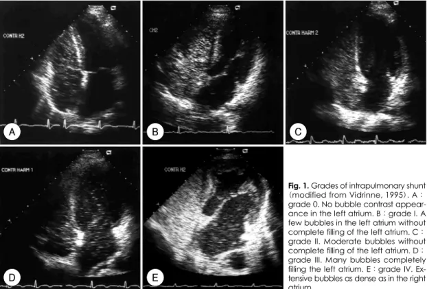
Fig. 1. Grades of intrapulmonary shunt (modified from Vidrinne, 1995). A:
grade 0. No bubble contrast appear- ance in the left atrium. B:grade I. A few bubbles in the left atrium without complete filling of the left atrium. C:
grade II. Moderate bubbles without complete filling of the left atrium. D:
grade III. Many bubbles completely filling the left atrium. E:grade IV. Ex- tensive bubbles as dense as in the right atrium.
A B C
D E
히 소실된 환자의 빈도는 환자 24명 중 19명으로, 79%
를 차지하였다(Table 4).
간이식 후 간 기능과 관련된 임상 지표들인 혈청 알 부민, 혈청 빌리루빈, 프로트롬빈 시간, 그리고 Child- Pugh 분류의 점수는 통계학적으로 의미 있는 변화를 보였고(p<0.001), 또한 PaO2와 alveolar-arterial O2
difference(A-aDO2)수치 역시 이식 전후에 통계학적 으로 의미 있는 차이를 보였다(PaO2:p=0.001, A- aDO2:p<0.001)(Table 5).
간이식 후 폐내 단락의 호전과 임상 지표들의 호전과 의 상관관계에 대한 분석 결과에서는 이식 후의 Child- Pugh 분류 점수의 호전만이 폐내 단락의 호전과 통계학 적으로 의미 있는 상관관계를 보였다(r=0.59, p=0.02) (Table 6).
고 찰
비가역적 급성 및 만성의 간질환 환자들에서 간폐 증 후군의 진단적 단서인 폐내 단락의 발견 빈도가 최근 심 초음파 기술의 발전으로 증가되었고, 또한 저 산소혈증 에 따른 호흡기계 증상이 발현되기 전의 준 임상적 폐 내 단락의 발견 역시 증가할 것으로 기대되었다. 본 저 자들의 연구에 의하면, 64명의 간이식을 기다리는 말기 간질환 환자들을 대상으로 시행한 harmonic imaging 을 이용한 조영 심 초음파 검사 결과, 54명(84%)에서 폐내 단락을 발견하였고 호흡기계 증상이 없고 동맥혈 산소분압이 70 mmHg 이상이었던 환자에서도 폐내 단 락의 발견 빈도가 35명(78%)으로 높은 빈도를 보였으 며 이는 비가역적 급만성 간질환 환자들에서 준 임상적 폐내 단락이 많음을 시사해 주었다.12)
이러한 폐내 단락의 원인은 최근까지 몇몇의 가설이 제시되었지만, 그 중 폐내 혈관 확장에 의한 것에 무게 가 실리고 있다.9)13) 폐내 혈관 확장은 정상인에서의 혈 관 수축 물질과 혈관 확장 물질의 균형 상태가, 간질환 환자들에서 간 기능의 저하로 이러한 물질들의 비정상 적인 대사로 인하여 균형이 깨어지면서 혈관 확장 물질 이 상대적으로 증가하여 폐내 혈관을 확장시킨다는 가 설이 가장 유력하며,8)13)14) 이러한 혈관 확장 물질로는 Table 3. Changes of grade of intrapulmonary shunt be-
fore and after liver transplantation (LT)
Pre-LT Post-LT
Grade 0 1 ( 4%) 20 (80%)
Grade I 10 (40%) 5 (20%)
Grade II 7 (28%) Grade III 7 (28%)
Table 4. Results of reversal of intrapulmonary shunt Intrapulmonary shunt after liver transplantation
Improvement Disappearance No change No. 24/24 (100%) 19/24 (79%) 0/24 (0%)
Table 5. Changes of clinical characteristics before and after liver transplantation (LT)
Pre-LT Post-LT p Albumin (g/dL) 2.7±0.5 4.3±0.4 <0.001 Bilirubin (mg/dL) 12.5±13 1.0±0.8 <0.001 Prothrombin time (sec) 23±6.8 14±1.0 <0.001 Score of child class. 8.5±1.6 5.2±0.5 <0.001 FVC (%) 84.6±16.3 89.0±12.0 0.24 FEV1 (%) 90.2±19.0 95.0±12.0 0.19 FEV1/FVC 80.7±5.6 81.1±5.9 0.45 PaO2 (mmHg) 89.4±8.7 101.5±13.6 0.001 PaCO2 (mmHg) 33.1±4.5 36.2±3.5 0.32 SaO2 (%) 97±1.7 97.7±1.3 0.75 A-aDO2 (mmHg) 16.6±9.3 4.5±11.5 0.001
Table 6. Correlation between improvement of clinical parameters and reversal of intrapulmonary shunt (LT)
Spearman’s correlation Mean±SD
r* p* r† p† Albumin (g/dL) 01.6±00.6 0.38 0.06 0.15 0.32 Bilirubin (mg/dL) 11.5±12.9 0.25 0.23 0.06 0.42 Prothrombin
time (sec) 09.0±07.3 0.41 0.06 0.24 0.21 Score of
child class. 03.4±01.6 0.59 0.002 0.59 0.02 FVC (%) 04.6±17.9 0.39 0.06 0.40 0.09 FEV1 (%) 05.0±17.4 0.35 0.09 0.30 0.16 FEV1/FVC 00.8±05.6 0.06 0.79 0.39 0.07 PaO2 (mmHg) 10.9±14.2 0.24 0.26 0.08 0.40 PaCO2 (mmHg) 01.0±04.3 0.09 0.68 0.24 0.21 SaO2 (%) 00.1±02.0 0.26 0.21 0.37 0.11 A-aDO2 (mmHg) 12.1±13.7 0.28 0.18 0.22 0.24
*:spearman’s bivariate correlation, †:spearman’s partial correlation
1
4
5 2
10 3
prostaglandin E2, I2, prostacycline, nitric oxide, glu- cagons, vasoactive intestinal peptide, platelet acti- vating factor 등이 거론되고 있다.
간질환 환자들에서 폐내 단락의 정도와 임상 지표들 과의 연관성에 대해서는 과거 문헌들에 의하면, 폐내 단 락의 정도는 간질환 환자들에서의 여러 가지 임상 지표 들인 간질환의 중증도, 복수, 간문맥 고혈압, 동맥혈 산 소 분압 등과 연관성이 없다고 보고하였지만,15-17) 이 들 연구는 폐내 단락을 진단하는데 있어서 예민도가 떨 어지는 폐관류 검사나 fundamental imaging의 조영 심 초음파 검사 기법을 사용한 것으로 준 임상적 폐내 단락이나 경한 폐내 단락의 발견에 제한점이 있었던 것 으로 사료된다. 과거 저자들의 연구에 의하면,12) 64명 의 간이식을 기다리는 비가역적 급만성 간질환 환자들 에서 폐내 단락이 있는 군과 없는 군 사이에서 Child- Pugh 분류의 점수가 두 군 간에 의미 있는 차이를 보였 고, 또한 폐내 단락의 정도는 Child-Pugh 분류의 점수 및 동맥혈 산소 분압과 유의한 상관관계를 보였다. 이는 폐내 단락의 정도가 간질환의 중증도와 직접적인 연관 성이 있는 것을 시사해 준다.
간이식 후 폐내 단락의 변화에 대한 보고는 몇몇 소 규모 연구들에서 이루어진 바 있다.18-22) Eriksson 등19) 은 6명의 말기 간질환 환자에서 간이식 후 A-aDO2의 호전을 5명의 환자에서 관찰하였고, Hobeika 등22)은 9명의 간이식 수술을 받은 간폐 증후군 환자들에서 4명 의 환자에서만 폐내 단락의 호전을 보고하였다. 그러나 이들 연구들은 대상환자 수가 적었다는 제한점이 있다.
아울러 간이식 전후 조영 심 초음파를 이용한 폐내 단락 의 변화에 대한 연구들은 몇몇의 증례 보고 외에는 드 물다.23) 또한 폐내 단락의 가역적 변화의 정도와 빈도, 그리고 이를 예측할 수 있는 인자들에 대해서는 아직 명 확한 보고가 없다. 본 연구에서는 급만성의 비가역적 간 질환 환자 24명 모두에서 간이식 후 폐내 단락의 호전 을 관찰하였다. 폐내 단락의 호전을 보였던 환자들의 평 균 추적 조영 심 초음파 검사 기간은 이식 후 4.3개월이 었고, 이식 전 Grade Ⅰ이었던 10명의 환자 중 3명에 서는 이식 후 평균 1.1개월째 추적 조영 심 초음파 검사 를 시행하였으나 이식 전과 폐내 단락의 변화가 없었다.
또한 이들 3명의 환자들은 수술 후 간 기능의 호전이 다른 21명의 환자들에 비해 경미하였으나, 3개월 뒤 추 적 조영 심 초음파 검사 및 혈액 검사를 시행한 결과, 3
명 모두에서 간 기능의 완전한 호전 및 폐내 단락의 소 실을 확인하였다. 이는 간이식 후 간 기능의 호전은 폐 내 단락의 가역적 변화와 밀접한 연관성이 있음을 설명 해 준다.
본 연구에서는 이식 후 폐내 단락의 호전은 이식 전 후의 Child-Pugh 분류의 점수 호전과 통계학적으로 의 미 있는 상관관계를 보였으며 이 역시 폐내 단락은 간 이식 후 간 기능이 호전됨에 따라 수주에서 수개월 내 에 소실된다는 것을 시사해 준다.
결론적으로 저자들은 비가역적 급만성 간질환 환자들 에서 폐내 단락이 간이식 후 대부분 호전되거나 소실되 는 것을 확인하였고, 이러한 폐내 단락의 가역적 변화 를 평가하는데 harmonic imaging의 조영 심 초음파 검 사가 매우 유용한 비침습적 검사 방법이라고 사료된다.
요 약
배경 및 목적:
조영 심 초음파 검사를 이용하여 비가역적 급만성 간 질환 환자들에서 간이식 전후의 폐내 단락의 변화와 이 러한 변화와 간이식 전후의 임상 지표들과의 연관성에 대해 알아보고자 하였다.
방 법:
간이식을 시행 받은 비가역적 급만성 간질환 환자 25 명(남:여=19:6, 평균연령 46±10세)을 대상으로 agitated saline를 이용하여 harmonic imaging의 조영 심 초음파 검사를 이식수술 전과 후에 각각 시행하였고, 폐내 단락의 정도는 미세기포의 조영되는 정도에 따라 Grade 0~Ⅳ로 분류하여 간이식 후 폐내 단락의 가역적 변화의 정도를 확인하였다. 대상 환자들의 임상적 지표 들로 간질환의 정도, 폐 기능 검사 및 동맥혈 가스 검사 결과 등을 확인하여 이들 지표들과 폐내 단락의 호전과 의 상관관계를 평가하였다.
결 과:
간이식 전 폐내 단락은 25명의 환자들 중 24명(96%) 에서 관찰되었고, 이식 후 폐내 단락은 24명의 환자들 중 5명(21%)에서만 관찰되었다. 이식 전 폐내 단락이 관찰되지 않았던 환자 1명을 제외한 24명의 환자들 모 두에서 간이식 후 폐내 단락의 호전을 보였고, 간이식 후 폐내 단락이 완전히 소실된 환자의 빈도는 24명의 환자들 중 19명으로, 79%를 차지하였다. 또한 간이식
후 폐내 단락의 호전은 이식 전후의 Child 분류의 점수 의 호전과 통계학적으로 의미 있는 상관관계를 보였다 (r=0.59, p=0.02).
결 론:
조영 심 초음파 검사는 간이식 전후 폐내 단락의 가 역적 변화를 확인하는데 매우 유용한 비침습적 검사 방 법이다. 폐내 단락은 간이식 후 대부분의 환자에서 호전 을 보이며, 이러한 호전은 child 분류의 점수 호전과 밀 접한 연관성을 보인다.
중심 단어:심 초음파;폐내 단락;간이식.
REFERENCES
1) Flückiger M. Vorkommen von trommelschagel formigen fin- gerendphalangen onhe chronische Veranderungen an der lungen oder am herzen. Wien Med Wochenschr 1884;34:1457.
2) Wolfe JD, Tashkin DP, Holly FE, Brachman MB, Genovesi MG. Hypoxemia of cirrhosis: detection of abnormal small pulmonary vascular channels by a quantitative radionuclide method. Am J Med 1977;63:746-54.
3) Krowka MJ, Dickson ER, Cortese DA. Hepatopulmonary syndrome: clinical observations and lack of therapeutic res- ponse to somatostatin analogue. Chest 1993;104:515-21.
4) Abrams GA, Jaffe CC, Hoffer PB, Binder HJ, Fallon MB.
Diagnostic utility of contrast echocardiography and lung perfusion scan in patients with hepatopulmonary syndrome.
Gastroenterology 1995;109:1283-8.
5) Nemec JJ, Davison MB, Marwick TH, Chimowitz MI, Stoller JK, Klein AL, Salcedo EE. Detection and evaluation of in- trapulmonary vascular shunt with “contrast Doppler” tran- sesophageal echocardiography. J Am Soc Echocardiogr 1991;
4:79-83.
6) Hind CR, Wong CM. Detection of pulmonary arteriovenous fistulae in patient with cirrhosis by contrast 2D echocardio- graphy. Gut 1981;22:1042-5.
7) Krowka MJ, Tajik AJ, Dickson ER, Wiesner RH, Cortese DA. Intrapulmonary vascular dilatations (IPVD) in liver transplant candidates: screening by two-dimensional con- trast enhanced echocardiography. Chest 1990;97:1165-70.
8) Hopkins WE, Waggoner AD, Barzilai B. Frequency and significance of intrapulmonary right-to-left shunting in end- stage hepatic disease. Am J Cardiol 1992;70:516-9.
9) Lange PA, Stoller JK. The hepatopulmonary syndrome. Ann Intern Med 1995;122:521-9.
10) Maddrey WC, van Thiel DH. Liver transpantation: an
overview. Hepatology 1988;8:948-59.
11) Vedrinne JM, Duperret S, Bizollon T, Magnin C, Motin J, Trepo C, Ducerf C. Comparison of transesophageal and tran- sthoracic contrast echocardiography for detection of an in- trapulmonary shunt in liver disease. Chest 1997;111:1236-40.
12) Kim BJ, Lee KJ, Hong JS, Lee SC, Gwon HC, Park SW, Kim JS, Kim DK, Lee SH, Hong KP, Park JE, Seo JD, Lee WR. Clinical characteristics and prevalence of intrapulmo- nary shunt detected by contrast echocardiography using harmonic imaging in liver transplant candidates [abstract].
Korean Circ J 2001;31(Suppl 10):II247.
13) Krowka MJ, Cortese DA. Hepatopulmonary syndrome: an evolving perspective in the era of liver transplantation. He- patology 1990;11:138-42.
14) Eriksson LS. Hypoxemia in patients with liver cirrhosis. Acta Gastroenterol Belg 1990;53:209-15.
15) Krowka MJ, Cortese DA. Pulmonary aspects of liver disease and liver transplantation. Clin Chest Med 1989;10:593-616.
16) Rodriguez-Roisin R, Agusti AG, Roca J. The hepatopulmo- nary syndrome:new name, old complexities. Thorax 1992;47:
897-902.
17) Krowka MJ, Cortese DA. Severe hypoxemia associated with liver disease: mayo clinic experience and the experimental use of almitrine bismesylate. Mayo Clin Proc 1987;62:164-73.
18) Schwarzenberg SJ, Freese DK, Regelmann WE, Gores PF, Boudreau RJ, Payne WD. Resolution of severe intrapulmonary shunting after liver transplantation. Chest 1993;103:1271-3.
19) Eriksson LS, Sőderman C, Ericzon BG, Eleborg L, Wahren J, Hedenstierna G. Normalization of ventilation/perfusion relationships after liver transplantation in patients with de- compensated cirrhosis: evidence for a hepatopulmonary syndrome. Hepatology 1990;12:1350-7.
20) Stoller JK, Moodie D, Schiavone WA, Vogt D, Broughan T, Winkelman E, Rehm PK, Carey WD. Reduction of intrapul- monary shunt and resolution of digital clubbing associated with primary biliary cirrhosis after liver transplantation.
Hepatology 1990;11:54-8.
21) Barry S, Pingleton S, Kindscher J, Eisenach J, Forster J. Com- parison of shunt fractions pre- and post- liver transplantation.
Transplant Proc 1993;25:1801.
22) Hobeika J, Houssin D, Bernard O, Devictor D, Grimon G, Chapuis Y. Orthotopic liver transplantation in children with chronic liver disease and severe hypoxemia. Transplantation 1994;57:224-8.
23) Hasegawa T, Kamiyama M, Azuma T, Sasaki T, Okada A, Miwatani T, Matsushita T. Reversal of intrapulmonary ar- teriovenous shunting detected by two-dimensional contrast- echocardiography after liver transplantation. Transplantation 2000;69:672-5.