© 2020 The Korean Ophthalmological Society
This is an Open Access article distributed under the terms of the Creative Commons Attribution Non-Commercial License (http://creativecommons.org/licenses /by-nc/3.0/) which permits unrestricted non-commercial use, distribution, and reproduction in any medium, provided the original work is properly cited.
Original Article
Pathologic myopia is a major cause of visual impairment especially in the Asian population [1]. In highly myopic eyes, posterior elongation of the sclera and the develop- ment of staphyloma result in retinal deformations, which lead to the development of myopic traction maculopathy (MTM). MTM is a critical complication that can threaten Purpose: To investigate prognostic factors related to the surgical outcome of vitrectomy in myopic traction mac-
ulopathy (MTM).
Methods: Medical records of patients with MTM who underwent pars plana vitrectomy with internal limiting membrane peeling and follow-up over 12 months were reviewed retrospectively. Best-corrected visual acuity (BCVA), fundoscopic examination and spectral-domain optical coherence tomography findings were evaluated postoperatively. Functional success was defined as visual acuity gain and anatomical success was defined as reduction or resolution of foveoschisis without complications.
Results: This study included 40 eyes of 36 patients. BCVA improved from 0.70 ± 0.44 to 0.63 ± 0.57 logarithm of minimum angle of resolution and central macular thickness decreased from 526.6 ± 132.1 to 277.8 ± 92.1 μm at final follow-up. Functional success was achieved in 24 (60.0%) eyes, and 33 (82.5%) eyes reached anatomical success. Presence of foveal detachment (FD) and higher category of myopic maculopathy were associated with both functional (p = 0.014, 0.021, respectively) and anatomical (p = 0.011, 0.022, respectively) failure. Longer preoperative axial length showed an association with functional failure but not with anatomical failure (p = 0.041). In multivariate analysis, FD was the only prognostic factor for both functional and anatomi- cal outcome (p = 0.041, 0.043, respectively). Preoperative BCVA (r2 = 0.259, p = 0.001), axial length (r2 = 0.172, p = 0.008), and myopic maculopathy category (r2 = 0.336, p < 0.001) showed significant correlation with final BCVA.
Conclusions: More severe myopic maculopathy and the presence of FD are associated with poorer functional and anatomical outcomes of pars plana vitrectomy in MTM. Better preoperative BCVA, shorter axial length, and less severe myopic maculopathy are correlated with better final BCVA.
Key Words: Myopic foveoschisis, Myopic traction maculopathy, Optical coherence tomography, Vitrectomy
Received: September 23, 2019 Final revision: October 25, 2019 Accepted: October 28, 2019
Corresponding Author: Kyu Hyung Park, MD, PhD. Department of Ophthalmology, Seoul National University Bundang Hospital, 82 Gu- mi-ro 173beon-gil, Bundang-gu, Seongnam 13620, Korea. Tel: 82-31-787- 7373, Fax: 82-31-787-4057, E-mail: [email protected]
Prognostic Factors Related with Surgical Outcome of Vitrectomy in Myopic Traction Maculopathy
Chung Young Kim, Min Seok Kim, Kyoung Lae Kim, Se Joon Woo, Kyu Hyung Park
Department of Ophthalmology, Seoul National University Bundang Hospital, Seoul National University College of Medicine, Seongnam, Korea
vision and is reported to occur in 9% to 34% of highly my- opic eyes [2,3]. MTM includes structural features such as vitreomacular traction (VMT), macular retinoschisis, lamellar hole, or foveal detachment (FD). VMT, remnant cortical vitreous layer, epiretinal membrane (ERM), in- trinsic noncompliance of inner limiting membrane, and in- flexibility of retinal vessels could contribute to the patho- genesis of MTM [4-7].
A study of the natural course of MTM indicated that more extensive macular retinoschisis had a higher proba- bility of progression, and retinoschisis was resolved after the surgical release of VMT [8]. Pars plana vitrectomy (PPV) with internal limiting membrane (ILM) peeling has been reported to be effective for the resolution of macular retinoschisis [7,9-11]. However, visual improvement was not significant in some patients and postoperative compli- cations, such as macular hole (MH), have also been report- ed [12,13].
Preoperative factors predicting surgical outcome in MTM are little known. In previous cases, preoperative vi- sual acuity and axial length have been reported to be cor- related with visual outcome [14-16] and photoreceptor layer defects or irregular choroidal surfaces were reported to limit visual recovery [17,18]. However, whether FD is a poor prognostic factor for surgical success is controversial [14,19]. For these reasons, more studies are required to elu- cidate the predictive factors of vitrectomy in MTM.
In this study, we investigated prognostic factors related with the functional and anatomical outcomes of vitrecto- my in MTM after more than one year of postoperative fol- low-up.
Materials and Methods
Subjects
This study was approved by the institutional review board of the Seoul National University Bundang Hospital (B-1904/534-102) and the study was conducted according to the tenets of the Declaration of Helsinki. Written in- formed consent was waived due to the retrospective nature of the study. The medical records of 56 eyes of 50 patients with MTM who underwent PPV from March 2007 to De- cember 2017, were retrospectively reviewed. Patients were excluded if they had a previous history of intraocular sur-
gery other than cataract extraction or coexistent retinal diseases, follow-up less than 12 months, axial length short- er than 26 mm, MH or myopic choroidal neovasculariza- tion. Consequently, 40 eyes of 36 patients who met the in- clusion criteria were included in the study. Functional success was defined as an improvement of visual acuity at the final visit compared with initial best-corrected visual acuity (BCVA). Anatomical success was defined as a re- duction or resolution of schisis without complications, such as retinal detachment, MH formation, or recurrence.
Surgical procedures
PPV was performed by 2 experienced retinal specialists (SJW and KHP). A 23/25-gauge transconjunctival suture- less vitrectomy was performed using an Accurus 800CS surgical system (Alcon, Fort Worth, TX, USA) or Constel- lation system (Alcon) with contact lens (Hoya, Tokyo, Ja- pan). Phacoemulsification with intraocular lens implanta- tion was performed before vitrectomy in patients with significant cataract. The peeling of the ILM was performed using end-gripping forceps (Alcon). Triamcinolone aceton- ide (1%; Hanmi Pharmaceutical, Seoul, Korea) or 0.05%
indocyanine green (Dong In Dang Pharmaceutical, Si- heung, Korea) were used for staining and peeling of ILM.
Foveal sparing ILM peeling was performed in 7 eyes and complete ILM peeling up to the temporal arcade was per- formed in the remaining 33 eyes. Gas tamponade with 14%
perfluoropropane or 18% sulfur hexafluoride was done in 19 eyes. Prednisolone acetate (1%; Pred Forte, Allergan, Ir- vine, CA, USA) and 0.5% levofloxacin (Cravit; Santen Pharmaceutical, Osaka, Japan), were topically instilled 4 times a day for 4 weeks.
Examinations
After the surgery, patients were followed up at postoper- ative 1, 4, 8, and 14 months and then every 6 months or an- nually. All patients underwent a comprehensive ophthal- mologic examination before and after the operation including measurement of BCVA, intraocular pressure (noncontact tonometer; KT-500, Kowa, Tokyo, Japan), slit- lamp biomicroscopy, and indirect ophthalmoscopy. The re- fractive errors were measured by auto-refractometer (KR- 8800; Topcon, Tokyo, Japan) and axial length by the IOL Master 500 (Carl Zeiss Meditec, Jena, Germany) before the
surgery.
Spectral-domain optical coherence tomography images were obtained (Spectralis; Heidelberg Engineering, Heidel- berg, Germany) with an eye-tracking system and automat- ic software to maintain the same position in serial scans.
Central macular thickness (CMT) was measured as the distance between the first signal from the vitreoretinal in- terface and the outer border of the retinal pigment epitheli- um.
Myopic maculopathy was classified as category 0, no macular lesion; category 1, tessellated fundus; category 2, diffuse chorioretinal atrophy; category 3, patch chorioreti- nal atrophy; and category 4, macular atrophy with or with- out lacquer crack, choroidal neovascularization, and Fuchs spot as plus lesions according to the international classifi- cation and grading system for myopic maculopathy [20].
For logistic regression analysis, myopic maculopathy cate- gories were divided into two groups, a group of categories
0, 1, 2 and a group of categories 3, 4.
Reviews of optical coherence tomography images and categorization of myopic maculopathy were performed by three investigators (CYK, MSK, and KLK) and inter-ob- server agreement was accomplished.
Statistical analyses
Statistical analyses were performed using IBM SPSS Statistics ver. 22 (IBM Corp., Armonk, NY, USA). Chang- es of BCVA and CMT at each postoperative follow-up were evaluated by paired t-test. Logistic regression analy- sis was performed to identify factors related with the func- tional and anatomical outcomes. Factors correlating with final BCVA were investigated by a linear regression mod- el. A p-value less than 0.05 was considered statistically significant.
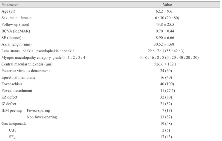
Table 1. Preoperative characteristics of study subjects (n = 40 eyes)
Parameter Value
Age (yr) 62.2 ± 9.6
Sex, male : female 6 : 30 (20 : 80)
Follow-up (mon) 43.8 ± 25.5
BCVA (logMAR) 0.70 ± 0.44
SE (diopter) -8.90 ± 6.66
Axial length (mm) 30.52 ± 1.68
Lens status, phakia : pseudophakia : aphakia 22 : 17 : 1 (55 : 42 : 3) Myopic maculopathy category, grade 0 : 1 : 2 : 3 : 4 0 : 8 : 16 : 8 : 8 (0 : 20 : 40 : 20 : 20)
Central macular thickness (μm) 526.6 ± 132.1
Posterior vitreous detachment 24 (60)
Epiretinal membrane 16 (40)
Foveoschisis 40 (100)
Foveal detachment 11 (27.5)
EZ defect 32 (80)
IZ defect 21 (52)
ILM peeling Fovea-sparing 7 (18)
Non fovea-sparing 33 (82)
Gas tamponade 19 (48)
C3F8 2 (5)
SF6 17 (43)
Values are presented as mean ± standard deviation or number (%).
BCVA = best-corrected visual acuity; logMAR = logarithm of the minimum angle of resolution; SE = spherical equivalent; EZ = ellipsoid zone; IZ = interdigitation zone; ILM = internal limiting membrane.
Results
Demographics and clinical characteristics
The demographics and preoperative clinical characteris- tics of the patients are presented in Table 1. Mean age was 62.2 ± 9.6 (range, 39.0 to 81.0) years and the patients were followed up for an average of 43.8 (range, 12.1 to 98.1) months after surgical treatment. Preoperative BCVA was 0.70 ± 0.44 logarithm of minimum angle of resolution (log- MAR). Mean spherical equivalent was -8.90 ± 6.66 (range, +0.25 to -23.63) diopters and axial length was 30.52 ± 1.68 (range, 27.39 to 34.12) mm. Eighteen (45%) patients had previous cataract surgery and combined cataract surgery was performed in 13 (36%) patients. Eight (20%), 16 (40%), 8 (20%), and 8 (20%) eyes were classified with categories 1, 2, 3, and 4 myopic maculopathies, respectively.
Parameters evaluated in optical coherence tomography are listed in Table 1. Mean CMT was 526.6 ± 132.1 (range, 234.0 to 878.0) μm preoperatively. Twenty-four eyes (60%) had posterior vitreous detachment, 16 eyes (40%) had ERM, 40 eyes (100%) had foveoschisis and 6 eyes (27.5%)
had FD, 32 eyes (80%) had ellipsoid zone (EZ) defect, and 21 eyes (52%) had interdigitation zone (IZ) defect in OCT.
Functional and anatomical outcome
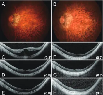
Representative fundus photographs and serial OCT im- ages at each postoperative follow-up are presented together with visual acuities (Fig. 1A-1H, 2A-2H, 3A-3H, 4A-4H).
Postoperative BCVA and their changes are listed in Table 2. Mean BCVA improved from 0.70 ± 0.44 at baseline to 0.63 ± 0.57 logMAR at the final follow-up although it was not statistically significant (Fig. 5A, 5B). Preoperative CMT was 526.6 ± 132.1 μm and it continuously reduced in every follow-up with statistical significance (p < 0.001) (Table 2 and Fig. 5). At the final follow-up, it was 277.8 ± 92.1 μm, which was decreased by 248.8 ± 153.8 μm. Func- tional success was achieved in 24 (60%) eyes. Thirty-three (82.5%) eyes reached anatomical success and 7 (17.5%) failed. Among the failed cases, MH developed in 3 cases, MH retinal detachment in 2 cases, and recurrence of fove- oschisis in 2 cases were documented. Seven cases were treated with repeated PPV with gas tamponade or silicone oil injection and 2 of them achieved functional success.
Fig. 2. A case of myopic traction maculopathy after pars plana vitrectomy with gas tamponade. (A) Preoperative and (B) final fundus images are presented. Serial optical coherence tomogra- phy images are arranged in chronological sequence: (C) preop- erative, (D) 1 month, (E) 4 months, (F) 8 months, (G) 14 months, and (H) final. Visual acuities at each postoperative follow-up are indicated at the bottom right side of the optical coherence tomog- raphy image.
A
C D E
B
F G
H
Fig. 1. A case of myopic traction maculopathy with foveal de- tachment after pars plana vitrectomy. (A) Preoperative and (B) final fundus images are presented. Serial optical coherence to- mography images are arranged in chronological sequence: (C) preoperative, (D) 1 month, (E) 4 months, (F) 8 months, (G) 14 months, and (H) final. Visual acuities at each postoperative fol- low-up are indicated at the bottom right of the optical coherence tomography image.
A
C D E
B
F G H
Fig. 4. A case of myopic traction maculopathy after pars plana vitrectomy with postoperative macular hole (*) development. (A) Preoperative and (B) final fundus images are presented. Serial optical coherence tomography images are arranged in chronolog- ical sequence: (C) preoperative, (D) 1 month, (E) 4 months, (F) 8 months, (G) 14 months, and (H) final. Visual acuities at each postoperative follow-up are indicated at the bottom right side of the optical coherence tomography image.
A
C D E
B
F G
H
Fig. 3. A case of myopic traction maculopathy after pars plana vitrectomy without gas tamponade. (A) Preoperative and (B) final fundus images are presented. Serial optical coherence tomogra- phy images are arranged in chronological sequence: (C) preop- erative, (D) 1 month, (E) 4 months, (F) 8 months, (G) 14 months, and (H) final. Visual acuities at each postoperative follow-up are indicated at the bottom right side of the optical coherence tomog- raphy image.
A
C D E
B
F G H
Table 2. Changes of BCVA and CMT after vitrectomy Preoperative Follow-up 1
(1.2 mon) Follow-up 2
(4.1 mon) Follow-up 3
(8.5 mon) Follow-up 4
(14.5 mon) Follow-up 5 (43.8 mon) BCVA (logMAR) 0.70 ± 0.44 0.82 ± 0.58 0.71 ± 0.59 0.65 ± 0.51 0.63 ± 0.45 0.63 ± 0.57 BCVA change
(logMAR) NA 0.16 ± 0.46 0.04 ± 0.49 -0.01 ± 0.51 -0.03 ± 0.44 -0.06 ± 0.51
p-value* NA 0.042 0.649 0.871 0.730 0.437
CMT (μm) 526.6 ± 132.1 397.4 ± 142.3 357.8 ± 102.6 323.9 ± 103.0 285.6 ± 79.8 277.8 ± 92.1 CMT change (μm) NA -133.9 ± 154.4 -173.2 ± 164.6 -208.8 ± 157.8 -240.0 ± 164.5 -248.8 ± 153.8
p-value† NA <0.001 <0.001 <0.001 <0.001 <0.001
Values are presented as mean ± standard deviation.
BCVA = best-corrected visual acuity; CMT = central macular thickness; logMAR = logarithm of the minimum angle of resolution; NA = not available.
*p-value of paired t-test between preoperative and postoperative BCVA; †p-value of paired t-test between preoperative and postoperative CMT.
Factors related with functional and anatomical outcome Factors associated with functional and anatomical out- come were identified using a logistic regression analysis.
Longer axial length (odds ratio [OR], 1.60; 95% confidence interval [CI], 1.02 to 2.50; p = 0.041), higher category of myopic maculopathy (OR, 5.00; 95% CI, 1.27 to 19.69; p =
0.021), existence of FD (OR, 7.00; 95% CI, 1.48 to 33.21; p
= 0.014) were significantly related with functional failure.
In multivariate analysis, FD (OR, 6.53; 95% CI, 1.09 to 39.32; p = 0.041) showed an association with functional failure (Table 3).
In univariate analysis investigating factors related with anatomical outcome, higher category of myopic maculopa-
thy (OR, 13.80; 95% CI, 1.47 to 130.1; p = 0.022) and FD (OR, 11.25; 95% CI, 1.75 to 72.50; p = 0.011) were found to be associated with anatomical failure. Multivariate analy- sis demonstrated significant association between FD and anatomical failure (OR, 8.01; 95% CI, 1.07 to 59.92; p = 0.043) (Table 4).
To find preoperative factors related with final BCVA, additional linear regression analysis was performed. Pre- operative BCVA (r2 = 0.259, p = 0.001), axial length (r2 = 0.172, p = 0.008) and myopic maculopathy category (r2 = 0.336, p < 0.001) showed significant correlation with final BCVA (Fig. 6A-6C).
Discussion
In this study, we investigated clinical factors related to the functional and anatomical outcomes of vitrectomy in MTM. The presence of FD and higher category of myopic maculopathy were associated with both functional and an- atomical failure. Longer preoperative axial length showed an association with functional failure but not with anatom- ical failure. In addition, preoperative BCVA, axial length, and myopic maculopathy category showed significant cor- relation with final BCVA.
Functional and anatomical success was obtained in 60%
and 82.5%, respectively. Previous studies on the surgical outcome of vitrectomy in MTM have reported visual acui- ty improvement in 42% to 100% and resolution of foveo- schisis in 73% to 100% [9,15,21,22]. MH development was the most common complication of PPV for MTM. Regard-
ing the rate of surgical success and complications, the re- sults of our study were comparable to those of previous studies. Preoperative BCVA showed positive correlation with final BCVA and it was consistent with previous re- ports [14-16]. Better preoperative BCVA implies a more preserved retinal neuronal function so greater visual reha- bilitation can be expected after surgical treatment.
Kumagai et al. [14] reported that patients with FD had significantly better improvement of visual acuity and can benefit most from the surgery. However, our study showed a preoperative FD was a predictive factor for poor func- tional and anatomical outcomes. In the study by Kumagai et al. [14], patients were divided into two groups according to the presence of FD; those with FD showed greater visu- al acuity gain than those without FD. However, preopera- tive BCVA was worse in the patients with FD and other confounding factors were not controlled. In a recent study by Hattori et al. [19], eyes with FD showed worse pre- and postoperative BCVA compared to those without FD (p = 0.036, 0.046), although logMAR gain was not significantly different (p = 0.437). In our study, we did not divide sub- jects into sub-groups by the presence of FD but included suspected candidate factors in a multivariate logistic mod- el. FD showed a significant correlation not only with func- tional but also anatomical outcome. The presence of FD should be considered for predicting the surgical outcome of MTM.
When EZ and IZ defects at final follow-up were ana- lyzed, prevalence rates of EZ and IZ defects were both 100% in eyes with preoperative FD but 51.7% and 48.3% in eyes without preoperative FD, respectively. The final pre- Fig. 5. Changes of (A) best-corrected visual acuity (BCVA) logarithm of minimum angle of resolution (logMAR) and (B) central macular thickness (CMT) at preoperative and different postoperative follow-up periods. *0.01 < p ≤ 0.05, **p ≤ 0.001.
BCVA (logMAR)
0.5 1.0 1.5
0 Preoperative 1 mon 4 mon 8 mon 14 mon Final
*
A
CMT (µm)
400 600 800
200 Preoperative 1 mon 4 mon 8 mon 14 mon Final
**
** **
** **
B
sentation of EZ and IZ defects was also significantly cor- related with preoperative FD (p = 0.004, 0.003, respective- ly). It is speculated that the preoperative FD and disruption of photoreceptors affect final visual outcome. Further studies investigating the association between the duration, severity of FD, and surgical outcome would be helpful in clarifying the predictive ability of FD in MTM.
Previous studies have suggested preoperative factors such as baseline BCVA, axial length, photoreceptor defect, irregular choroidal surface, and FD were associated with surgical outcomes in MTM patients [14-17]. This study
demonstrated that the eyes with a higher category of myo- pic maculopathy are less likely to achieve functional and anatomical success after vitrectomy. The severity of myo- pic degeneration is determined in part by the progressive distension of the posterior pole, therefore the structural de- struction in the inner and outer retina by greater distension is estimated to induce more severe irreversible damages precluding visual recovery.
Among the 40 eyes, postoperative MH developed in 3 eyes (7.5%) and MH retinal detachment occurred in 2 eyes (5%) as postoperative complications. Preoperative BCVA Table 3. Factors associated with functional outcome of vitrectomy in myopic traction maculopathy
Univariate Multivariate
OR (95% CI) p-value OR (95% CI) p-value
Age 1.05 (0.98–1.13) 0.154 NA NA
Preoperative BCVA 0.94 (0.22–4.01) 0.935 NA NA
Preoperative CMT 1.00 (1.00–1.01) 0.575 NA NA
Preoperative AXL 1.60 (1.02–2.50) 0.041 1.51 (0.92–2.47) 0.100
Preoperative MM category 5.00 (1.27–19.69) 0.021 2.78 (0.59–13.19) 0.197
Foveal detachment 7.00 (1.48–33.21) 0.014 6.53 (1.09–39.32) 0.041
EZ defect 2.54 (0.63–10.17) 0.188 NA NA
IZ defect 1.41 (0.39–5.13) 0.602 NA NA
Fovea-sparing ILM Peeling 1.15 (0.22–6.02) 0.865 NA NA
Gas tamponade 1.80 (0.50–6.46) 0.367 NA NA
OR = odds ratio; CI = confidence interval; NA = not available; BCVA = best-corrected visual acuity; CMT = central macular thickness;
AXL = axial length; MM = myopic maculopathy; EZ = ellipsoid zone; IZ = interdigitation zone; ILM = internal limiting membrane.
Table 4. Factors associated with anatomical outcome of vitrectomy in myopic traction maculopathy
Univariate Multivariate
OR (95% CI) p-value OR (95% CI) p-value
Age 0.99 (0.91–1.08) 0.876 NA NA
Preoperative BCVA 4.60 (0.73–28.73) 0.103 NA NA
Preoperative CMT 1.00 (0.99–1.01) 0.745 NA NA
Preoperative AXL 1.46 (0.85–2.52) 0.173 NA NA
Preoperative MM category 13.80 (1.47–130.1) 0.022 10.02 (0.95–105.7) 0.055
Foveal detachment 11.25 (1.75–72.50) 0.011 8.01 (1.07–59.92) 0.043
EZ defect 4.42 (0.48–40.98) 0.191 NA NA
IZ defect 0.98 (0.19–5.11) 0.983 NA NA
Fovea-sparing ILM Peeling 0.75 (0.08–7.44) 0.806 NA NA
Gas tamponade 3.39 (0.57–2010) 0.178 NA NA
OR = odds ratio; CI = confidence interval; NA = not available; BCVA = best-corrected visual acuity; CMT = central macular thickness;
AXL = axial length; MM = myopic maculopathy; EZ = ellipsoid zone; IZ = interdigitation zone; ILM = internal limiting membrane.
(0.70 logMAR), CMT (529.2 μm), and the axial length (29.85 mm) of these eyes did not show significant differ- ence compared with those without MH development. How- ever, the prevalence of the EZ defect (100%) and FD (60%) was higher. Sayanagi et al. [18] reported higher prevalence of the EZ defect in FD and Gao et al. [13] demonstrated that the EZ defect is a risk factor for development of full-thickness MH after vitrectomy for myopic foveoschi- sis. The weakness of the inner retinal surface, combined with a disrupted inner segment/outer segment junction, in- creases its vulnerability to trauma during ILM peeling and contributes to MH development. The technical modifica- tion of ILM peeling, which spares foveal ILM was then in- troduced and demonstrated better anatomical and func- tional outcomes with decreased MH development [23,24].
In our study, fovea-sparing ILM was performed in 7 eyes (18%) and none of them developed MH. Fovea-sparing ILM peeling thus may be one of the surgical options to re- duce iatrogenic MH, especially in eyes with an EZ defect.
Our study has some limitations, mostly inherent in a ret- rospective study design. The sample size was small and follow-up period was variable. Due to the lack of standard- ized surgical indication for MTM, each surgeon has made surgical decisions individually and this was susceptible to selection biases.
In conclusion, a higher category of myopic maculopathy and the presence of FD are associated with functional and anatomical failure of PPV in MTM. Better preoperative BCVA, shorter axial length, and lower category of myopic maculopathy are associated with better final BCVA.
Conflict of Interest
No potential conflict of interest relevant to this article was reported.
References
1. Verkicharla PK, Ohno-Matsui K, Saw SM. Current and predicted demographics of high myopia and an update of its associated pathological changes. Ophthalmic Physiol Opt 2015;35:465-75.
2. Panozzo G, Mercanti A. Optical coherence tomography findings in myopic traction maculopathy. Arch Ophthalmol 2004;122:1455-60.
3. Baba T, Ohno-Matsui K, Futagami S, et al. Prevalence and characteristics of foveal retinal detachment without macu- lar hole in high myopia. Am J Ophthalmol 2003;135:338-42.
4. Johnson MW. Myopic traction maculopathy: pathogenic mechanisms and surgical treatment. Retina 2012;32:S205- 10.
5. VanderBeek BL, Johnson MW. The diversity of traction mechanisms in myopic traction maculopathy. Am J Oph- thalmol 2012;153:93-102.
6. Ikuno Y, Gomi F, Tano Y. Potent retinal arteriolar traction as a possible cause of myopic foveoschisis. Am J Ophthal- mol 2005;139:462-7.
7. Panozzo G, Mercanti A. Vitrectomy for myopic traction maculopathy. Arch Ophthalmol 2007;125:767-72.
8. Shimada N, Tanaka Y, Tokoro T, Ohno-Matsui K. Natural Fig. 6. Association between (A) preoperative best-corrected visual acuity (BCVA) logarithm of minimum angle of resolution (logMAR), (B) axial length (mm), (C) myopic maculopathy category and final BCVA (logMAR).
Final BCVA (logMAR)
1.0 1.5 2.0 2.5
0.5 0
r2 = 0.336 p < 0.001
Myopic maculopathy category
1 2 3 4
C
Final BCVA (logMAR)
1.0 1.5 2.0
0.5 0
r2 = 0.259 p = 0.001
Pre-BCVA (logMAR)
0 0.5 1.0 1.5 2.0
A
Final BCVA (logMAR)
1.0 1.5 2.0
0.5 0
r2 = 0.172 p = 0.008
Axial length (mm)
28 30 32 34
B
course of myopic traction maculopathy and factors associ- ated with progression or resolution. Am J Ophthalmol 2013;156:948-57.
9. Kobayashi H, Kishi S. Vitreous surgery for highly myopic eyes with foveal detachment and retinoschisis. Ophthal- mology 2003;110:1702-7.
10. Futagami S, Inoue M, Hirakata A. Removal of internal limiting membrane for recurrent myopic traction maculop- athy. Clin Exp Ophthalmol 2008;36:782-5.
11. Kanda S, Uemura A, Sakamoto Y, Kita H. Vitrectomy with internal limiting membrane peeling for macular retinoschi- sis and retinal detachment without macular hole in highly myopic eyes. Am J Ophthalmol 2003;136:177-80.
12. Gaucher D, Haouchine B, Tadayoni R, et al. Long-term fol- low-up of high myopic foveoschisis: natural course and surgical outcome. Am J Ophthalmol 2007;143:455-62.
13. Gao X, Ikuno Y, Fujimoto S, Nishida K. Risk factors for development of full-thickness macular holes after pars pla- na vitrectomy for myopic foveoschisis. Am J Ophthalmol 2013;155:1021-7.
14. Kumagai K, Furukawa M, Ogino N, Larson E. Factors cor- related with postoperative visual acuity after vitrectomy and internal limiting membrane peeling for myopic foveo- schisis. Retina 2010;30:874-80.
15. Hwang JU, Joe SG, Lee JY, et al. Microincision vitrectomy surgery for myopic foveoschisis. Br J Ophthalmol 2013;97:879- 84.
16. Figueroa MS, Ruiz-Moreno JM, Gonzalez del Valle F, et al.
Long-term outcomes of 23-gauge pars plana vitrectomy
with internal limiting membrane peeling and gas tampon- ade for myopic traction maculopathy: A Prospective Study.
Retina 2015;35:1836-43.
17. Shin JY, Yu HG. Visual prognosis and spectral-domain op- tical coherence tomography findings of myopic foveoschi- sis surgery using 25-gauge transconjunctival sutureless vit- rectomy. Retina 2012;32:486-92.
18. Sayanagi K, Ikuno Y, Soga K, Tano Y. Photoreceptor inner and outer segment defects in myopic foveoschisis. Am J Ophthalmol 2008;145:902-8.
19. Hattori K, Kataoka K, Takeuchi J, et al. Predictive factors of surgical outcomes in vitrectomy for myopic traction maculopathy. Retina 2018;38:S23-30.
20. Ohno-Matsui K, Kawasaki R, Jonas JB, et al. International photographic classification and grading system for myopic maculopathy. Am J Ophthalmol 2015;159:877-83.
21. Taniuchi S, Hirakata A, Itoh Y, et al. Vitrectomy with or without internal limiting membrane peeling for each stage of myopic traction maculopathy. Retina 2013;33:2018-25.
22. Ikuno Y, Sayanagi K, Ohji M, et al. Vitrectomy and inter- nal limiting membrane peeling for myopic foveoschisis.
Am J Ophthalmol 2004;137:719-24.
23. Shimada N, Sugamoto Y, Ogawa M, et al. Fovea-sparing internal limiting membrane peeling for myopic traction maculopathy. Am J Ophthalmol 2012;154:693-701.
24. Ho TC, Yang CM, Huang JS, et al. Long-term outcome of foveolar internal limiting membrane nonpeeling for myo- pic traction maculopathy. Retina 2014;34:1833-40.