J O U R N A L O F
Veterinary Science
*Corresponding author
Tel: +82-43-261-3372; Fax: +82-43-261-3224 E-mail: [email protected]
Evaluation of partial cranial cruciate ligament rupture with positive contrast computed tomographic arthrography in dogs
Sungyoung Han
1, Haengbok Cheon
1, Hangmyo Cho
1, Juhyung Kim
1, Ji-Houn Kang
1, Mhan-Pyo Yang
1, Youngwon Lee
2, Heechun Lee
3, Dongwoo Chang
1,*
1
Veterinary Medical Center, College of Veterinary Medicine, Chungbuk National University, Cheongju 361-763, Korea
2
Department of Veterinary Medicine, College of Veterinary Medicine, Chungnam National University, Daejeon 305-764, Korea
3
Department of Veterinary Medicine, College of Veterinary Medicine, Gyeongsang National University, Jinju 660-701, Korea
Computed tomographic arthrography (CTA) of four cadaveric canine stifles was performed before and after partial cranial cruciate ligament rupture in order to verify the usefulness of CTA examination for the diagnosis of partial cranial cruciate ligament rupture. To obtain the sequential true transverse image of a cranial cruciate ligament, the computed tomography gantry was angled such that the scanning plane was parallel to the fibula. True transverse images of cranial cruciate ligaments were identified on every sequential image, beginning just proximal to the origin of the cranial cruciate ligament distal to the tibial attachment, after the administration of iodinated contrast medium. A significant decrease in the area of the cranial cruciate ligament was identified on CTA imaging after partial surgical rupture of the cranial cruciate ligament. This finding implies that CTA can be used for assessing partial cranial cruciate ligament ruptures in dogs.
Keywords: arthrography, computed tomography, cruciate ligament, dog, rupture
Introduction
Most ligament injuries in canine stifle joints involve some kind of cranial cruciate ligament rupture, including partial rupture [9]. This results in severe instability and predisposes the joint to degenerative changes [7].
The cranial cruciate ligament is attached to a fossa on the caudal aspect of the medial side of the lateral femoral condyle. It courses cranially, medially, and distally across the intercondylar fossa and attaches to the cranial intercondyloid area of the tibia [1]. The cranial drawer test
is diagnostic of cranial cruciate ligament injuries. A positive test result implies craniocaudal movement beyond the 0 mm to 2 mm mobility found in a normal stifle joint.
However, if a partial tear is present, the cranial drawer sign may reveal only 2 mm to 3 mm of instability when the test is done with the stifle flexed and no instability with the stifle in extension [13]. In addition, one study found that 12 of 25 dogs with partial rupture of the cranial cruciate ligament had no detectable cranial drawer sign in response to manipulation of the involved stifle [9]. Hence, it is not surprising that veterinarians encounter difficulties in diagnosing partial ruptures of the cranial cruciate ligament.
Echography is a useful technique in the evaluation of intra-articular proliferation of reactive fibrotic tissue of unstable stifle joints affected by cranial cruciate ligament rupture as a result of chronic synovitis [6]. However, ultrasonographic examination is not an accurate test for cranial cruciate ligament rupture evaluation [6]. To overcome the diagnostic limitations of ultrasonographic examination for the detection of cranial cruciate ligament rupture in one study, the stifle was imaged via dynamic intra-articular saline injection. The investigators concluded that ultrasonographic examination of stifle joints had potential as a diagnostic tool for assessing cranial cruciate ligament rupture [10]. Nevertheless, ultrasonographic examinations are highly operator-dependent, and a great deal of flexibility is often required for good images to be obtained.
Arthroscopy and magnetic resonance imaging (MRI) may be useful diagnostic procedures for confirming the diagnosis, although arthroscopy is invasive and MRI is expensive [3,4].
The advantages of computed tomographic arthrography
(CTA) over MRI include increased availability of equipment,
shorter examination time, and decreased imaging artifacts
[4,12]. Dual-detector helical CTA is as sensitive and specific
as MRI in identifying stifle intraarticular ligamentous
Fig. 1. Lateral radiograph of the stifle showing the relationship between stifle angle and the cranial cruciate ligament. Metal landmarks implanted in the cranial cruciate ligament (arrow) are shown. The cranial cruciate ligament and the fibula cross at a right angle when the stifle is flexed at 90 degrees. Cr: cranial, Cd:
caudal, F: femur, T: tibia.
Fig. 2. Photograph of the canine cadaveric stifle joint illustrating the cranial cruciate ligament. Experimental cranial cruciate ligament rupture is identified (arrow). L: lateral, M: medial.
pathology [8,11]. However dual-detector helical computed tomography (CT) is not yet readily available for use in canine patients.
Recently, the diagnostic utility of single-detector CTA for identifying ligamentous structures in the normal canine stifle has been investigated and has been established as a repeatable imaging protocol [8]. The ligamentous structures of the normal canine stifle are easily identified using the CTA protocol described.
The purpose of this study was to optimize the CTA protocol to obtain sequential true transverse images of cranial cruciate ligaments and to evaluate the effectiveness of CTA for detecting partial cranial cruciate ligament rupture in dogs.
Materials and Methods Animals
All experimental procedures were approved by the Institutional Animal Care and Use Committee (Chungbuk National University, Korea). Four hind limbs obtained from 4 mongrel dogs (body weights 20 to 30 kg) euthanized for reasons unrelated to this study were used for CTA investigation. The average age was 16 months. All dogs had body condition scores (BCS) of 3 on the 5-point BCS system [5]. Radiographs and synovial fluid examination of the stifle were performed to confirm the absence of abnormal findings.
The specimens were disarticulated at the hip joint, and all soft tissues distal to the hip joint were preserved.
CT protocol
Each limb was mounted on a custom-made v-shape positioner, with the cranial surface of the limb apposed to the CT couch. The stifle was flexed visually at a 90 degree
angle. All data were collected using a fourth-generation CT scanner (Picker IQ; Philips Medical Systems, USA). After acquisition of lateral pilot images, the stifle angle was readjusted to 90 degrees with the built-in goniometer (Fig.
1). To obtain the true transverse image of the cranial cruciate ligament, the CT gantry was angled such that the scanning plane was parallel to the fibula. Two-millimeter thick, contiguous transverse pre-arthrography CT images were obtained from just caudal to the femoral epicondyle to just cranial to the femoral epicondyle. All scans were performed using a bone algorithm, 85 mA, 130 kVp, and field of view of 50 mm.
CTA protocol
A 21-gauge needle was directed midway between the cranial point of the patella and the tibial tuberosity and just medial to the patella [2]. Digital pressure was applied to the caudal aspect of the joint opposite the point of entry into the joint. Iohexol (Omnipaque 300; Nycomed, USA) 150 mg I/ml was injected into the joint at a volume of 0.3 ml/cm of the medial to lateral thickness of the joint [2,6]. The joint was manipulated and massaged to assure even distribution of the material. The limb was repositioned on the CT couch as before, and the CT acquisition protocol was repeated.
Surgical procedure
After CTA scans of the intact stifle joint were performed, the cranial cruciate ligaments were partially transected by lateral stifle arthrotomy in a routine manner [12]. Partial transection of the cranial cruciate ligament was performed locally at the craniomedial band 2 mm distal to the tibial insertion (Fig. 2). After partial transection was performed, an extracapsular technique involving lateral imbricating sutures was used to ensure sealing of the stifle joint.
Residual air within the joint space was removed using the
21-gauge needle. After the procedure was completed,
CTAs were done in the same manner.
Computed tomographic arthrography and partial cranial cruciate ligament 397
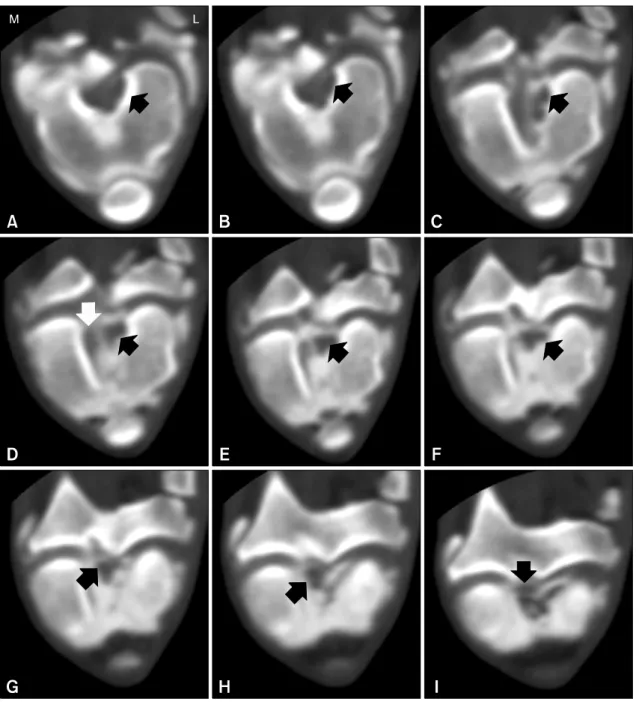
Fig. 3. Two-millimeter sequential transverse computed tomographic arthrography images were scanned parallel to the fibula, femoral attachment (A), and tibial attachment (I). The cranial cruciate ligament (black arrow) transverse images and caudal cruciate ligament sagittal images (white arrow) were clearly identified. L: lateral, M: medial.
Image analysis
Using Visus Image Analysis software (Ista-Video Test;
Foresthill Products, USA), the sequential transverse images of the cranial cruciate ligament were evaluated.
Pre-operative images were compared with post-operative images in each cadaver.
Results
Nine sequential transverse cranial cruciate ligament
images were obtained (Fig. 3). The initial transverse CTA
image of the intact cranial cruciate ligament at the tibial
attachment revealed a comma shape. The middle stage
revealed a round shape. The final CTA transverse image
obtained at the attachment of the cranial cruciate ligament
to the menisci was eclipse shaped. Total cranial cruciate
ligament slices involved the femoral attachment to the
tibial attachment. The mean number of slices for five
cadavers was 5.7. Five slices were obtained from cadaver
4, while six slices were obtained from the other cadavers.
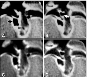
Fig. 4. Comparison of the pre-operative conventional view (A) and tracing view (C) with the post-operative conventional view (B) and tracing view (D). The transverse area of the cranial cruciate ligament image (black arrow) was decreased by 25% on the post-operative image. The small gas artifact (white arrow) was considered a normal finding. L: lateral, M: medial.
Table 1. Transverse area (mm
2) before and after partial cranial cruciate ligament rupture
Cadaver No.
Slice No.
1 2 3 4 5 6
1
2
3
4 Pre Post
% Pre Post
% Pre Post
% Pre Post
%
0.33 0.34 104 0.54
0.5 91 0.55 0.47 85 0.5 0.46
93
0.48 0.47 99 0.66 0.52 78 0.58 0.56 94 0.56 0.51 90
0.46 0.45 99 0.68 0.62 90 0.68 0.23*
33 0.55 0.46*
83
0.57 0.25*
44 0.71 0.52*
73 0.7 0.17*
25 0.5 0.33*
65
0.56 0.1*
17 0.74 0.49*
66 0.8 0.71
88 0.58
0.5 87
0.56 0.3*
59 0.71 0.64 90
- - - 0.66 0.61 93
Pre: pre-operative computed tomographic arthrography, Post:post-operative computed tomographic arthrography. *The slice in which the partial cranial cruciate ligament rupture was done, % = post-operative value/pre-operative value ×100.
Images at the same anatomical location were compared before and after surgery. Defect lesions were identified on post-surgery CTA images (Fig. 4). Air artifact were also identified after the procedure was complete.
In the pre-operative period, the cranial cruciate ligament area range was 0.3∼0.6 mm
2/kg for cadaver 1, 0.5~0.7 mm
2/kg for cadaver 2, 0.6∼0.8 mm
2/kg for cadaver 3, and 0.5∼0.7 mm
2/kg for cadaver 4. In the post-operative period, decreases in the area of the cranial cruciate ligament defect in cadaver 1 were 56% in the fourth slice, 83% in the fifth slice, and 41% in the sixth slice; in cadaver 2 they were 27% in the fourth slice and 34% in the fifth slice; in cadaver 3 they were 67% in the third slice and 75% in the fourth slice; in cadaver 4 they were 17% in the third slice and 35%
in the fourth slice (Table 1). The decreases in non-defect lesion area ranged from -4% to 15%.
In cadaver 2, the area of the lesion in the second slice decreased by 22%; this finding may have been due to contrast medium infiltrating the cranial cruciate ligament.
Discussion
We sought to determine the diagnostic utility of single detector CTA for identifying ligamentous structures in the normal canine stifle and to establish a repeatable imaging protocol. The ligamentous structures were easily identified using the CTA protocol described. Multiplanar reconstructions were helpful for the evaluation of cranial cruciate ligaments, medial and lateral menicsi, long digital extensor
tendons, and popliteal tendons.
We tried to orient reconstruction planes to parallel the axis of the cranial cruciate ligament in order to evaluate its entire length. The obliquity required to achieve this was variable within and between dogs. Subtle differences in limb positioning were not resolved. In addition, there were limitations in describing the total cranial cruciate ligament appearance. In an earlier study, the stifle position had no definite angle and was extended caudally at random. The CT gantry was angled such that the scanning plane was parallel to the tibial plateau. For this reason, transverse cranial cruciate ligament images were obtained atypically [8]. For successful examination to occur, each patient needed to be in a fixed position while scanning for constant images of the cranial cruciate ligament occurred. During the primary examination, cranial cruciate ligament tension was observed in various positions. We found that, when the stifle angle was 90 degrees, the assumption line was horizontal to the cranial cruciate ligament and vertical to the fibula. For this reason, the CT gantry was angled such that the scanning plane was parallel to the fibula. As a result, we obtained sequential transverse CTA images of the cranial cruciate ligament.
In the present study, an average of 5.7 slices were taken from some regular-distant cross sections of whole images in four cadavers. In the pre-operative stage, the cross sections of the cranial cruciate ligament progressed from
‘comma’, to ‘round’, to ‘eclipse’, in sequence from the
femoral attachment to the tibial attachment, and the
Computed tomographic arthrography and partial cranial cruciate ligament 399