CASE REPORT
간세포암종의 고주파 열 치료 후 발생한 장-농양 누공에 대해 Histoacryl 색전술로 성공적으로 치료한 1예
김지연, 권용환, 이상직, 장세영, 양해민, 전성우, 권영오
경북대학교 의과대학 내과학교실
Abscesso-Colonic Fistula Following Radiofrequency Ablation Therapy for Hepatocellular Carcinoma; A Case Successfully Treated with Histoacryl Embolization
Ji Yeon Kim, Young Hwan Kwon, Sang Jik Lee, Se Young Jang, Hae Min Yang, Seong Woo Jeon and Young Oh Kweon Department of Internal Medicine, Kyungpook National University School of Medicine, Daegu, Korea
Hepatocellular carcinoma (HCC) is one of the most common malignant neoplasms occuring worldwide. Although surgical resection still remains the treatment of choice for HCC, radiofrequency ablation (RFA) has emerged as reliable alternatives to resection.
It is less invasive and can be repeated after short intervals for sequential ablation in case of multiple lesions. The most common complication of RFA is liver abscess, and bile duct injury such as bile duct stricture has been reported. This is a case report of a rare complication of abscesso-colonic fistula after RFA for HCC. The case was treated by percutaneous abscess drainage and antibiotics and occlusion of abscesso-colonic fistula with n-butyl-2-cyanoacrylate embolization. (Korean J Gastroenterol 2011;58:270-274)
Key Words: Carcinoma; Hepatocellular; Radiofrequency catheter ablation; Fistula; Abscess
Received June 29, 2011. Revised August 1, 2011. Accepted August 5, 2011.
CC This is an open access article distributed under the terms of the Creative Commons Attribution Non-Commercial License (http://creativecommons.org/licenses/
by-nc/3.0) which permits unrestricted non-commercial use, distribution, and reproduction in any medium, provided the original work is properly cited.
교신저자: 권영오, 700-721, 대구시 중구 동덕로 200, 경북대학교병원 내과
Correspondence to: Young Oh Kweon, Department of Internal Medicine, Kyungpook National University Hospital, 200, Dongduck-ro, Jung-gu, Daegu 700-721, Korea.
Tel: +82-53-420-5518, Fax: +82-53-426-8773, E-mail: [email protected] Financial support: None. Conflict of interest: None.
INTRODUCTION
Hepatocellular carcinoma (HCC) is a major health problem worldwide, with an estimated incidence ranging between 500,000 and 1,000,000 new cases annually. It is highly prev- alent in the Asia-Pacific region and Africa, and is increasing in Western countries. It is the fifth most common cancer in the world, and the third most common cause of cancer-re- lated death.1,2
Liver resection and liver transplantation remain the op- tions that give the best chance of a cure. But, the selection of patients with HCC for partial hepatectomy has evolved into a complex task that incorporates information regarding tu-
mor extent, the severity of the underlying liver disease, the liv- er functional reserve and the general medical condition of the patient.3,4
Recent evidence suggests that local ablative therapy may offer comparable survival results in patients with small HCC, and preserved liver function. Radiofrequency ablation (RFA) for the treatment of HCC is currently widely practiced around the world. Randomized trials justify RFA for small HCC (single tumor ≤5 cm or three or less tumors ≤3 cm).5 RFA can be performed in a minimally invasive manner, but even when performed using a minimally invasive route, a wide spectrum of complications has been reported since its clinical ap- plication. We report a patient who suffered a rare complica-
Fig. 1. CT shows 1.6 cm sized hepatocellular carcinoma Hepato- cellular carcinoma in segment V. Black arrow indicates the close proximity of the colon to HCC.
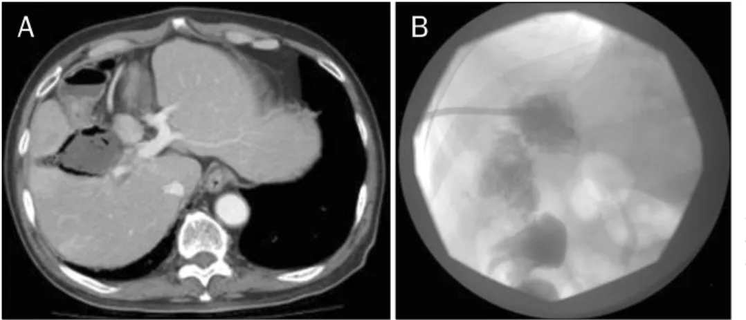
Fig. 2. (A) CT showed 6 cm sized air-bubble containing liver abscess and communication with hepatic flexure of colon. (B) Contrast study through percutaneous drainage ca- theter demonstrated communication between the abscess cavity and the ascending colon.
tion of abscesso-colonic fistula after RFA and successfully treated with n-butyl-2-cyanoacrylate (NBCA, Histoacryl; B.
Braun, Melsungen, Germany) embolization.
CASE REPORT
A 76-year-old male with a history of recurred HCC, measur- ing 1.6 cm in diameter located in the segment V was treated with RFA (Fig. 1). He was asymptomatic after the procedure and was discharged on the fifth day after RFA. Ten days later, he presented with watery diarrhea (2-3 times a day), inter- mittent febrile sensation and chills. The patient also had fe- ver, anorexia and nausea. The patient had a history of chronic hepatitis C, type 2 diabetes mellitus, cerebral infarction and open cholecystectomy for gallbladder empyema. Five years ago, he was diagnosed to have HCC on serial CT scan, and
then transarterial chemoembolization (TACE) was per- formed. About three years later, he developed intrahepatic recurrence and received TACE, and it was successful. The pa- tient was disease-free for the subsequent 20 months.
On physical examination, the abdomen was noted for a right subcostal line of surgical scar due to open chol- ecystectomy which was done ten years ago. There was no ten- derness, abdominal distension, or hepatosplenomegaly.
Laboratory results revealed ALP of 174 IU/L (normal:
35-129), GGT of 97 U/L (normal: 8-61), albumin of 2.9 mg/dL (3.2-4.8), white blood cells of 16,180/μL (normal: 4,800- 10,800), and CRP of 7.74 mg/dL (normal: 0-0.4). Serum AST, ALT, bilirubin, PT, and partial thromboplastin time was within normal limits.
A dynamic CT scan of the abdomen revealed liver abscess communicating with hepatic flexure of colon (Fig. 2A). Ultra- sonography-guided percutaneous drainage of the liver ab- scess was performed and thick dark-yellowish pus was drained. Injection of contrast via the needle demonstrated the presence of abscesso-colonic fistula (Fig. 2B).
The patient underwent parenteral nutrition for emptying of gastric contents and broad spectrum antibiotics were ad- ministered for Pseudomonas aeroginosa isolated and cul- tured from abscess cavity fluid. Three weeks later, the pa- tient’s symptoms improved and drainage of abscess cavity fluid significantly decreased, but the fistula remained on seri- al exams.
1.5 mL of NBCA and iodized oil (10 mL Lipiodol Ultra-Fluid;
Laboratoire Andre Guerbet, Aulnay-sous-Bois, France) were mixed at a 1:2 ratio and injected at a volume suited to the size of the enteric leak while filling and sealing the fistula tract and remnant abscess cavity via percutaneous catheter. Follow-
Fig. 4. CT scan obtained 3 months after removal of percutaneous catheter drainage. CT showed resolution of liver abscess and communication with colon.
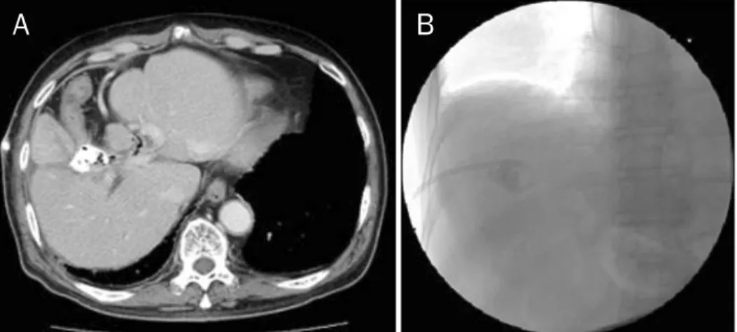
Fig. 3. (A) Hepatic abscess was filled with histoacryl. (B) Percutaneous tu- bography revealed no evidence of fistula between abscess and colon.
up CT scan and percutaneous tubography showed absence of abscess cavity and communication between abscess and- colon (Fig. 3A, B). Three weeks later, the percutaneous cathe- ter was removed and he was discharge from hospital. At fol- low-up 3 months after discharge, the patient had no symp- tom and CT scan revealed no liver abscess and fistula (Fig.
4).
DISCUSSION
HCC is usually associated with liver cirrhosis in patients with chronic viral hepatitis status, rendering resection not possible in many cases even for small HCC. RFA has gained wide acceptance as a safe alternative to surgery in the man- agement of early HCC and metastatic liver tumors.6,7
Patients are required to have either a single tumor smaller than 5 cm or as many as three nodules smaller than 3 cm each, no evidence of vascular invasion or extrahepatic spread, performance status test of 0, and liver cirrhosis in Child-Pugh class A or B.8,9 In the setting of metastatic dis- ease, percutaneous ablation is generally indicated for non- surgical patients with colorectal cancer oligometastases iso- lated to the liver.10
It produces complete necrosis of the tumor and achieves a satisfactory survival rate with low recurrence rate on long term follow-up. The main advantages of RFA include 1) it is minimally invasive with acceptable morbidity, 2) it enables excellent local tumor control, 3) it has promising long-term survival, and 4) it is a multimodal approach.11 Despite the benefits, RFA entails some risks as revealed by post-RFA complications. Complications such as hepatic failure, intra- peritoneal bleeding, abscess, bile duct injury, and tumor seeding are very serious and can be life threatening.12,13 Kong et al.14 recently reported thirty-seven (10%) major com- plications among 255 patients which included 13 cases of hepatic failure, 10 cases of hydrothorax requiring drainage, 3 cases of tumor seeding, one case of upper gastrointestinal bleeding, one case of intrahepatic abscess, one case of bile duct injury, one case of cardiac arrest, and 5 cases of hyperglycemia.
The thermal energy generated by RFA, when not insulated by the visceral peritoneum covering the liver, may spread into surrounding organs. Perforation of the gastrointestinal tract has been reported as a serious complication specific to RFA, occuring with an overall incidence of 0.1-0.2%.12,15 Postoper- ative adhesion of abdominal organs may remain a major problem in patients undergoing RFA. Livraghi et al.12 noted
that six of seven perforations occurred in patients with a his- tory of colonic resection, and fibrotic adhesions between the liver and gastrointestinal tract. Adhesions between the liver and gastrointestinal tract appear to increase the risk of per- foration.
Although occurrence of biliary-colonic fistula after chol- ecystectomy or secondary to malignancy was mentioned in previous review,15,16 abscesso-colonic fistula following RFA has not been reported in the literature so far. While the colon is typically hydrodissected during RFA to reduce the risk of in- jury, it is possible to inadvertently puncture the bowel with an applicator or extension of the ablation zone. Perforation may be followed by fistula and abscess formation.17 In our case, the possible mechanism of bowel injury could be caused by 1) direct perforation of the bowel by sharp electrodes which penetrated through the liver capsule during the procedure, and 2) thermal damage by heat generated and conducted to the colon adjacent to the subcapsular liver tumor, resulting in delayed perforation of the colonic wall. This could have been enhanced by adhesions between the liver and the colon following previous surgery. Leakage of colonic content track- ing into the liver and the subsequent ascending infection re- sulted in liver abscess. There is no consensus on the optimal treatment of abscesso-colonic fistula, and most of the liter- ature comes from case reports. Percutaneous image- guided drainage was required in addition to intravenously ad- ministered antibiotics and colonoscopic suture of the perfo- ration site. If it was not sufficient for treatment, surgical inter- vention was performed on the basis of surgical preference.12 The liver including abscess cavity, was partially resected and the fistulous orifice of the colon was closed.18
NBCA is a tissue monomer that instantly polymerizes upon contact with body fluids at neutral pH. It is used routinely in gastroenterology for control of bleeding gastic varix and plas- tic surgery for sutureless closure of surgical incisions, in neu- rosurgery for obliteration of bleeding aneurysm, and in gen- eral surgery for closure of refractory post-operative fistula.
Endoscopic treatment of postoperative fistulas with NBCA was reported in both the upper and lower gastrointestinal tract, as well as in case of pancreatic and biliary fistulas.19,20 This report describes the first case of abscesso-colonic fistu- la due to RFA that was treated injection of NBCA. NBCA (one or two 0.5 mL doses) and iodized oil were mixed at a 1:2 ratio rather than a 1:1 ratio to avoid adherence. The required vol-
ume was estimated from the contrast volume required to fill the tract. The catheter and delivery syringe were thoroughly flushed with 5% dextrose solution to avoid premature poly- merization; then, NBCA was injected.21 The catheter should be removed quickly to avoid adherence but in this case, the drainage catheter was not removed because the amount of drainage fluid was still more than 10 mL/day and the drain- age fluid was still discolored, although the drainage fluid was clearer than at the initial presentation. Follow-up CT scan and percutaneous tubography showed absence of abscess cavity and communication between abscess and colon, so the per- cutaneous drainage catheter was withdrawn.
The close proximity of the bowel to the targeted lesion at a subcapsular location is not favorable for percutaneous RFA and adhesions between the liver and gastrointestinal tract because of previous surgery appear to increase the risk of perforation. So a better understanding and careful assess- ment of the tumor and the approach of the procedure are im- portant in preventing patient morbidity and death.
REFERENCES
1. Lau WY. Management of hepatocellular carcinoma. J R Coll Surg Edinb 2002;47:389-399.
2. Llovet JM, Burroughs A, Bruix J. Hepatocellular carcinoma.
Lancet 2003;362:1907-1917.
3. Lau WY. The history of liver surgery. J R Coll Surg Edinb 1997;42:303-309.
4. Lau WY. A review on the operative techniques in liver resection.
Chin Med J (Engl) 1997;110:567-570.
5. Ng KK, Poon RT. Radiofrequency ablation for malignant liver tumor. Surg Oncol 2005;14:41-52.
6. Khan MR, Poon RT, Ng KK, et al. Comparison of percutaneous and surgical approaches for radiofrequency ablation of small and medium hepatocellular carcinoma. Arch Surg 2007;142:
1136-1143.
7. Guglielmi A, Ruzzenente A, Valdegamberi A, et al. Radiofre- quency ablation versus surgical resection for the treatment of hepatocellular carcinoma in cirrhosis. J Gastrointest Surg 2008;
12:192-198.
8. Lencioni R, Cioni D, Crocetti L, Bartolozzi C. Percutaneous abla- tion of hepatocellular carcinoma: state-of-the-art. Liver Transpl 2004;10(2 Suppl 1):S91-97.
9. Lencioni R, Crocetti L. A critical appraisal of the literature on local ablative therapies for hepatocellular carcinoma. Clin Liver Dis 2005;9:301-314.
10. Gillams AR, Lees WR. Radio-frequency ablation of colorectal liv- er metastases in 167 patients. Eur Radiol 2004;14:2261-2267.
11. Rhim H, Lim HK. Radiofrequency ablation of hepatocellular car- cinoma: pros and cons. Gut Liver 2010;4 Suppl 1:S113-S118.
12. Livraghi T, Solbiati L, Meloni MF, Gazelle GS, Halpern EF, Goldberg SN. Treatment of focal liver tumors with percutaneous radio-frequency ablation: complications encountered in a multi- center study. Radiology 2003;226:441-451.
13. Giorgio A, Tarantino L, de Stefano G, Coppola C, Ferraioli G.
Complications after percutaneous saline-enhanced radiofre- quency ablation of liver tumors: 3-year experience with 336 pa- tients at a single center. AJR Am J Roentgenol 2005;184:
207-211.
14. Kong WT, Zhang WW, Qiu YD, et al. Major complications after ra- diofrequency ablation for liver tumors: analysis of 255 patients.
World J Gastroenterol 2009;15:2651-2656.
15. Mulier S, Mulier P, Ni Y, et al. Complications of radiofrequency coagulation of liver tumours. Br J Surg 2002;89:1206-1222.
16. Munene G, Graham JA, Holt RW, Johnson LB, Marshall HP Jr.
Biliary-colonic fistula: a case report and literature review. Am Surg 2006;72:347-350.
17. Uppot RN, Silverman SG, Zagoria RJ, Tuncali K, Childs DD,
Gervais DA. Imaging-guided percutaneous ablation of renal cell carcinoma: a primer of how we do it. AJR Am J Roentgenol 2009;
192:1558-1570.
18. Satoh H, Matsuyama S, Mashima H, Imoto A, Hidaka K, Hisatsugu T. A case of hepatocolic fistula after percutaneous drainage for a gas-containing pyogenic liver abscess. J Gas- troenterol 1994;29:782-785.
19. Petersen B, Barkun A, Carpenter S, et al; Technology Assess- ment Committee, American Society for Gastrointestinal Endo- scopy. Tissue adhesives and fibrin glues. Gastrointest Endosc 2004;60:327-333.
20. Seewald S, Brand B, Groth S, et al. Endoscopic sealing of pancre- atic fistula by using N-butyl-2-cyanoacrylate. Gastrointest En- dosc 2004;59:463-470.
21. Bae JH, Kim GC, Ryeom HK, Jang YJ. Percutaneous embolization of persistent biliary and enteric fistulas with Histoacryl. J Vasc Interv Radiol 2011;22:879-883.