Korean J Gastroenterol Vol. 57, No. 4, 258-261 DOI: 10.4166/kjg.2011.57.4.258
CASE REPORT
Korean J Gastroenterol, Vol. 57 No. 4, April 2011 www.kjg.or.kr
초음파유도하 경피적 배액술에 의해 성공적으로 치유된 비장피막하 출혈을 동반한 췌장가성낭종 1예
김영일, 박선영, 이정현, 기원주, 박창환, 김현수, 최성규, 류종선
전남대학교 의과대학 내과학교실
A Case of Pancreatic Pseudocyst with a Large Subcapsular Splenic Hematoma Treated Successfully by Ultrasonography-guided Percutaneous Drainage
Young-Il Kim, Seon-Young Park, Jeong-Hyeon Lee, Won-Ju Kee, Chang-Hwan Park, Hyun-Soo Kim, Sung-Kyu Choi and Jong-Sun Rew Department of Internal Medicine, Chonnam National University Medical School, Gwangju, Korea
A subcapsular splenic hematoma is a very rare hemorrhagic complication of pancreatitis. We report here on a case of pseudocyst with a large subcapsular splenic hematoma in a 43-year-old man who presented with severe left flank pain for one week.
Despite the initial conservative treatment consisting of pain control, bowel rest, intravenous fluids and antibiotics, the pain was not relieved. An abdominal computed tomography (CT) was performed, and it showed a pseudocyst that was increasing in size with a large subcapsular splenic hematoma measuring 6×13 cm compared to the images at admission. Ultrasonography (US)-guided percutaneous drainage was performed without any complications, and splenectomy was avoided. After the discharge, the patient remained asymptomatic for eight months. We suggest that percutaneous drainage of a large subcapsular hematoma complicating pancreatitis might be a useful treatment option in selected patients. (Korean J Gastroenterol 2011;57:258-261) Key Words: Subcapsular splenic hematoma; Pancreatitis; Pseudocyst; Drainage
Received April 28, 2010. Revised June 29, 2010. Accepted July 2, 2010.
CC This is an open access article distributed under the terms of the Creative Commons Attribution Non-Commercial License (http://creativecommons.org/licenses/
by-nc/3.0) which permits unrestricted non-commercial use, distribution, and reproduction in any medium, provided the original work is properly cited.
교신저자: 최성규, 501-757, 광주시 동구 학동 8번지, 전남대학교 의과대학 내과학교실
Correspondence to: Sung-Kyu Choi, Department of Internal Medicine, Chonnam National University Medical School, 8, Hak-dong, Dong-gu, Gwangju 501-757, Korea.
Tel: +82-62-220-6296, Fax: +82-62-228-1330, E-mail: [email protected] Financial support: None. Conflicts of interest: None.
INTRODUCTION
A pancreatic pseudocyst is a relatively common complica- tion of acute and chronic pancreatitis, and it may be asso- ciated with many splenic complications including massive hemorrhage into pseudocyst, sepsis with splenic infarction, and splenic vein thrombosis.1 However, a subcapsular splen- ic hematoma is a very rare hemorrhagic complication of pancreatitis.2-7 We describe herein a case of pancreatic pseudocyst with a large subcapsular splenic hematoma suc- cessfully treated with US-guided percutaneous drainage.
CASE REPORT
A 43-year-old man presented to the emergency room at Chonnam National University Hospital with severe left flank pain for one week duration that was increasing in intensity.
The patient had no history of recent trauma. He quit drinking alcohol after two episodes of pancreatitis during the past 19 months. During the second episode of pancreatitis occurred 11 months after the first episode, a contrast-enhanced ab- dominal CT scan showed multiple pseudocysts with variable sizes at the tail of the pancreas compressing adjacent struc-
Kim YI, et al. A Case of Pancreatic Pseudocyst with a Large Subcapsular Splenic Hematoma 259
Vol. 57 No. 4, April 2011
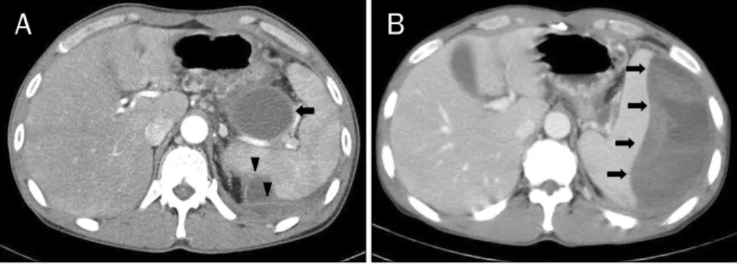
Fig. 1. Computed tomographic (CT) scan images during the second episode of pancreatitis and in the emergency room. (A) Variable sized multiple pseudocysts in the tail of the pancreas compressed the adjacent structures including splenic vein, gastric fundus (arrow), and splenic parenchyma (arrow heads). (B) A CT scan showed increased size of the pseudocyst with a large subcapsular splenic hematoma (arrows) measuring 5×12 cm in the tail of pancreas.
tures including the splenic vein, splenic parenchyma and gastric fundus (Fig. 1A). A drainage procedure was not per- formed because he recovered after the conservative treat- ment consisting of pain control, bowel rest, intravenous flu- ids, and antibiotics.
When the patient arrived at the emergency room 12 months after recovery from the second episode of pan- creatitis, his blood pressure, pulse rate and body temper- ature were 100/70 mmHg, 84 beats per minute and 37.0oC, respectively. On physical examination, there was marked ab- dominal tenderness and rebound tenderness at the left up- per quadrant of the abdomen. The laboratory findings showed that the liver function tests, renal function tests and electrolytes were within normal limit. However, there was a leukocytosis (16,200/mm3), increased C-reactive protein (18.2 mg/dL), and decreased hemoglobin (11.7 g/dL) and hematocrit (35.9%). The serum amylase and lipase levels were 113 U/L and 71 U/L, respectively, which were slightly above the normal range. An abdominal CT was performed to rule out a surgical abdomen, and showed increased size of the pseudocyst with a newly developed large subcapsular splenic hematoma in the tail of pancreas measuring 5×12 cm (Fig. 1B).
During the first 5 days of hospitalization, the pain was sig- nificantly relieved after initial treatment with intravenous an- algesics, antibiotics, fluids and bowel rest. However, on the sixth day, the patient complained of a sudden onset of severe left flank pain not relieved by analgesics, and the hemoglobin levels were decreased (9.7 g/dL). However, there was no hypotension. Another abdominal CT was performed, and it
showed an increase in the size (6×13 cm) of the pseudocyst at the tail of the pancreas with a large subcapsular splenic hematoma (Fig. 2A, 2B). Immediate surgical treatment was considered, but the patient refused due to the fear of complications. Therefore, US-guided percutaneous drainage of the hematoma was performed, immediately. About 300 mL of dark colored fluids was drained via a catheter, initially.
There were no procedure-related complications.
After the percutaneous drainage, the pain subsided dramatically. Because the amount of the drained fluid de- creased markedly and the size of pseudocyst with a sub- capsular splenic hematoma was markedly decreased on a follow-up abdominal CT scan (Fig. 2C, 2D), the catheter was removed three weeks after the procedure. There were no oth- er complications or symptoms after the catheter was re- moved, and the patient was discharged and has remained asymptomatic at the 8-month follow-up.
DISCUSSION
In the present case, a subcapsular splenic hematoma complicating pancreatic pseudocyst at the tail of the pan- creas was successfully treated with US-guided percutaneous drainage, and surgical procedures such as a splenectomy and distal pancreatectomy were avoided. Prior to the present case, three cases have been reported to be successfully treated by percutaneous drainage of a subcapsular splenic hematoma. Table 1 shows the three cases.3,5,7 The patients in the cases were all male, had no history of trauma. They all had a history of recent episodes of acute or chronic pan-
260 김영일 등. 다량의 비장피막하 출혈을 동반한 췌장가성낭종 1예
The Korean Journal of Gastroenterology
Fig. 2. CT scan images on hospital day 6 and 3 weeks after US-guided drainage. (A and B) increased size (approximately 6×13 cm) of the psedocyst with subcapsular splenic hematoma is shown (arrows and arrow heads). (C and D) CT images 3 weeks after US-guided percutaneous drainage, the size of the pseudocyst with subcapsular splenic hematoma was markedly decreased. A drainage catheter is shown (arrow head).
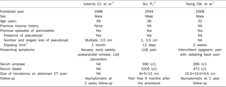
Table 1. Cases of Successful Percutaneous Drainage of Subcapsular Splenic Hematoma in the Literature
Vyborny CJ, et al.3 Siu TL.5 Tseng CW, et al.7 Published year
Sex Age (year)
Previous trauma history Previous episodes of pancreatitis Presence of pseudocyst
Number and largest size of pseudocyst Elapsing timea
Presenting symptoms
Serum amylase Serum lipase
Size of hematoma on abdomen CT scan Follow-up
1988 Male 58 None
Yes Yes Multiple, 2.5 cm
1 month Nausea, early satiety, postprandial emesis, LUQ
discomfort NA NA NA Asymptomatic at 2 years follow-up
2004 Male 38 NA Yes Yes 1, 3.5 cm
11 days LUQ pain
366 U/L 1505 U/L 8×5×13 cm Pain free 6 months after
the procedure
2008 Male 32 NA Yes NA NA 2 weeks
Intermittent epigastric pain with radiating back pain
266 U/L 473 U/L 15.0×13.0×9.5 cm Asymptomatic at 1 year
follow-up LUQ, left upper quadrant; NA, not available.
aElapsing time between the recent episode of pancreatitis and the identification of a subcapsular splenic hematoma.
creatitis, and two of them had single or multiple pancreatic pseudocysts previously. After the percutaneous drainage, there were no procedure-related complications and recurrence. The clinical course of the case reported here was similar to the previous reports.
Although splenic complications such as splenic vein thrombosis, intra-splenic pseudocysts, splenic rupture, in-
farction, necrosis and splenic hematoma may occur during the course of acute or chronic pancreatitis, because of the close anatomical location of the pancreatic tail to the hilum of spleen,8 hemorrhagic complications of pancreatitis involv- ing the spleen are very rare.2-7
There have been several possible mechanisms suggested that might explain how pancreatitis result in hemorrhagic
Kim YI, et al. A Case of Pancreatic Pseudocyst with a Large Subcapsular Splenic Hematoma 261
Vol. 57 No. 4, April 2011
splenic complications. A direct erosion of pancreatic en- zymes into the splenic parenchyma and subsequent dis- rupted splenic hilar vessels by pancreatic enzymes may lead to intra-splenic bleeding or subcapsular hematoma.6,9 Mechanical effects of intra-splenic pseudocysts may also cause a subcapsular splenic hematoma.10 Furthermore, pancreatic exudative materials may dissect into the sub- capsular space directly, and then, cause hemorrhage from the affected splenic parenchymal surface.3 In the case re- ported here, there was a history of two episodes of pan- creatitis, and a CT scan performed during the second episode showed variable sized pseudocysts at the tail of the pancreas compressing the splenic vein and parenchyma. Therefore, we assume that direct erosion into the splenic parenchyma or disruption of the splenic vein might have led to a sub- capsular splenic hematoma in this case. In addition, a sub- capsular splenic hematoma in this case might have resulted from pancreatitis; this patient, like another case,6 had no his- tory of trauma but marked elevations of the amylase and li- pase levels. Furthermore, the time between the recent epi- sode of pancreatitis and the identification of the subcapsular hematoma was 12 months in this case, longer than previous cases,3,5,7 suggesting possible chronic slow progression of the subcapsular hematoma.
Because the patient in this case was hemodynamically stable, had no signs of infection, refused surgical treatment due to the fear of complications, US-guided percutaneous drainage was performed to alleviate the abdominal pain and decompress the expanding subcapsular splenic hematoma.
Vyborny et al.3 suggested that patients with a slowly develop- ing (over weeks or months) post-traumatic hematoma and hematomas not associated with other complicating features of co-existent pancreatitis might be candidates for percuta- neous drainage. The hematoma in this case had these features. Prompt relief of symptoms, a short recovery time, avoidance of rupture and spleen preservation are the bene- fits of percutaneous drainage.3,5,7
Despite the benefits of percutaneous drainage, many in- vestigators have recommended surgical treatment such as the resection of the pseudocyst by splenectomy and, if neces- sary, distal pancreatectomy because of the risk of splenic rupture, infection and recurrence with drainage alone.4,8,11-13 Kuramitsu et al.2 suggested that a large splenic hematoma, larger than 5 cm, complicating pancreatitis should be treated
with pressure reduction by percutaneous drainage at an ear- ly stage or a laparotomy, even if the clinical course had tempo- rarily improved. However, the serious complications asso- ciated with surgical treatment such as bleeding and in- fections,12 and distal pancreatitis may predispose patients to the development of diabetes mellitus.4
In conclusion, US-guided percutaneous drainage was a useful treatment option for the subcapsular splenic hema- toma complicating a pancreatic pseudocyst in a patient with multiple episodes of pancreatitis. This procedure is less in- vasive and preserves the spleen.
REFERENCES
1. Habashi S, Draganov PV. Pancreatic pseudocyst. World J Gastroenterol 2009;15:38-47.
2. Kuramitsu T, Komatsu M, Ono T, et al. Ruptured subcapsular giant hematoma of the spleen as a complication of chronic pancreatitis. Intern Med 1995;34:564-568.
3. Vyborny CJ, Merrill TN, Reda J, Geurkink RE, Smith SJ. Subacute subcapsular hematoma of the spleen complicating pan- creatitis: successful percutaneous drainage. Radiology 1988;
169:161-162.
4. Malka D, Hammel P, Lévy P, et al. Splenic complications in chron- ic pancreatitis: prevalence and risk factors in a medical-surgical series of 500 patients. Br J Surg 1998;85:1645-1649.
5. Siu TL. Percutaneous drainage of spontaneous subcapsular haematoma of the spleen complicating chronic pancreatitis.
Surgeon 2004;2:52-55.
6. Patel VG, Eltayeb OM, Zakaria M, Fortson JK, Weaver WL.
Spontaneous subcapsular splenic hematoma: a rare complica- tion of pancreatitis. Am Surg 2005;71:1066-1069.
7. Tseng CW, Chen CC, Chiang JH, Chang FY, Lin HC, Lee SD.
Percutaneous drainage of large subcapsular hematoma of the spleen complicating acute pancreatitis. J Chin Med Assoc 2008;71:92-95.
8. Lankisch PG. The spleen in inflammatory pancreatic disease.
Gastroenterology 1990;98:509-516.
9. Greenstein A, DeMaio EF, Nabseth DC. Acute hemorrhage asso- ciated with pancreatic pseudocysts. Surgery 1971;69:56-62.
10. Warshaw AL, Chesney TM, Evans GW, McCarthy HF. Intrasplenic dissection by pancreatic pseudocysts. N Engl J Med 1972;287:
72-75.
11. Sitzmann JV, Imbembo AL. Splenic complications of a pancre- atic pseudocyst. Am J Surg 1984;147:191-196.
12. Heider R, Behrns KE. Pancreatic pseudocysts complicated by splenic parenchymal involvement: results of operative and per- cutaneous management. Pancreas 2001;23:20-25.
13. Agnifili A, Gianfelice F, Gola P, et al. Subcapsular splenic hema- toma complicating chronic relapsing pancreatitis. Case report.
Eur J Surg 1991;157:63-65.