* Department of Periodontology, Research Institute of Periodontal Regeneration, College of Dentistry, Yonsei University, Seoul, Korea
Abstract
The augmentation of hard and soft tissue deficiencies in replacement of missing anterior single tooth is critical to the esthetic outcomes. When considering the selection of an augmentation method, the morphology of the osseous defect may act as an important factor. The fewer the bony walls remain, the greater the need for autogenous block graft technique might be expected.
The use of autogenous bone graft represents the “gold standard”for bone augmentation procedures, which provides adequate volume of the bone reconstruction and the bone quality does not show a difference from the density of the native hard tissue in implant to bone contact. The biomechanical distribution of stress occurs primarily where the bone is in contact with the implant and increased density of interfacial bone provides better distribution and transmission of stress. Ideally, block bone graft should provide crestal bone with adequate density to withstand the implant loading. In spite of gains in the volume and the density of the deficient bone, there might be some disadvantages in esthetic aspects like recession of adjacent gingival margin and loss of interdental papilla when proper and careful management of the gingival flap is not prepared.
The cases in this report show horizontal and vertical hard tissue augmentation of alveolar ridge using autogenous block bone graft in replacement of missing anterior single tooth and gains in the width of the volume retained in the grafted area. However, some losses in the height of grafted hard tissue and some limitations of esthetic results were also found.
In conclusion, stability in maintenance of the vertical level in augmented area depends on the adjacent alveolar bone height. Barrier membrane serves as a preservation device for the grafted area protecting the autograft from postoperative resorption. In addition, careful handling of the soft tissue flap using fine surgical instruments and primary wound closure with tension-free sutures are important factors for soft tissue healing and prevent the barrier membrane from exposing, which in turn provides lower risks in further bone resorption or gingival recession.
K
Keeyy wwoorrddss :: block bone grafts, alveolar ridge augmentation, complication
Vertical and Horizontal Augmentation of Alveolar Ridge Using Autogenous Block Bone Graft for Anterior Single Tooth Replacement:
Benefits & Complications
Seung-Bum Lee*, Gyung-Joon Chae*, Ui-Won Jung*, Chang-Sung Kim*, Kyoo-Sung Cho*, Jung-Kiu Chai*, Chong-Kwan Kim*, Seong-Ho Choi*
상실된 단일 전치부 치조제에서 블록 자가골을 이용한 수직적, 수평적 재건 : 득과 실
이승범*, 채경준*, 정의원*, 김창성*, 조규성*, 채중규*, 김종관*, 최성호*
* 연세대학교 치과대학 치주과학교실, 치주 조직 재생 연구소
골과 임프란트 간에 발생하는 골 유착을 통해 악골을 고정원으로 사용하는 임프란트 시술 은 상실치 수복의 보편적인 치료방법으로 사 용되고 있다1). 하지만 임프란트 이식시 불충분한 골량은 성공률을 감소시키므로2), 장기적이고, 안정적인 예후를 위한 충분한 골량과 양호한 골질의 확보가 선행되어야 한다. 이를 위해 골유도 재생 술식이 상용되고 있다. 특 히 상악 전치부에 있어 외상이나 만성적 치주질환으로 인해 치아주변에 나타나는 심한 치조제 결손은 임프란 트 지지골의 재건은 물론, 심미적 요구의 충족과 발음상 문제의 해결이라는 측면에서도 수복되어야 한다3). 더불 어 부적절한 전치부 유도에 의해 발생하는 모멘트 힘이 최소화하도록 결손된 치조제를 재건해 줌으로써, 임프 란트 지대주가 보철학적 측면에서 생체역학적 지지를 얻도록 해야 한다4~6).
성공적인 치조제의 수평적, 수직적 재건은 차단막을 포 함하여, 블록 자가골과 분쇄 자가골을 함께 사용한 술식 이 이용되어져 왔다7~9). 블록 자가골 획득을 위한 공여 부는 구강내 공여부과 구강외 공여부로 나눌 수 있다.
그러나, 전신마취를 통한 수술의 부담으로 인하여, 많은
부의 재건뿐만 아니라, 치조능 결손부에 층판처럼 덧붙 여 적절한 부피를 확보할 수 있다3,10,12). 그러나, 차단막 이 노출되거나, 치간 유두 부위를 보존하지 못하는 경우 처럼 판막 조작이 미숙할 경우 연조직 재건에 실패하여 오히려 부가적인 치은퇴축이 일어날 수 있으며, 그로 인 하여 심미성이 요구되는 전치부위에 부적절한 결과를 초래할 수 있다.
이에 본 증례는 상실된 단일 전치부 치조제에서 블록 자 가골을 사용할 경우 수직적, 수평적으로 얻을 수 있는 골의 재건적 측면과 치간 유두 및 치은연의 높이를 통한 심미적 측면의 득과 실을 관찰하며, 이를 통해 본 술식 을 행할 경우 고려하여야 할 사항을 알아보고자 하는 바 이다.
증
증례례발발표표
CASE 1
Ⅱ
Fig. 1. 환자 초진시 임상사진(정면) Fig. 2. 발치 4개월 후 임상사진(교합면)
original article
2005년 4월, 43세 남자 환자가 위 앞니 잇몸이 부어서 고름이 나온다는 주소로 연세대학교 치과대학 병원 치 주과를 내원하였다. 증상은 5개월 전부터 지속되었다.
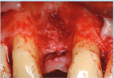
내원 당시 상악우측 측절치 부위 치은은 심한 부종과 발 적을 보였으며, 3도의 동요도와 치근단 1/3부위까지 치 은 퇴축을 보이고 있었다(Fig.1). 상악 우측 측절치의 발 치 후 3개월 동안 전악에 걸친 치주 치료를 시행하였고, 발치부위 치조제는 수평적, 수직적 골흡수로 Misch 가 용골분류 C-w28)의 양상을 보였다(Fig 2). 컴퓨터단층 촬영(Computer tomography)상으로 순,구개측 골의
단면을 파악한 후, 상악 우측 측절치 부위에 판막을 전 층으로 거상하였다(Fig 3). 수혜부의 크기를 측정하고, 수혜부의 피질골에 천공을 시행한 후, 동측 후구치 부위 에서 Piezosurgery (Piezosurgery, mectrion, Italy) 로 블록 자가골을 획득하였다(Fig 4). 획득한 블록 자가 골 은 수 혜 부 의 골 결 손 부 를 완 전 히 수 복 하 도 록 miniscew(Salvin Titanium Fixation Screw kit, Salvin, USA)로 고정시켰고(Fig 5), 주변은 흡수성 합 성골 synthetic resorbable bone substitute)인 MBCPTM(Macroporous biphasic calcium
Fig. 3. 치은 판막 거상 후 사진(정면) Fig. 4. 우측 하악 제2대구치 후방 공여부 사진
(블록 자가골 획득 전)
Fig. 5. 수혜부에 블록 자가골 위치 후 사진(정면) Fig. 6. 수술 2주후 차단막 노출사진 (정면)
Fig. 9. 임프란트 식립 직후 봉합사진(정면) Fig. 10. 임프란트 식립 5개월 후 사진(정면)
Fig. 11. 임프란트 식립 5개월 후 사진(교합면)
phosphate, Biomatlante, France)로 채운 후, 상부는 Gore-tex (Gore-tex, Nobel Biocare, USA)차단막 으로 덮었다. 증가된 치조능은 1차 봉합을 위해 치은 판 막에 장력이 생기지 않도록 당긴 후 봉합하였다.
치조능을 덮은 차단막 일부가 겹쳐진 상태에서 1차 봉 합되었고, 2주 후 겹쳐진 차단막 부위를 중심으로 노출 이 생겼다(Fig 6). 염증이 진행되지 않도록 주기적으로 관리하였으며, 4주 후 차단막의 제거를 위해 치은 판막 을 거상하였다. 노출된 부위 이외의 차단막은 건전한 상 태였으므로 노출된 부위의 차단막만을 제거한 후, 부족
original article
Fig. 13. 환자 발치 2개월 후 임상사진(임시치아 장착상태 의 정면)
Fig. 12. 환자 발치 2개월 후 임상사진(임시치아 미장착상 태의 교합면)
Fig. 14. 치은 판막 거상 후 사진(정면) Fig. 15. 우측 하악 제2대구치 후방 공여부 사진(블록 자가
골 획득 전) 한 연조직은 경구개로부터 획득한 결합조직을 이용하여
1차 봉합하였다(Fig 7). 4개월 후 충분한 부피의 골량은 얻었으나, 상악 우측 중절치 원심면에 치은 퇴축을 동반 한 치간 유두 소실이 있어났다(Fig 8).
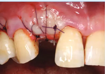
6개월 후 임프란트 식립을 위해 miniscrew(Salvin Titanium Fixation Screw kit, Salvin, USA)와 나 머지 차단막을 제거하였고, 직경 3.5mm, 길이 13mm 의 Replace select (Replace, Nobel Biocare, Sweden) 임프란트를 식립하였다. 봉합 시 치간 유두부 위의 재생을 위해 치은 판막을 당겨서 봉합하였다(Fig
9). 식립 5개월 후 수평적으로는 양호하나 수직적으로 다소 부족한 골의 양상이 보였다. 또한 우측 중절치 원 심면의 치간 유두 부위 소실 및 치은 퇴축이 관찰되었다 (Fig 10, 11).
CASE 2
2005년 10월, 36세 남자 환자가 전체적으로 이가 좋지 않아서 부드러운 것만 먹는다는 주소로 연세대학교 치 과대학병원 치주과에 내원하였다. 증상은 1년 전부터 지
을 포함하는 골소실을 보였다. 상악 우측 중절치의 발치 후 2개월 동안 전악에 걸친 치주 치료를 시행하였고, 예 후가 불량한 치아(hopeless tooth)의 추가적인 발치가 진행되었다. 전치부의 발치된 치조제는 수평적, 수직적 골흡수로 Misch 가용골 분류 C-h28)의 양상을 보였다 (Fig 12, 13). 컴 퓨 터 단 층 촬 영 (Computer tomography)상으로 순, 구개측 골 단면을 파악한 후,
재건하기로 하였다(Fig 14). 수혜부의 크기를측정하고, 수혜부의 피질골을 천공한 후, 동측 후구치 부위에서 Piezosurgery (Piezosurgery, mectrion, Italy)로 블 록 자가골을 획득하였다(Fig 15). 획득한 블록 자가골은 수 혜 부 의 골 결 손 부 를 완 전 히 수 복 하 도 록 miniscrew(Salvin Titanium Fixation Screw kit, Salvin, USA)로 고정시켰고, 주변은 흡수성 합성골
Fig. 16. 술 후 봉합사진(정면) Fig. 17. 이식술 6개월 후 사진(교합면)
Fig. 18. 임프란트 식립 후 봉합사진(정면)
original article
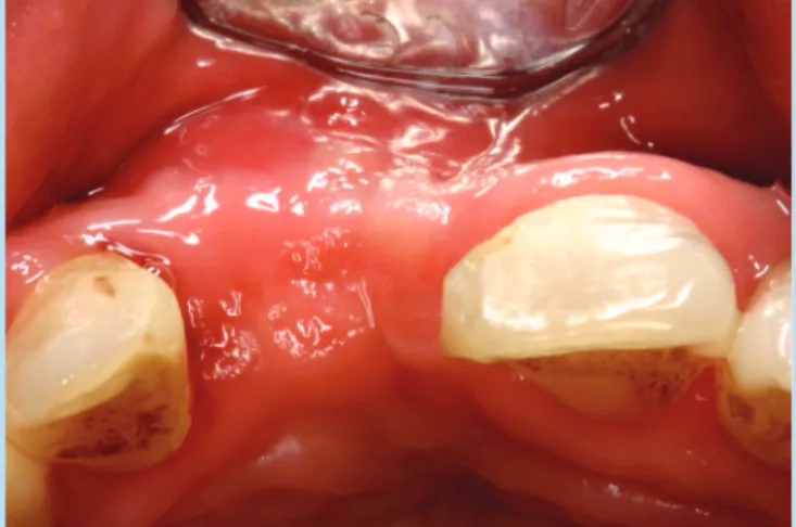
(synthetic resorbable bone substitute)인 MBCPTM 로 채운 후, 상부는 Gore-tex (Gore-tex, Nobel Biocare, USA) 차단막으로 덮었다. 이식 자가골 부피 로 증가된 치조능의 1차 봉합을 위해 치은 판막에 장력 이 생기지 않도록 당긴 후 봉합하였다(Fig 16). 6개월 후 임프란트 식립을 위한 충분한 수평적, 수직적 골량은 획득하였으나, 상악 우측 측절치 근심면에 치은 퇴축을 동반한 치간 유두의 소실을 보였다(Fig 17).
임프란트 식립을 위해 miniscrew(Salvin Titanium Fixation Screw kit, Salvin, USA)와 차단막을 제거 하였고, 직경 4.3mm, 길이 13cm의 Replace select (Replace, Nobel Bcocare, Sweden)임프란트를 식립 하였다(Fig. 18). 2차 수술시 상악 우측 측절치 근심면 의 치은 퇴축 부위에 추가적인 연조직 이식을 계획하고 있다.
총
총괄괄 및및 고고찰찰
전치부 상실된 단일 치아부위에 있어서, 소실된 경조직 과 연조직의 재건은 심미적인 결과를 도모할 수 있다4,5). 이때 경조직 결손의 형태는 골 증대술식을 결정짓는 중 요한 고려요소가 되며, 골 증대술식을 위한 이식골의
“gold standard”는 자가골로 평가되고 있다9,13). 따라 서, 골 벽이 적은 경우는 블록 자가골 이식의 강력한 적 응증이 되며, 이렇게 형성된 충분한 골량과 치밀한 골질 은 임프란트와의 접촉에 있어서 수여부의 원래 경조직 과 크게 다르지 않다. 따라서 식립된 임프란트의 하중을 균등하게 분산할 수 있게 된다4,14). 이는 블록 자가골을 이용한 상악 치조제 고경의 증가 후 임프란트 식립시 나 타나는 98.3%의 성공률로 입증된다15). 그러나 적절하고 숙련된 치은 판막의 조작이 뒷받침되지 않는다면, 인접
치은 조직의 퇴축과 치간 유두의 소실로 심미성의 개선 에는 한계를 보이게 된다.
1. 공여부
구강내 공여부에서 채득한 블록 자가골은 발생의 기원 이 동일한 막내골로써, 구강외 장골에서 채취한 블록 자 가골인 연골내골에 비해 이식 후 수평적 부피의 흡수가 적고, 재 혈관화가 빠르며, 수혜부에 대한 생착률이 높
다16~20). 따라서, 이식 후 치조능은 적은 수축량으로 유사
한 수직적 높이를 유지할 수 있으며, 골괴사 없이 생활 력을 유지할 수있다13,21,22). Proussaefs 등13)은 6개월 후 단지 17%의 흡수를 보이는 것으로 이를 입증하였다. 또 한, 전신 마취나 입원 없이 하악골 후구치 부위의 외사 선과 치조능에서 채득하므로 환자의 불편함과 통증을 최소화할 수 있고, 공여부로의 접근도가 좋아서 임상 현 장에서 비교적 간단히 시술할 수 있다23). 본 증례의 경우 에서도 블록 자가골 채득 과정과 채득 후 치유기간 동안 에 있어서 환자의 별다른 불편 호소는 없었다.
2. 수혜부
블록 자가골의 이식 전에 수혜부 표면의 피질골을 천공 시킨다. 이는 이식골이 하방의 골수와 교통하도록 하여, 혈류 공급을 조기에 안정화 시킴으로서 이식골의 생착 을 촉진시키고 초기의 골부피가 잘 유지되도록 한다1). 이를 위해 블록 자가골의 해면골 부위가 수혜부에 직접 맞닿도록 위치시킨다8).
치조제 골 증대술의 결과 수평적으로 4~7mm의 증가 와 수직적으로 2~3mm증대를 보였다17,24). 최근 4~5mm의 증가를 보고하고 있으나25), 술 후 76.3%의 흡수율을 보였다는 연구도 있다26). 결과적으로 수직적 골증대 보다 수평적 골증대의 획득이 용이하며, 수직적
Ⅲ
직적 골결손을 보이고 있다.
4개월 후 차단막의 노출이 시작된 곳도 동일 부위로서, 중절치 치근 원심면쪽 치조능을 덮은 차단막이 겹쳐진 것과 더불어 노출의 원인이 된 것으로 사료되고 있다.
임프란트 식립을 위한 블록 자가골 이식 6개월 후, 치은 판막의 재거상시‘V’형태의 골결손이 잔존해 있는 것을 확인할 수 있었으며, 이후 치은 퇴축은 개선되지 않았 다. 증례 2의 경우도, 상악 우측 측절치 치근 근심면에 원심면에서부터 이어진 수직적 골결손이 존재하였다.
술후 발생한 측절치 근심면의 치은 퇴축은 6개월 후에 도 개선되지 않았다.
3. 차단막
비흡수성 차단막을 이용하여 블록 자가골의 이식부위를 격리시킴으로써 이식된 골부피의 감소를 최소화하는 것 은 이미 여러 명의 연구자에 의해 입증되었다. 즉, 차단 막은 치은 점막에서 이주된 비골원성 세포가 골 증대를 위해 형성된 공간으로 침입하는 것을 막기위한 물리적 방어막으로 작용하게 된다8). 또한 차단막 없이 이식술을 시행한 경우 50% 정도의 흡수를 보인 반면, 차단막을 사용한 경우에는 이식골의 흡수를 막아서 재건된 치조 제의 부피를 유지시키게 된다27). 하지만, 장력 없이 치은 판막을 봉합하는 적절한 조작이 필요하며, 차단막의 노 출시 추가적인 이식골의 흡수와 치은 퇴축이 발생할 수 있다. 본 증례 1에서도, 차단막의 조기 노출로 1개월 후 차단막의 일부를 제거하였다.
이는 이식골 흡수를 동반한 치은 퇴축을 초래하였으며, 이후 연조직 이식술로는 회복할 수 없었다.
란트의 성공률을 보장하고, 보철적으로 장기적인 예후 를 보장하지만 주위 치아의 치조골 상태와 적절한 연조 직의 조작이 동반되지 않는다면 심미성에 있어서 만족 할 수 없는 결과를 나타낼 수 있다.
Reference
1. Buser D, Dula K, Belser U, Hirt HP, Berthold H. Localized ridge augmentation using guided bone regeneration. 1. Surgical procedure in the maxilla. Int J Periodontics Restorative Dent. 1993;13(1):29-45.
2. Lekholm U, Ericsson I, Adell R, Slots J. The condition of the soft tissues at tooth and fixture abutments supporting fixed bridges. A microbiological and histological study. J Clin Periodontol. 1986 Jul;13(6):558-62.
3. Misch CM, Misch CE, Resnik RR, Ismail YH. Reconstruction of maxillary alveolar defects with mandibular symphysis grafts for dental implants: a preliminary procedural report. Int J Oral Maxillofac Implants. 1992 Fall;7(3):360-6.
4. Misch CE. Contemporary Implant Dentistry. St. Louis, MO: Mosby- Year Book; 1993: 427-431, 575-597.
5. Taylor T, in discussion, Sullivan D, Kay H, Schwarz M, et al. Esthetic problems in the anterior maxilla. Int J Oral Maxillofac Implants.
1994;9(suppl):74.
6. Katona TR, Goodacre CJ, Brown DT, Roberts WE. Force-moment systems on single maxillary anterior implants: effects of incisal guidance, fixture orientation, and loss of bone support. Int J Oral Maxillofac Implants. 1993;8(5):512-22.
7. Buser D, Dula K, Hirt HP, Schenk RK. Lateral ridge augmentation using autografts and barrier membranes: a clinical study with 40 partially edentulous patients. J Oral Maxillofac Surg. 1996 Apr;54(4):420-32.
8. Buser D, Dula K, Belser UC, Hirt HP, Berthold H. Localized ridge augmentation using guided bone regeneration. II. Surgical procedure in
original article
the mandible. Int J Periodontics Restorative Dent. 1995 Feb;15(1):10- 29.
9. Misch CM, Misch CE. The repair of localized severe ridge defects for implant placement using mandibular bone grafts. Implant Dent. 1995 Winter;4(4):261-7. Review.
10. Misch CM. Ridge augmentation using mandibular ramus bone grafts for the placement of dental implants: presentation of a technique. Pract Periodontics Aesthet Dent. 1996 Mar;8(2):127-35; quiz 138.
11. Misch CM. Comparison of intraoral donor sites for onlay grafting prior to implant placement, Int J Oral Maxillofac Implants. 1997 Nov- Dec;12(6):767-76.
12 Pikos MA. Buccolingual expansion of the maxillary ridge. Dent Implantol Update. 1992 Nov;3(11):85-7.13. Proussaefs P, Lozada J, Kleinman A, Rohrer MD. The use of ramus autogenous block grafts for vertical alveolar ridge augmentation and implant placement: a pilot study. Int J Oral Maxillofac Implants. 2002 Mar-Apr;17(2):238-48.
14. Misch CE, Dietsh F. Bone-grafting materials in implant dentistry.
Implant Dent. 1993 Fall;2(3):158-67. Review.
15. Buser D, Ingimarsson S, Dula K, Lussi A, Hirt HP, Belser UC. Long- term stability of osseointegrated implants in augmented bone: a 5-year prospective study in partially edentulous patients. Int J Periodontics Restorative Dent. 200 2 Apr;22(2):109-17.
16. Smith JD, Abramson M. Membranous vs endochondrial bone autografts. Arch Otolaryngol. 1974 Mar;99(3):203-5. No abstract available.
17. Zins JE, Whitaker LA. Membranous versus endochondral bone:
implications for craniofacial reconstruction. Plast Reconstr Surg. 1983 Dec;72(6):778-85.
18. Borstlap WA, Heidbuchel KL, Freihofer HP, Kuijpers-Jagtman AM.
Early secondary bone grafting of alveolar cleft defects. A comparison between chin and rib grafts. J Craniomaxillofac Surg. 1990 Jul;18(5):201-5.
19. Lin KY, Bartlett SP, Yaremchuk MJ, Fallon M, Grossman RF, Whitaker LA. The effect of rigid fixation on the survival of onlay bone grafts: an experimental study. Plast Reconstr Surg. 1990 Sep;86(3):449-56.
20. Kusiak JF, Zins JE, Whitaker LA. The early revascularization of membranous bone. Plast Reconstr Surg. 1985 Oct;76(4):510-6.
21. Proussaefs P, Lozada J, Rohrer MD. A clinical and histologic evaluation of a block onlay graft in conjunction with autogenous particulate and inorganic bovine mineral (Bio-Oss): a case report. Int J Periodontics Restorative Dent. 2002 Dec;22(6):567-73.
22. De Carvalho PS, Vasconcellos LW, Pi J. Influence of bed preparation on the incorporation of autogenous bone grafts: a study in dogs. Int J Oral Maxillofac Implants. 2000 Jul-Aug;15(4):565-70.
23. Schwartz-Arad D, Levin L. Intraoral autogenous block onlay bone grafting for extensive reconstruction of atrophic maxillary alveolar ridges. J Periodontol. 2005 Apr;76(4):636-41.
24. Urist MR. Surface-decalcified allogeneic bone (SDAB) implants. A preliminary report of 10 cases and 25 comparable operations with undecalcified lyophilized bone implants. Clin Orthop Relat Res. 1968 Jan-Feb;56:37-50.
25. Proussaefs P, Lozada J. The use of intraorally harvested autogenous block grafts for vertical alveolar ridge augmentation: a human study. Int J Periodontics Restorative Dent. 2005 Aug;25(4):351-63.
26. Simon BI, Von Hagen S, Deasy MJ, Faldu M, Resnansky D. Changes in alveolar bone height and width following ridge augmentation using bone graft and membranes. J Periodontol. 2000 Nov;71(11):1774-91.
27. Ten Bruggenkate CM, Kraaijenhagen HA, van der Kwast WA, Krekeler G, Oosterbeek HS. Autogenous maxillary bone grafts in conjunction with placement of I.T.I. endosseous implants. A preliminary report. Int J Oral Maxillofac Surg. 1992 Apr;21(2):81-4.
28. Misch CE. Divisions of available bone in implant dentistry. Int J Oral Implantol. 1990;7(1):9-17. No abstract available.
교신저자 : 최성호
서울특별시 서대문구 신촌동 134 연세대학교 치과대학 치주과학교실 우편번호 : 120-752
이메일: [email protected]