375
Effect of Fluoride Treatment after Bleaching with Hydrogen Peroxide exposed to Plasma Arc
Sun-Young Chung
1, Young-Eun Lee
2, Sang-Hun Ahn
1,2, Hae-Young Yang
1,3, Eun-Suk Jeon
1,4, Youn-Hee Choi
1and Keun-Bae Song
1†1
Dept. of Preventive Dentistry, School of Dentistry, Kyungpook National University, Daegu, 700-412, Korea
2
Dept. of Dental Hygiene, Daegu Health College, Daegu, 702-722, Korea
3
Dept. of Dental Hygiene, Daegu Science College, Daegu, 702-723, Korea
4
Dept. of Dental Hygiene, Choonhae College of Health Scienes, Ulsan, 689-784, Korea
고농도 과산화수소와 플라즈마 아크를 이용한 미백 치료에 있어서 불소의 효과
정선영1·이영은2·안상헌1,2·양해영1,3·전은숙1,4·최연희1·송근배1†
1
경북대학교 치의학전문대학원 예방치과학교실,
2대구보건대학 치위생과,
3
대구과학대학 치위생과,
4춘해보건대학 치위생과
Abstract This study evaluated whether fluoride treatment can affect recovery of the irregularity of enamel surface after tooth whitening with a high concentration of hydrogen peroxide (HP) activated by plasma arc light. A total of 36 bovine teeth stained with coke were used in this experiment. The specimens were classified into following three groups (two different commercial plasma arc groups and a control group without light curing source): (1) 35% HP gel only, (2): 35%
HP gel and Plasma arc A, and (3) 35% HP gel and Plasma arc B. To measure color changes and surface morphologies before and after the bleaching, colorimeter and scanning electron microscopy were used, respectively. When the specimens were bleached with hydrogen peroxide and plasma arc lights, the bleaching effect was greater than when only hydrogen peroxide gels were used (Kruskal-Wallis test, p<0.05). In addition, plasma arc B showed the more color changes than plasma arc A (Bonferroni post-hoc test, p<0.05). The surfaces of the teeth treated with fluoride gel after the whitening treatment came to be smooth. Therefore, the results of this study suggested that the fluoride application for patients who got tooth whitening therapy with a high concentration of hydrogen peroxide gels activated by plasma arc light will be effective to recover rough enamel surfaces.
Key words Fluoride, Hydrogen peroxide, Plasma arc, Remineralization, Tooth bleaching
Introduction
Tooth whitening, an option of cosmetic therapy has become a popular remedy in dentistry for aesthetics, which are raised by patients’ demands for white teeth and an attractive smile. Tooth bleaching have been considered to be a safe, effective, minimally invasive, and non- destructive treatment to remove the discoloration of the tooth
1,2).
Tooth discoloration is attributable to extrinsic staining
on the surface of the teeth and intrinsic staining within the structure of the tooth. Extrinsic stains may be caused by foods and drinks, such as tea, coffee and cokes which con- tain dark pigments as well as mouthwashes containing the antibacterial chlorhexidine and iron tables. On the other hand, intrinsic stains may caused by fluoride from tooth- paste and fluoride drops or the use of tetracycline antibi- otic during a period of dental development
3).
In general, there are two different ways to whitening the teeth: one is the in-office procedures commonly known as
‘power bleaching’ and the other is ‘home bleaching’ or the
‘night guard vital bleaching’ technique
4-7). Power bleach- ing is the term given to accelerate tooth whitening proce- dures using either a xenon plasma arc-curing light or a
†
Corresponding author
Tel: 053-660-6870
Fax: 053-423-2947
E-mail: [email protected]
laser
6). Home bleaching commonly uses a relatively low level of whitening agents applied to the teeth for at least 2 weeks
5).
When hydrogen peroxide (HP) gels placed on a tooth surface, the oxidizing agent diffuses into enamel and den- tin, and then the free radicals create lighter intermediates by oxidizing the long-chained, dark-colored chromophore molecules to intermediates
7). The recent bleaching agents for a professional application are based on 35~50% HP with photosensitive components
8-10)that act as starters to initiate and catalyze the reaction when exposed to light sources. There are several different types of light activatin sources, and among the newest light sources are lasers, light emitting diodes, plasma arc lamps and halogen lamps. The theoretical advantage of a light source is its ability to heat the hydrogen peroxide, thereby increasing the rate of decomposition of oxygen to form oxygen free radicals and enhancing the release of stain-containing molecules
11).
The effect of HP on teeth and surrounding tissues are still controversial. However, previous studies suggest that hydroxyl radicals are extremely reactive and those have been shown to degrade components of connective tissue.
These toxic chemical by-products may be related to the side effects such as periodontal tissue destruction, root resorption, increasing of tooth sensitivity, mineral loss from enamel and reduction in micro-hardness of resin restorative materials
12,13). These adverse effects could be accelerated by photo curing devices, such as the plasma arc lamp by generating a high voltage pulse that creates hot plasma between two electrodes in a xenon-filled bulb
14).
Fluoride application is one of the protective methods to recover the mineral loss from enamel by tooth whitening.
Application of fluoride greatly enhances the rate of rem- ineralization of demineralized enamel and dentin
14,15). Therefore, it is necessary to assess the efficacy of preven- tive remedies such as fluoride application for mineral loss after tooth bleaching with a high concentration of hydro- gen peroxide gels exposed to light source, especially plasma arc lamp, by employing a variety of methodologi- cal frameworks.
The aim of this study was to evaluate the effectiveness of teeth bleaching that only 35% HP was applied and that both 35% HP and plasma arc light were applied and to examine that fluoride application can recover rough enamel surfaces after whitening procedures with high con- centrations of bleaching agents by using scanning elec- tron microscopy.
Materials and Methods
1. Preparation of Samples
A total of 36 bovine teeth without no caries and no enamel cracking were randomly selected. Those were sec- tioned horizontally at the point of 5 mm under cementoe- namel junction (CEJ), thereafter the pulp tissues were extirpated. The teeth stored in 0.1% tymol (Sigma-Ald- rich Corp., St. Louis, USA) at 4
oC before the experiment.
The teeth were embeded in an acrylic plastic mold 30 mm in diameter and 15 mm of in depth. Specimens were highly polished using an automatic polisher, Labopol-1 (Struers Inc., Ballerup, Denmark) with sandpaper (800 grit, 1000 grit, 1500 grit and 2000 grit) and diamond paste (6 µm , 3 µm, 1 µm, 0.25 µm and 0.1 µm) for the surface standardization. All of the specimens kept in wet or moist conditions at every step in the experiment. The specimens were artificially stained with commercial coke
4)(Coca- Cola Enterprises, Inc. Atlanta, USA) in which carbonic acid gas was eliminated by stirring for 2 hours. The spec- imens were dipped in coke for 24 hours. Coke as well as tea or coffee that we usually consume in daily life are one of the sources leading to tooth discoloration. To reflect the origin of exposure as usual, coke was used to stain bovine tooth samples, even though there are some methods to dye natural or artificial tooth samples.
2. Treatment with 35% Hydrogen Peroxide and Plasma arc Lights
Plasma arc lamps used in this study are recently devel- oped methods. The plasma arc light system has filters that are able to narrow the spectrum of visible light to a band centered at 470 nm and light produced by plasma arc lamps is different from that generated by halogen lamps.
Rather than relying upon a heated tungsten filament, plasma arc lamps work by application of high voltage cur- rent across two closely placed electrodes, resulting in a light arc between the electrodes. These lights have an evergy level of 900 mV, which is much higher than halo- gen lights
16). Two different commercial plasma arc lamps were used in this study design and a control group with- out any light source was prepared to compared the results of the experiment. So, a total of 36 pre-treated specimens randomly classified into the following three groups.
1) Group 1 (n=12): 35% HP gel (Oratech Inc., Spring-
field, USA) with a normal room light. The bleaching gels
were stirred every 10 seconds with the tip of the syringe
for the optimal effectiveness. The bleaching gels were
applied for 20 minutes under the room light and the pro- cedures were repeated three times.
2) Group 2 (n=12): 35% HP gel with Plasma arc A (Remedent Inc., Deurle, Belgium)
The bleaching gels were applied to the specimen with Plasma arc A for 20 minutes: the specimen was exposed to the Plasma arc A for 90 seconds with a 30 second-rest- ing, and this procedure was repeated 10 times. The light was positioned according to the manufacturer's instruc- tions and these procedures were repeated three times.
3) Group 3 (n=12): 35% HP gel with Plasma arc B (Rolence Enterprise Inc., Chungli, Taiwan)
The bleaching gels were applied to the specimen with Plasma arc B for 20 minutes and the procedures were repeated three times. Other conditions were same as the Group 2.
3. Treatment with Fluoride
To evaluate the effectiveness of the fluoride application after tooth whitening, 1.23% acidulated phosphate fluo- ride (APF) gel (Pascal Co., Bellevue, USA) was applied to 60 seconds with microbrush based on the results of the previous studies
17,18)and an instruction of the manufac- turer . Then, APF gel was removed 60 seconds after fluo- ride application using cotton pellets.
4. Measurement of Color Change
To evaluate color changes, a colorimeter (Nippon Den- shoku industries co. Ltd., Tokyo, Japan) was used before and after bleaching procedures. At the baseline, three des- ignated points were measured for each specimen. The average of each sample was calculated using the CIE Lab uniform color scale. The colors of all specimens were compared to a white standard, a pressed powder tablet of barium sulfate. Magnitude of the color difference was rep- resented by a single number ( E), which was calculated using the following equation:
∆E*={(∆L*)2+(∆a*)2+(∆b*)2}1/2
L*: lightness, a*: redness-greenness, and b*: yellow- ness-blueness.
5. Observation of Surface Change
Changes of the surface morphology were examined by using Scanning Electron Microscopy (SEM) (Hitachi Co., Tokyo, Japan). All specimens were dried at 60
oC in one hour at the same time and then sputter-coated with plati- num (180-200 Å thickness) under high vacuum in an ion
sputter (Eiko Engineering Co., Tokyo, Japan) for observa- tion of surface changes after bleaching (10,000x magnifi- cation).
5. Statistical Analyses
As the values of color changes did not show the normal distribution, non-parametric tests were used to analyze magnitude of the color difference. Kruskal-Wallis test was performed to examine the color changes among the three groups. Bonferroni post-hoc analysis was also undertaken to assess the difference between the plasma arc groups.
The repeated measures ANOVA was done to test any dif- ference of the color changes at each measurement time.
All statistical analyses were performed by SPSS 17.0 (SPSS Inc., Chicago, USA). Significance level was set at 0.05.
Results
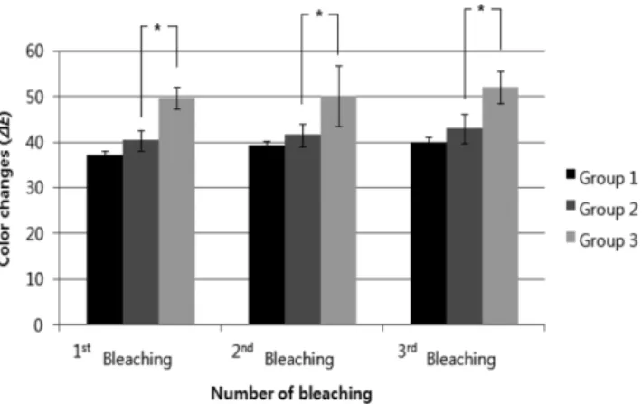
1. Color Changes before and after Tooth Bleaching Figure 1 shows color changes ( ∆E) before and after 1
st, 2
ndand 3
rdbleaching. The difference was significant among groups (Kruskal-Wallis test, p<0.05): 35% HP gel only (Group 1), 35% HP gel exposed to plasma arc A (Group 2) and 35% HP gel exposed to plasma arc B (Group 3). The color changes were greater when the spec- imens were exposed to HP gels and plasma arc B com- pared to those were exposed to HP gels and plasma arc A (Bonferroni post-hoc test, p<0.05). The values of color changes rose with the increase in the number of times of bleaching, yet it was not statistically significant.
Fig. 1.
Color changes ( ∆E) of 35% Hydrogen Peroxide group and Plasma Arc groups: Group 1 with 35% HP gel only;
Group 2 with 35% HP gel exposed to plasma arc A; and Group
3 with 35% HP gel exposed to plasma arc B. The significant
differences were shown between Group 2 and Group 3 at each
bleaching time (Bonferroni post-hoc test, p<0.05).
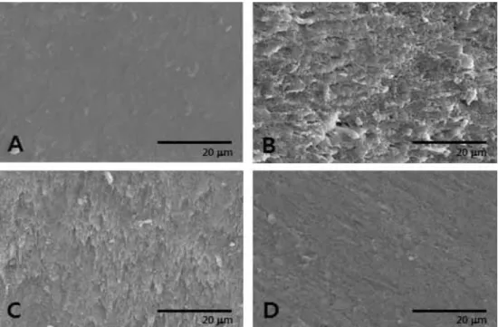
2. Effect of Fluoride Treatment after Bleaching Figure 2 shows the enamel surfaces of the specimen observed by SEM after bleaching in the four different con- ditions. The roughest surface was shown when only 35%
HP gel was applied. The second roughest surface was made by both 35% HP gel and plasma arc A, yet applica-
tion of 35% HP gel and plasma arc B led to a relatively smoother surface than that of the same concentration of HP with plasma arc A (Fig. 2).
Figure 3 shows the enamel surfaces of the specimen observed by SEM after application of fluoride in the four different conditions. The enamel surfaces came to be
Fig. 2.
Surface morphology after bleaching procedures without fluoride application under the four different conditions: A, non- bleaching; B, 35% HP gel only for control (Group 1); C, 35% HP gel and plasma arc A (Group 2); and D, 35% HP gel and plasma arc B (Group 3).
Fig. 3.
Surface morphology after application of fluoride under the four different conditions: A, fluoride application after non-
bleaching; B, fluoride application after 35% HP gel treatment (Group 1); C, fluoride application after 35% HP gel and plasma arc A
treatment (Group 2); and D, fluoride application after 35% HP gel and plasma arc B treatment (Group 3).
smoother after fluoride application compared to before fluoride application. Surfaces of the specimen treated with 35% HP only was similar to those with non-bleaching and samples treated with HP gels and both plasma arc A and B light sources became smoother than before fluoride appli- cation (Fig. 3).
Discussion
The mechanism of the tooth whitening is related to the oxidation-reduction reaction between the whitening agents and the discolored substances. When the whitening agents enter into deep dentinal tubules and reach darkened sub- strates, a bleaching therapy can succeed
7). Light activation which catalyzes the dissociation of hydrogen peroxide accelerates the reaction
19). Most of the patients prefer to bleach teeth because of less chair time, more comfortable treatment and immediate results
20).
Hydrogen peroxide and two types of plasma arc light sources were used to bleach bovine teeth specimen in this study. Bleaching with both hydrogen peroxide and plasma arc showed the greater bleaching effect than hydrogen per- oxide only. This result indicates that a light source not only promotes a tooth whitening reaction but also it has its own bleaching effect. Previous study demonstrated that the light by itself appeared to have a bleaching effect
21). Certain light activated peroxide bleaching products in par- ticular contain materials such as carotene and manganese sulphate and may be able to assist energy transfer from light to peroxide gels and discolored substances
22-24). It was also reported that hydrogen peroxide activated by light sources for intracoronal bleaching presented a simi- lar efficacy to walking bleaching
25).
When both HP gel and plasma arc lights were applied to the specimens, the effect of tooth whitening was greater than when HP gels were only applied. Moreover, the dif- ference of the color changes were larger when plasma arc B was combined with HP gel than when plasma arc A was combined with HP gel. These indicate that the light emis- sion source affects the effectiveness of the bleaching. Two key factors in determining final tooth bleaching efficacy are the concentration of peroxides and duration of application
26,27). However, these factors were fixed in this study so that the types of the light source brought about the magnitude of the color changes in specimens.
The result of the present study showed that uneven sur- faces after tooth whitening using 35% HP only were almost recovered by application of the fluoride. However,
the cracks on the enamel surfaces were not completely regained in Group 2 and Group 3 (Fig. 3). Application of the fluoride to the tooth specimen exposed to the high concentration of HP activated by light sources made the surface of the sample smooth, which supported the result of the previous study that reported that fluoride has poten- tial for remineralization of the demineralized enamel
28). So, it is possible to report that fluoride treatment protects the enamel surface of the teeth from damage by bleaching procedures.
This study has some limitations. First, the light by itself appears to have a bleaching effect
21), even though it is still controversial. If plasma arc light source has its own bleaching effect, duration of exposure time by light sources could be one of the factors related to the whiten- ing effect like concentration of the peroxide. However, the duration for exposure to light was fixed in this study. Sec- ond, a certain concentration of HP was used in this study;
the concentration of the bleaching agents is one of the key factors determining the effectiveness of the tooth whiten- ing though. Further study is needed to find the optimal concentration of fluoride that would provide the best clin- ical results without the side-effects. Moreover, it is neces- sary to examine that the surface changes and micro- hardness of teeth related to different whitening products in the presence of high fluoride concentrations. Finally, the staining material used in this study was coke, while other studies which were mentioned above utilized various dif- ferent substances for teeth staining such as black tea, cof- fee, and red wine. These substances can promote extrinsic stains, which are possible to affect results of the bleaching.
Henceforth, several bleaching systems have been devel- oped as patients’ esthetic needs and dentists’ efforts to find the safety way for tooth whitening. To extend the tooth whitening markets, the way to relieve the side- effects from bleaching should be developed and advanced.
It is necessary to go forward coupling the esthetic benefits of whitening with the preventive benefits of fluoride for safe therapy.
Summery
The purpose of this study was to examine the effective-
ness of teeth bleaching that only 35% HP was applied and
that both 35% HP and plasma arc light were applied and
to evaluate the effectiveness of fluoride treatment to
recover an irregularity of enamel surface after tooth whit-
ening with a high concentration of hydrogen peroxide
activated by plasma arc light. A total of 36 specimens were classified into following three groups: (1) 35% HP gel only, (2): 35% HP gel and Plasma arc A, and (3) 35%
HP gel and Plasma arc B. To measure color changes and surface morphologies before and after the bleaching, col- orimeter and SEM were used, respectively. Kruskal-Wal- lis test, Bonferroni post-hoc analysis and the repeated measures ANOVA was done with SPSS 17.0. The results of this study are following:
1. When the specimens were bleached with hydrogen peroxide and plasma arc lights, the bleaching effect was greater than when only hydrogen peroxide gels were used (Kruskal-Wallis test, p<0.05).
2. Plasma arc B showed the more color changes than plasma arc A (Bonferroni post-hoc test, p<0.05).
3. The surfaces of the teeth treated with fluoride gels after the whitening treatment came to be smooth.
Therefore, the results of this study suggested that the fluoride application for patients who got tooth whitening therapy with a high concentration of hydrogen peroxide gels activated by plasma arc light might be effective to protect an irregularity of enamel surfaces after bleaching.
References
1. Nathanson D: Vital tooth bleaching: sensitivity and pulpal considerations. J Am Dent Assoc 128(Suppl): 41S-44S, 1997.
2. Dahlstrom SW, Heithersay GS, Bridges TE: Hydroxyl radical activity in thermo-catalytically bleached root-filled teeth.
Endod Dent Traumatol 13(3): 119-125, 1997.
3. Yoon HY et al.: Comparison of Tooth Bleaching Effect between Low Concentration Hydrogen Peroxide with Plasma Arc and Only High Concentration Hydrogen Peroxide. J Korean Res Soc Dent Materials 37(1):21-28, 2010.
4. Sung MK et al.: Effect of 10% Carbamide peroxide bleaching on the artifically discolored enamel. J Korean Acad Dent Health 29(3): 241-249, 2005.
5. Haywood VB: Nightguard vital bleaching: current concepts and research. J Am Dent Assoc 128(Suppl): 19S-25S, 1997.
6. Goldstein RE: In-office bleaching: where we came from, where we are today. J Am Dent Assoc 128(Suppl): 11S-15S, 1997.
7. Carrasco LD et al.: Effect of internal bleaching agents on dentinal permeability of non-vital teeth: quantitative assessment.
Dent Traumatol 19(2): 85-89, 2003.
8. Lee HJ et al.: Effect of fluoridated 10% carbamide peroxide on enamel surface change and whitening. J Dent Hyg Sci 10(2): 95-100, 2010.
9. Gu HJ, Song KB: The bleaching effect of plasma arc and
35% carbamide peroxide and its influence on the enamel surface. J Dent Hyg Sci 9(5): 525-530, 2009.
10. Lee KH et al.: 35% hydrogen peroxide gel in the whitening effect and enamel changes. J Dent Hyg Sci 8(4): 255-260, 2008.
11. Gurgan S, Cakir FY, Yazici E: Different light-activated in- office bleaching systems: a clinical evaluation. Laser Med Sci 25: 817-822, 2010.
12. Hannig C et al.: Effect of bleaching on subsurface micro- hardness of composite and a polyacid modified composite.
Dent Mater 23(2): 198-203, 2007.
13. Lee KH et al.: Mineral loss from bovine enamel by a 30%
hydrogen peroxide solution. J Oral Rehabil 33(3): 229-233, 2006.
14. Turssi CP et al.: Permeability of enamel following light- activated power bleaching. Gen Dent 54(5): 323-326, 2006.
15. Kwon YH et al.: Change of enamel after Er:YAG and CO
2laser irradiation and fluoride treatment. Photomed Laser Surg 23(4): 389-394, 2005.
16. Radzi Z et al.: Light curing units: tips for orthodontists.
Annal Dent Univ Malaya 11: 13-23, 2004.
17. Shim YS, Choi WY: The effect of fluoride and casein phosphopeptide-amorphous calcium phosphate (CPP-ACP) application on the color and microhardness of bleached enamel. J Korean Soc Dent Hyg 10(3): 473-481, 2010.
18. Gu HY et al.: Effect of fluoride application after tooth bleaching using the diode laser. J Korean Acad Dent Health 32(2): 160-169, 2008.
19. Smigel I: Laser tooth whitening. Dent Today 15(8): 32-36, 1996.
20. Benjamin SD: Dental lasers: Part 3. Use of dental lasers on hard tissue. Pract Proced Aesthet Dent 14(5): 422-424, 2002.
21. Tavares M et al.: Light augments tooth whitening with peroxide. J Am Dent Assoc 134(2): 167-175, 2003.
22. Luk K, Tam L, Hubert M: Effect of light energy on peroxide tooth bleaching. J Am Dent Assoc 135(2): 194-201, 2004.
23. Sulieman M: An overview of bleaching techniques: 3. In- surgery or power bleaching. Dent Update 32(2): 101-108, 2005.
24. Wetter NU, Barroso MC, Pelino JE: Dental bleaching efficacy with diode laser and LED irradiation: an in vitro study.
Lasers Surg Med 35(4): 254-258, 2004.
25. Carrasco LD et al.: Efficacy of intracoronal bleaching techniques with different light activation sources. Int Endod J 40(3): 204-208, 2007.
26. Joiner A: The bleaching of teeth: a review of the literature. J Dent 34(7): 412-419, 2006.
27. Joiner A: Review of the effects of peroxide on enamel and dentine properties. J Dent 35(12): 889-896, 2007.
28. Gladwell J, Simmons D, Wright Jt: Remineralization Potential of a Fluoridated Carbamide Peroxide Whitening Gel. J Esthet Restor Dent 18(4): 206-212, 2006.
(Received May 12, 2011; Revised August 16, 2011;
Accepted August 20, 2011)