Assessment of the Location of the
Peroneus Longus Tendon in the Cuboid Groove Using 3D Isotropic Fast
Spin-Echo MRI
INTRODUCTION
Peroneal tendon injuries are common but frequently overlooked as a source of chronic ankle pain, and only 60% of peroneal tendon injuries are accurately diagnosed at the initial office visit (1). The three broad categories of peroneal tendon lesions include:
This is an Open Access article distributed under the terms of the Creative Commons Attribution Non-Commercial License (http://creativecommons.org/licenses/
by-nc/4.0/) which permits unrestricted non-commercial use, distribution, and reproduction in any medium, provided the original work is properly cited.
Received: August 16, 2019 Revised: September 27, 2019 Accepted: October 24, 2019 Correspondence to:
Hye Jung Choo, M.D.
Department of Radiology, Inje University Busan Paik Hospital, 75, Bokji-ro, Busanjin-gu, Busan 47392, Korea.
Tel. +82-51-890-6549 Fax. +82-51-896-1085
E-mail: [email protected]
Copyright © 2020 Korean Society of Magnetic Resonance in Medicine (KSMRM)
Original Article
Purpose: To investigate normal location of the peroneus longus tendon (PL) in the cuboid groove by evaluating it between ankles with no significant abnormality (asymptomatic group) and those with retromalleolar PL dislocation (dislocation group) using three-dimensional isotropic fast spin-echo (3D-FSE) magnetic resonance imaging (MRI) of the ankle.
Materials and Methods: Thirty-six and 32 3D-FSE ankle MRI were assigned to the asymptomatic group and the dislocation group, respectively. Using multiplanar reformatted 3D-FSE, qualitative PL location (i.e., outside, overlying, and inside in relation to the cuboid groove), quantitative PL location (i.e., distance between the proximal margins of PL and cuboid groove), and cuboid groove size were measured in lateral, middle, and medial levels of the cuboid groove.
Results: In the asymptomatic group, 64%, 42%, and 11%, respectively, had the outside or overlying-located PL in lateral, middle, and medial levels of the cuboid groove and the quantitative location gradually decreased from lateral to medial level.
Qualitative and quantitative PL locations were not significantly different between the asymptomatic group and dislocation group. Cuboid groove size showed significant negative correlation with quantitative PL location in both groups.
Conclusion: Outside- or overlying-located PL in lateral and middle levels of the cuboid groove would be a normal finding, regardless of PL status at the retromalleolar level.
Keywords: Peroneus longus tendon; Dislocation; Cuboid; Magnetic resonance imaging;
Three-dimensional
Ji Yoon Lee1, Hye Jung Choo1, Sun Joo Lee1, Joon-Yong Jung2, Dong Wook Kim1, Jin Wook Baek1, Young Jin Heo1, Heui-Chul Gwak3
1Department of Radiology, College of Medicine, Inje University, Busan Paik Hospital, Busan, Korea
2Department of Radiology, Seoul St. Mary’s Hospital, College of Medicine, The Catholic University of Korea, Seoul, Korea
3Department of Orthopedic Surgery, College of Medicine, Inje University, Busan Paik Hospital, Busan, Korea
tendinitis and tenosynovitis, tendon subluxation and dislocation, and tendon tear (2). Peroneus longus tendon (PL) subluxation and dislocation, although uncommon causes of lateral ankle pain, can be significantly disabling (3). While subluxation and dislocation in the retromalleolar level are relatively well studied, to the best of our knowledge, only two studies have investigated the location of the PL in the cuboid groove depending on the foot position. However, the results were conflicting. One study using ultrasound (US) and magnetic resonance imaging (MRI) concluded that the PL showed a tendency to be overlying the cuboid tuberosity in dorsiflexion and glided entirely within the cuboid groove in plantar-flexion (4). Another study using US concluded that the PL location did not significantly change depending on the ankle-foot position and “overlying” location was the most common type, regardless of the ankle-foot position (5).
The recently developed three-dimensional isotropic fast spin echo (3D-FSE) applied to the ankle, provides images with thin slice thickness and multiplanar reformation after single acquisition. The image quality and diagnostic accuracy of 3D-FSE is comparable to conventional two- dimensional images and is well suited for evaluating the complex musculoskeletal structure (6-8). Thus, the 3D-FSE with multiplanar reformation, dedicated to the cuboid groove, would allow for a more precise evaluation of the PL location in the cuboid groove. However, because MR usage was limited in this dynamic study, we postulated that a retromalleolar PL dislocation would exert some degree of positive or negative tension on the PL, providing an alternative theory for the position change of the ankle-foot.
Thus, we aimed to investigate the normal PL location in the cuboid groove by comparing ankles with no significant abnormality with those with a retromalleolar PL dislocation, using 3D-FSE and its multiplanar reformation, and to evaluate the size of the cuboid groove to determine the factors influencing PL location in the cuboid groove.
MATERIALS AND METHODS
This study was designed as a retrospective study. The Institutional Review Boards approved the study and informed consents were waived given the retrospective nature of the study and the use of anonymized data.
Participants
Seven-hundred and forty-four consecutive ankle MRI including a 3D-FSE sequence taken January 2014-November
2018 were reviewed to select normal or nearly normal ankles (asymptomatic group) and ankles with dislocation of the PL from the retromalleolar fossa (dislocation group) by a musculoskeletal radiologist with 11 years of experience.
The patients with inflammatory arthritis, operation history at the ankle-foot, and younger than age 18 were excluded.
Finally, 36 ankles of 33 patients (18 females, 15 males;
mean age, 45 years; age range, 18-71 years) were included into an asymptomatic group and 32 ankles of 31 patients (15 females, 16 males; mean age, 45 years; age range, 18- 71 years) were included in the dislocation group. In the dislocation group, 23 patients were surgically confirmed with PL dislocation. Other patients have not undergone surgery (n = 3) or have undergone surgery without description about PL findings in surgical notes (n = 6).
Major findings from the MRI in the asymptomatic group were normal ankle (n = 19), ganglion (n = 8), joint effusion (n
= 2), adventitial bursitis (n = 3), and mild soft tissue edema (n = 4). Major MRI findings in the dislocation group, aside from retromalleolar PL dislocation, were calcaneal fracture (n = 12), lateral ankle ligament tear (n = 2), and mild joint effusion (n = 2).
MRI Acquisition
MRI was performed using 3T MR scanners (Achieva 3.0T TX, Philips Medical System, Best, the Netherlands;
MAGNETOM Skyra, Siemens Healthcare, Erlangen, Germany) with a chimney ankle coil, which placed the ankles in a neutral position (i.e., 0° plantar position). A 3D-FSE sequence was obtained with 0.5-mm slice thickness in the orthotropic sagittal plane. The parameters of this sequence were summarized in Table 1 for each MR scanner.
Table 1. Parameters of Three-Dimensional Fast Spin Echo MRI of the Ankle
Achieva Skyra
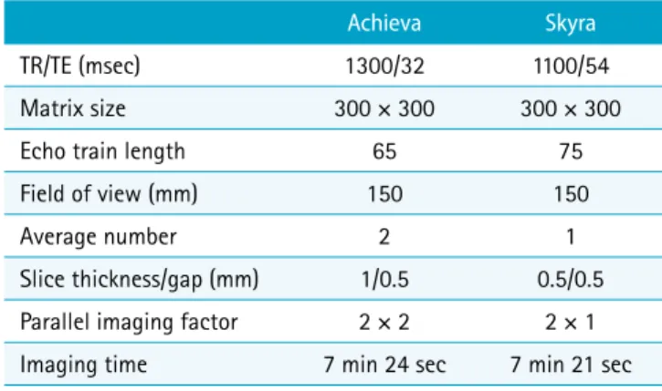
TR/TE (msec) 1300/32 1100/54
Matrix size 300 × 300 300 × 300
Echo train length 65 75
Field of view (mm) 150 150
Average number 2 1
Slice thickness/gap (mm) 1/0.5 0.5/0.5
Parallel imaging factor 2 × 2 2 × 1
Imaging time 7 min 24 sec 7 min 21 sec
TE = echo time; TR = repetition time
Image Analysis
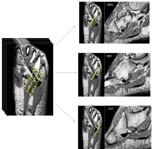
To evaluate the exact PL location in the cuboid groove and the cuboid groove size, the orthotropic sagittal plane of 3D-FSE images were multiplanar reformatted into the oblique sagittal, oblique axial, and oblique coronal planes according to the orientation of the cuboid groove with 1-mm slice thickness and no interslice gap using Aquarius iNtuition software (Version 4.4.12, TeraRecon, Inc., San Mateo, CA, USA). Among the three planes, the oblique sagittal plane that is perpendicular to the long axis of the cuboid groove was used for analysis of PL location and cuboid groove size because this plane showed the accurate
location relationship between the cuboid groove and the PL. The PL location and cuboid groove size were evaluated in the three levels of the cuboid groove (i.e., lateral, middle, and medial levels). In the cuboid groove, the lateral level indicated the most lateral margin; the medial level, the most medial margin; the middle level, the midway between the lateral and medial levels (Fig. 1).
Peroneus Longus Tendon Location
PL location was analyzed qualitatively and quantitatively.
For qualitative analysis, the PL location was categorized into three types: an inside-located type, defined as the whole
Fig. 1. Differentiation of the three levels of the cuboid groove. The peroneus longus tendon (PL) location and cuboid groove size are evaluated on the oblique sagittal images of the lateral, middle, and medial levels of the cuboid groove. On the oblique axial image, the three yellow-dotted lines indicate the lateral, middle, and medial levels of the cuboid groove.
1st MT = 1st metatarsal; LAT = lateral; MED = medial; MID = middle
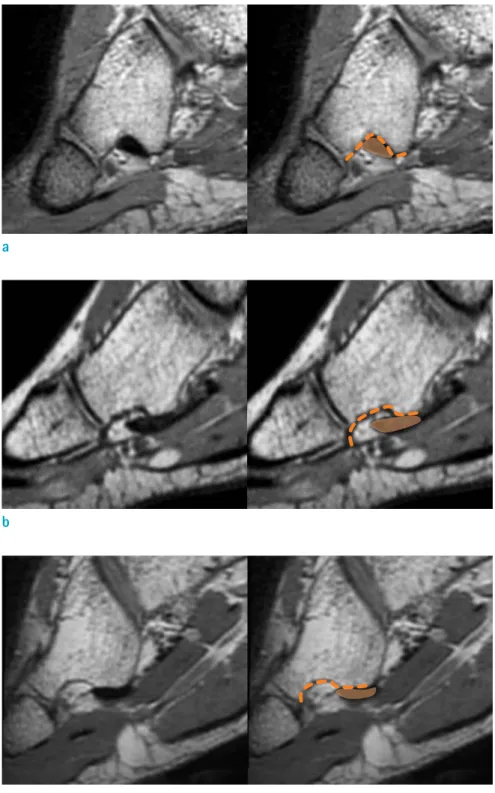
cross-sectional area of the PL on the oblique sagittal plane located inside of the cuboid groove; an overlying-located type, as some part of the PL cross-sectional area located proximal to the cuboid groove; and an outside-located type, as the entire cross-sectional area of the PL located proximal to the cuboid groove (5) (Fig. 2). For quantitative analysis of
the PL location, the distance between the proximal margin of the cross-sectional area of the PL and the proximal border of the cuboid groove on the oblique sagittal image was measured (Fig. 3). A positive measurement (+) indicated that the proximal margin of the cross-sectional area of the PL was located more proximal than the proximal border of
a
b
c
Fig. 2. Mimetic diagram of the categorization for the qualitative location of the PL . (a) The images show an inside-located type, wherein the whole cross-sectional area of the PL is inside the cuboid groove. (b) The images show an overlying-located type, wherein some part of cross- sectional area of the PL is outside the groove. (c) The images show an outside-located type, wherein the entire cross-sectional area of the PL is outside the groove.
the cuboid groove. A negative measurement (-) indicated that the proximal end of the cross-sectional area of the PL was distal to the proximal border of the cuboid groove (5).
Cuboid Groove Size
The depth and width of the cuboid groove was measured on each of the three oblique sagittal images that were relevant to the lateral, middle, and medial levels of the cuboid groove. The width of the cuboid groove was defined as the distance between the most inferior contours of the cuboid tuberosity and the anterior edge of the cuboid bone.
The depth of the cuboid groove was defined as the distance between the deepest point of the cuboid groove and an imaginary line, crossing the most inferior contours of the
cuboid tuberosity and the anterior edge of the cuboid bone (Fig. 4).
Statistical Analysis
Qualitative data for the location of the PL were compared using Fisher’s exact test. To determine the relationship between the quantitative PL location and cuboid groove size, Pearson’s correlation coefficient was used. All continuous data were tested for normality using the Kolmogorov-Smirnov test (the data did not show a normal distribution). The quantitative data for the location of the PL and the depth and width of the cuboid groove between the groups were compared using Mann-Whitney U test. In all test, P values less than 0.05 were significant.
Statistical analyses were performed using SPSS (IBM SPSS Statistics version 24.0; IBM Corporation, Armonk, NY, USA).
RESULTS
PL LocationIn the asymptomatic group, the proportion of ankles with inside-located PL was 36% in the lateral level of the cuboid groove and increased to 58% and 89% in middle and medial levels, respectively. When compared with the dislocation group, the qualitative PL location was not significantly different in all three levels of the cuboid groove (P = 0.461, 0.806, and 1.000 in lateral, middle, and medial levels, respectively) (Fig. 5). The proportion of ankles with inside- located PL increased as PL goes through the cuboid groove from the lateral to medial level in the dislocation group.
The dislocation group showed similar tendency and seemed to have a higher proportion of inside-located PL than the Fig. 3. Measurement for the quantitative location of the PL.
On the oblique sagittal plane, the distance (double-headed arrow) between the proximal margin of cross-sectional area of the PL (orange solid line) and the proximal border of the cuboid groove (orange dotted line) is measured.
a b
Fig. 4. Measurement for the width and depth of the cuboid groove. (a) The cuboid groove width (double-headed arrow) indicates the distance between the most inferior contour of the cuboid tuberosity and the anterior edge of the cuboid. (b) Its depth indicates the distance between the deepest point of the cuboid groove and the imaginary line, crossing the most inferior contour of the cuboid tuberosity and the anterior edge of the cuboid.
asymptomatic group in all three levels, but the difference was not statistically significant (Figs. 6, 7).
The results of the quantitative PL location were similar to those of the qualitative PL location. The quantitative PL location in the asymptomatic group decreased in the lateral, middle, and medial levels of the cuboid groove (median:
6.5 mm, 0 mm, and 0 mm, respectively), suggesting the gradual closeness of PL to the cuboid groove from lateral to medial cuboid grooves. There was no statistically significant difference of quantitative PL location between the asymptomatic group and the dislocation group in all three levels of the cuboid groove (P = 0.152, 0.306, and 0.722 in lateral, middle, and medial levels, respectively) (Table 2).
In the lateral level, the median values of the quantitative PL location showed positive measurements in the asympto- matic group and the dislocation group (6.55 mm and 3.80 mm, respectively), meaning the proximal margin of the cross-sectional area of the PL showed a tendency to be more proximal than the proximal border of the cuboid groove (Table 2). The tendency seems to correlate with
a b
c d
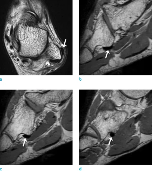
Fig. 6. An age 19 male with PL dislocation from the retromalleolar fossa. (a) On the axial T2-weighted spin-echo image, the PL (arrow) is anterolateral aspect of the lateral malleolus (arrow). (b-d) On the oblique sagittal reformatted 3D-FSE, the PL (arrow) is ‘inside-located’ in the lateral (b), middle (c) and medial levels of the cuboid groove (d).
Fig. 5. Bar graph showing PL location in the asymptomatic group and the dislocation group. The proportions of ankles with the inside-located PL increase as the PL goes through the cuboid groove from the lateral to medial level in the asymptomatic group and the dislocation group, but there is no statistically significant difference.
the result that the sum of the proportion of overlying and outside-located PL in the lateral level is more than 50% in the asymptomatic group and the dislocation group. In the middle and medial levels, the median measurement value was zero in the asymptomatic group and the dislocation group, meaning the proximal end of the cross-sectional area of the PL met the proximal border of the cuboid groove (Table 2). These features seem to correlate with the result
that the proportion of inside-located PL is more than 50%
in the asymptomatic group and the dislocation group in the middle and medial levels.
Cuboid Groove Size
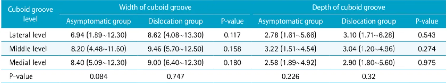
The width and depth of cuboid groove were not significantly different between the asymptomatic group and the dislocation group and among the three levels of cuboid grooves (Table 3).
Relationship between PL Location and Cuboid Groove Size
The quantitative location of the PL showed significant negative correlation with the width of the cuboid groove in the all three levels of cuboid groove and depth of the cuboid groove in lateral and middle levels, in the asymptomatic group and the dislocation group (Table 4). Particularly, the width of the cuboid groove in the lateral level showed the stronger negative correlation with the PL location.
Table 2. Quantitative Peroneus Longus Tendon Location Cuboid groove
level Asymptomatic
group Dislocation
group P-value Lateral level 6.55 (0.00~12.9) 3.80 (0.00~15.9) 0.152 Middle level 0 (-1.44~11.7) 0 (-5.26~4.90) 0.306 Medial level 0 (-3.79~3.20) 0 (-3.70~4.30) 0.722
P-value < 0.001 < 0.001
Unit: millimeter (mm) Data are median (range)
a b
c d
Fig. 7. An age 19 female with PL dislocation from the retromalleolar fossa. (a) On the axial T2-weighted spin-echo image, the PL (arrow) is dislocated from the retromalleolar fossa (arrow). (b-d) The oblique sagittal reformatted 3D-FSE demonstrates the outside-located PL (arrows) at the lateral level of cuboid groove (b), which is glided entirely into the cuboid groove in its middle and medial levels (c, d).
This result indicates that the ankles with the PL close to the cuboid groove tended to have a wider and deeper inlet of the cuboid groove, whereas the ankles with the PL distant from the cuboid groove tended to have a narrower and shallower inlet of the cuboid groove (Figs. 6, 7).
DISCUSSION
In this study, 36% of asymptomatic ankles in a neutral position had an inside-located PL at the lateral level of the cuboid groove, whereas 58% and 89% had in the middle and medial levels, respectively. This pattern of PL location at the cuboid groove was also demonstrated in ankles with a retromalleolar PL dislocation. These results may suggest that retromalleolar PL location, regardless of ankle-foot position change, does not affect the PL location at the cuboid groove. In both groups, the quantitative PL location was significantly and negatively correlated with the cuboid groove size, indicating that ankles with a wider and deeper cuboid groove had an inside-located PL at the cuboid groove, and those with a narrower and shallower cuboid groove had an outside or overlying-located PL at the cuboid groove. The cuboid groove size could be a factor influencing the PL location at the cuboid groove. However, in this
cross-sectional study, we could not determine if the size of the cuboid groove is causative or compensatory to the PL location in the cuboid groove. The proportion of ankles with an inside-located PL was slightly higher in the dislocation group than in the asymptomatic group, at all levels of the cuboid groove, although the differences were statistically insignificant. However, considering that the sizes of the cuboid grooves were slightly larger in the dislocation group than in the asymptomatic group, the insignificant difference in quantitative PL location between the two groups is not unreasonable.
To the best of our knowledge, only two published studies have investigated the location of the PL in the cuboid groove; also, it is debatable if the PL location in the cuboid groove is dependent on the ankle-foot position (4, 5).
Stone et al. (4) evaluated the location of the PL in the cuboid groove in two ankle-foot positions, dorsiflexion and plantar-flexion, using dynamic US and conventional MRI.
They demonstrated the dislocation of PL during dorsiflexion, but in the plantar-flexion position, the PL was inside- located. Choo et al. (5) evaluated the PL location in five ankle-foot positions: neutral, dorsiflexion, plantar-flexion, supination, and pronation. They concluded that PL location did not significantly change depending on the ankle-foot position, and that approximately only half of the PL was Table 4. Relationship between Peroneus Longus Tendon Location and Cuboid Groove Size
Cuboid groove level Width of cuboid groove Depth of cuboid groove
Asymptomatic group Dislocation group Asymptomatic group Dislocation group
Lateral level Coefficient -0.807 -0.644 -0.458 -0.555
P-value < 0.001 < 0.001 0.005 0.001
Middle level Coefficient -0.455 -0.419 -0.500 -0.409
P-value 0.005 0.017 0.002 0.020
Medial level Coefficient -0.471 -0.415 -0.259 -0.065
P-value 0.004 0.018 0.128 0.724
Table 3. Width and Depth of Cuboid Groove Cuboid groove
level
Width of cuboid groove Depth of cuboid groove
Asymptomatic group Dislocation group P-value Asymptomatic group Dislocation group P-value Lateral level 6.94 (1.89~12.30) 8.62 (4.08~13.30) 0.117 2.78 (1.61~5.66) 3.10 (1.71~6.28) 0.543 Middle level 8.20 (4.48~11.60) 9.46 (5.70~12.50) 0.158 3.22 (1.51~4.54) 3.04 (1.20~4.96) 0.274 Medial level 8.40 (5.09~12.30) 9.00 (6.40~12.30) 0.180 2.58 (1.89~4.92) 2.90 (1.80~5.60) 0.975
P-value 0.084 0.747 0.226 0.32
Unit: millimeter (mm)
constantly placed inside the cuboid groove, regardless of the ankle-foot position. This study used ankle MRI with a chimney coil, similar to the dorsiflexion group in the study by Stone et al. (4). However, the MRI technique in our study was different from those used by Stone et al. (4).
They evaluated the PL location on the orthotropic planes of conventional two-dimensional FSE, with a 3-mm thickness (4). However, the cuboid groove is obliquely present in the coronal and sagittal planes. Thus, this study evaluated the PL on reformatted oblique sagittal images of 3D-FSE, perpendicular to the cuboid groove axis, with a 1-mm thickness, a superior method to evaluate the PL location in the cuboid groove. However, unfortunately, we could not evaluate the PL location by 3D-FSE MRI in various ankle- foot positions because of the retrospective design of this study. Instead, we verified if a retromalleolar dislocation of the PL could change the PL tension to some degree. This condition was compared with the asymptomatic ankle in a neutral position, and we found no significant difference in the PL location in the cuboid groove between the two groups.
Although the PL location in the cuboid groove using 3D-FSE was not compared with that using the routine orthogonal 2D MRI, we believe that the out-side located PL from the cuboid groove will be also found on the orthogonal 2D MRI and this feature should not be interpreted as a pathology.
Our study has limitations. First, the ankle MRI data that made up the asymptomatic group were not obtained from healthy volunteers. However, patients in the asymptomatic group had either insignificant abnormalities such as mild joint effusion or ganglions or no abnormalities based on MRI findings. Second, the volume of data in this study was minimal. Third, the PL location was not evaluated on MRI in the various positions of ankle-foot because of the retrospective study design. Although the recent prospective US study already demonstrated the insignificant movement of PL in the cuboid groove during dynamic US (5), ankle MRI obtained in the various positions of the foot-ankle would have provided more objective results. Fourth, inter- reader agreement and reproducibility for qualitative and quantitative measurements of PL location were not analyzed because of the unavailability of a secondary reader.
In conclusion, we investigated the location of the PL in the cuboid groove in normal or nearly normal ankles and in ankles with a retromalleolar PL dislocation using 3D-FSE MRI. Our study suggests that the outside- or overlying- located PL at the lateral and middle levels of the cuboid
groove would be a normal finding, regardless of PL status at retromalleolar level. Width and depth of the cuboid groove strongly correlated with the PL location in the cuboid groove.
Acknowledgments
This work is supported by Musculoskeletal MR Study Group, Korean Society of Magnetic Resonance in Medicine, 2018. This material is based upon work supported by the Ministry of Trade, Industry & Energy (MOTIE, Korea) under the Industrial Technology Innovation Program.
NO.10062728.
REFERENCES
1. Dombek MF, Lamm BM, Saltrick K, Mendicino RW, Catanzariti AR. Peroneal tendon tears: a retrospective review. J Foot Ankle Surg 2003;42:250-258
2. Heckman DS, Gluck GS, Parekh SG. Tendon disorders of the foot and ankle, part 1: peroneal tendon disorders. Am J Sports Med 2009;37:614-625
3. Philbin TM, Landis GS, Smith B. Peroneal tendon injuries. J Am Acad Orthop Surg 2009;17:306-317
4. Stone TJ, Rosenberg ZS, Velez ZR, Ciavarra G, Prost R, Bencardino JT. Subluxation of the peroneus long tendon in the cuboid tunnel: is it normal or pathologic? An ultrasound and magnetic resonance imaging study. Skeletal Radiol 2016;45:357-365
5. Choo HJ, Lee SJ, Huang BK, Resnick DL. The location of the peroneus longus tendon in the cuboid groove:
sonographic study in various positions of the ankle-foot in asymptomatic volunteers. Skeletal Radiol 2018;47:1277- 1284
6. Choo HJ, Lee SJ, Kim DW, Jeong HW, Gwak H. Multibanded anterior talofibular ligaments in normal ankles and sprained ankles using 3D isotropic proton density-weighted fast spin-echo MRI sequence. AJR Am J Roentgenol 2014;202:W87-94
7. Stevens KJ, Busse RF, Han E, et al. Ankle: isotropic MR imaging with 3D-FSE-cube--initial experience in healthy volunteers. Radiology 2008;249:1026-1033
8. Ulbrich EJ, Zubler V, Sutter R, Espinosa N, Pfirrmann CW, Zanetti M. Ligaments of the Lisfranc joint in MRI:
3D-SPACE (sampling perfection with application optimized contrasts using different flip-angle evolution) sequence compared to three orthogonal proton-density fat-saturated (PD fs) sequences. Skeletal Radiol 2013;42:399-409