Influence of zirconia and lithium disilicate tooth- or implant-supported crowns on wear of antagonistic and adjacent teeth
Martin Rosentritt, Frederik Schumann, Stephanie Krifka, Verena Preis*
Department of Prosthetic Dentistry, UKR University Hospital Regensburg, Regensburg, Germany
PURPOSE. To investigate the influence of crown material (lithium-disilicate, 3Y-TZP zirconia) and abutment type (rigid implant, resin tooth with artificial periodontium) on wear performance of their antagonist teeth and adjacent teeth. MATERIALS AND METHODS. A mandibular left first molar (#36) with adjacent human teeth (mandibular left second premolar: #35, mandibular left second molar: #37) and antagonistic human teeth (maxillary left second premolar: #25, maxillary left first molar: #26, maxillary left second molar: #27) was prepared simulating a section of the jaw. Samples were made with extracted human molars (Reference), crowned implants (Implant), or crowned resin tooth analogues (Tooth). Crowns (tooth #36; n = 16/material) were milled from lithium-disilicate (Li, IPS e.max CAD) or 3Y-TZP zirconia (Zr, IPS e.max ZirCAD, both Ivoclar Vivadent).
Thermal cycling and mechanical loading (TCML) in the chewing simulator were applied simulating 15 years of clinical service. Wear traces were analyzed (frequency [n], depth [µm]) and evaluated using scanning electron pictures. Wear results were compared by one-way-ANOVA and post-hoc-Bonferroni (α = 0.05). RESULTS. After TCML, no visible wear traces were found on Zr. Li showed more wear traces (n = 30-31) than the reference (n = 21). Antagonistic teeth #26 showed more wear traces in contact to both ceramics (n = 27-29) than to the reference (n = 21). Strong wear traces (> 350 µm) on antagonists and their adjacent teeth were found only in crowned groups. Abutment type influenced number and depth of wear facets on the antagonistic and adjacent teeth. CONCLUSION. The clinically relevant model with human antagonistic and adjacent teeth allowed for a limited comparison of the wear situation. The total number of wear traces and strong wear on crowns, antagonistic and adjacent teeth were influenced by crown material. [J Adv Prosthodont 2020;12:1-8]
KEYWORDS: Zirconia; Ceramics; Crowns; Implant; Wear
INTRODUCTION
Full-contour ceramic crowns made of lithium disilicate or zirconia have become very popular due to good esthetics and biocompatibility, as well as their cost-effective comput- er-aided design and manufacturing (CAD/CAM). These ceramics have proven their suitability for monolithic single crowns in many previous studies.1-3 Ongoing modifications
in ceramic structure and composition (e.g. leucite, lithium silicate, lithium disilicate or lithium-aluminosilicate) offer a wide range of glass-ceramic materials for individual applica- tions according to the clinical requirements of strength (<
500 MPa) and translucency (> 50%). Currently available 3Y-, 4Y- and 5Y- Y2O3 tetragonal zirconia polycrystals (Y-TZP) differ in strength (500 - 1200 MPa) and translucen- cy (< 50%), but show comparable hardness (HV: 1200 - 1300). Hardness and other material properties vary between zirconia (HV: 1200 - 1300), lithium disilicate (HV: 600), and human tooth enamel (HV: 300 - 400).4,5
However, hard surfaces might not cause increased wear without considering the surface state. Smooth polished and glazed zirconia surfaces did not show high antagonistic wear rates.6-8 Despite of identified influencing factors (e.g. surface roughness, hardness, occlusal crown design), wear behavior of ceramic restorations in the complex mastication system has not yet been fully understood. The complex interaction between indirect restorations and natural teeth in this sys-
Corresponding author:
Verena Preis
Department of Prosthetic Dentistry, UKR University Hospital Regensburg 93042 Regensburg, Germany
Tel. +4994194416073: e-mail, [email protected]
Received June 19, 2019 / Last Revision January 22, 2020 / Accepted January 23, 2020
© 2020 The Korean Academy of Prosthodontics
This is an Open Access article distributed under the terms of the Creative Commons Attribution Non-Commercial License (http://creativecommons.
org/licenses/by-nc/4.0) which permits unrestricted non-commercial use, distribution, and reproduction in any medium, provided the original work is properly cited.
tem may influence physiological chewing or even provoke craniomandibular dysfunctions. Many previous in vitro stud- ies focused on wear without including the complex influ- ences of the jaw situation.9,10 Some studies at least evaluated human enamel as antagonistic material.8,11-17 The type of abutment (resilient tooth support or rigid implant situation) may have an impact on wear performance of the crown itself but also on the wear of antagonistic and adjacent teeth. It seems advantageous to design an in vitro model that approximates the in vivo situation with human antagonistic and adjacent teeth in a mastication device simulating rele- vant clinical parameters (chewing cycles, forces, frequency;
thermocycles). Such a clinically approximated two-body wear simulation may allow for comparative evaluation of wear facets and maximum wear on ceramic crown materials and adjacent or antagonistic human teeth, although it may not fully reproduce in vivo wear processes. A comparison to clinical data would be wishful, but clinical wear studies are rare1,18-21 and valid in vivo methods of evaluation are miss- ing.22
The present pilot study aimed to test the suitability of an in vitro jaw model for wear testing of dental crowns. The hypothesis was that crown material (lithium disilicate, zirco- nia) and type of abutment (rigid implant, resin tooth with artificial periodontium) have an influence on the wear per- formance (traces, depth, superficial damages) of molar crowns and their antagonistic and adjacent teeth.
MATERIALS AND METHODS
200 extracted human teeth (stored in 0.5% chloramine solu- tion for no longer than four weeks) were selected to mimic 40 models of a clinical posterior situation. For each model, a premolar (mandibular left second premolar: #35) and two molars (mandibular left first molar: #36 and mandibular left second molar: #37) were embedded in resin blocks (Palapress Vario, Kulzer, Hanau, Germany) to simulate a section of the lower jaw. Corresponding human tooth antagonists were selected for the upper jaw (maxillary left second premolar:
#25, maxillary left first molar: #26, and maxillary left sec- ond molar: #27). The variability of human premolars and molars were respected by preselecting teeth with compara- ble size and shape and by randomly dividing the teeth to the subgroups. The teeth and their antagonists were positioned
in a clinically relevant occlusal contact situation (maximum intercuspidation) using a dental articulator (Artex, Amann- Girrbach, Pforzheim, Germany). Every lower tooth had antagonistic contacts to only one upper tooth. The contact points were adjusted and controlled with articulating paper.
Five groups (see Table 1) with eight models per group were prepared. Two clinically relevant situations (implant- and resin tooth-supported) for restoring tooth the mandibu- lar left first molar (#36) in the lower jaw, as well as two ceramic crown materials (lithium disilicate, zirconia), were investigated. A group with intact human teeth served as ref- erence.
In the two implant (I) groups, the mandibular left first molars (#36) were represented by implant analogues (Straumann, Freiburg, Germany; titanium grade IV, implant diameter 4.1 mm, implant length 12 mm, abutment length 4 mm, 8°), which were rigidly positioned in resin blocks (Palapress Vario) in order to simulate a posterior implant situation.
In the two tooth (T) groups, teeth #36 were represented by identical resin tooth replicas (Palapress Vario), which were flexibly positioned in resin blocks (Palapress Vario) in order to simulate an artificial periodontium. The prepara- tion design was based on ceramic guidelines with a circular reduction of 1 mm and occlusal anatomical reduction of about 1.5 mm (height ~ 6 mm, angle ~ 6°, rounded edges).
The 1 mm circumferential deep shoulder with rounded inner angles was at an isogingival height of the tooth cervix.
In the reference (R) group, eight unprepared extracted human teeth #36 with an artificial periodontium were posi- tioned in resin blocks (Palapress Vario).
The resilience of the tooth periodontium (for all human teeth and replaced resin teeth) was simulated by coating the natural or resin roots of the teeth with a 1 mm polyether layer (Impregum, 3M, Neuss, Germany). For achieving a constant layer, the roots were dipped in a wax bath, which was replaced by polyether in the second fabrication pro- cess.23,24
Mean tooth mobility was 80 µm in the axial direction, 280 µm in the buccal direction, and 130 µm in the oral direction.
In the implant and tooth groups, the implant analogues or resin tooth replicas were restored with lithium disilicate (Li) or zirconia (Zr) crowns, respectively. Therefore, implants and resin teeth were digitalized (Cerec Omnicam, Dentsply Sirona, Bensheim, Germany) and full-contour molar crowns were milled (Cerec, MC XL, Dentsply Sirona; juvenile, spac- er 80 µm, contact thickness 25 µm, edge reinforcement 50 µm). Ceramic materials were 3Y-TZP zirconia (IPS e.max ZirCAD, Ivoclar Vivadent, Schaan, Liechtenstein; Mo 1 C15L, LOT: S20285) and lithium disilicate (IPS e.max CAD, Ivoclar Vivadent; LT A2/C14, LOT: T23748) (n = 8 per material per group, Table 1). Zirconia crowns were sintered (Cercon heat plus, Degudent, Hanau, Germany; 1500°C, 2 hours). Lithium disilicate crowns were crystalized and glazed together (820°C; 10 min). All crowns were polished (silicon polisher, diamond paste, EVE Ernst Vetter, Keltern, Table 1. Study overview (5 groups)
Groups Abutment
Implant I Resin Tooth T Crown
material
Zirconia I-Zr T-Zr
Lithium disilicate I-Li T-Li
Reference Human Tooth -- R (human tooth
reference)
Germany) before applying a glaze layer and firing in the ceramic furnace (Programat EP 5000, IPS e.max Ceram glaze, Ivoclar Vivadent) following the respective firing pro- tocol provided by the manufacturer. Following this proce- dure, a consistently smooth and comparably thick glaze lay- er was achieved. In pretests, the thickness of the glaze layer was determined to range between 100 µm and 120 µm.
The inner surfaces of the zirconia crowns were sand- blasted (aluminium oxide, 100 µm, 1.5 bar) and the lithium disilicate crowns were etched for 20 seconds (IPS Ceramic Etching Gel HF, Ivoclar Vivadent). Primer (Monobond-Plus, Ivoclar Vivadent, 60 s) was applied. All crowns on resin teeth were adhesively bonded (Heliobond, Syntac Adhesive, Variolink 2 Catalyst, Variolink Base, Ivoclar Vivadent; Elipar Trilight, 3M). Implants were sandblasted (aluminium oxide, 100 µm, 1.5bar) and primed (Monobond-Plus, 60 s) before bonding the crowns (Multilink Hybrid Abutment, Ivoclar Vivadent; Elipar Trilight, 3M).
Upper and lower restoration models were stored in dis- tilled water until simulation. Thermal cycling and mechani- cal loading (TC: 6 × 3.000 cycles with changing tempera- tures between 5°C/55°C, distilled water; ML: 100 N for 3.6
× 106 cycles; f = 1.6 Hz; mouth opening 2 mm; lateral movement 2 mm; chewing simulator EGO, Regensburg, Germany) with online failure-control were performed to simulate and control fatigue failures. Chewing simulation parameters were based on literature data regarding zirconia and ceramic restorations, simulating ten to fifteen years of oral service.25,26
After simulation, all teeth and crowns were optically examined (light microscope, Vision Engineering, Woking, UK, 4× magnification) documenting the frequency and location of all wear traces in the area of the contact points.
A 3D color laser scanning microscope (VK-X100 Series, Keyence Corporation, Osaka, Japan) was used for quantita- tive evaluation of the occlusal surfaces. The depth of the respective wear facet was measured, whereby both the deep- est point and the average depth were determined. Assuming annual wear rates of about 30 - 35 µm,27 wear depths below 350 µm (after 10 - 15 years of simulation) were graded
“normal”. Wear facets deeper than 350 μm were graded
“strong”. Exemplary scanning electron micrographs (Quanta FEG 400, FEI Company, Hillsboro, OR, USA; 10 kV, low vacuum, working distance: 30 mm) of tooth #36 and antagonistic tooth #26 in every group were used to investigate crowns and antagonistic surfaces for wear phe- nomena, defects, or cracks.
Calculations and statistical analyses were performed using SPSS 23.0 for Windows (SPSS Inc., Chicago, IL, USA). Means and standard deviations were calculated and analyzed using one-way analysis of variance (ANOVA) guid- ed by Bonferroni-test for post-hoc analyses where appropri- ate. The level of significance (α) was set to 0.05.
RESULTS
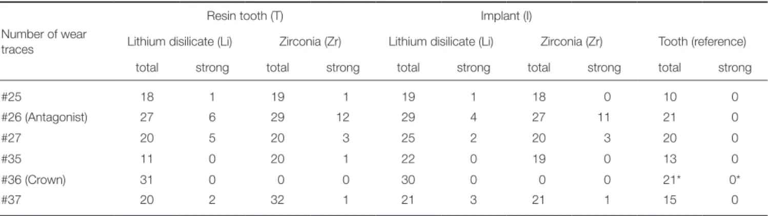
The number of wear traces was analyzed for all teeth of the upper and lower jaw. Table 2 gives an overview of the total number of wear traces in the different groups (sum of all wear facets of the eight tooth representatives in every mod- el). The number of strong wear facets (> 350 μm) is sepa- rately presented.
For the maxillary left second premolar (#25), in total, around twice the number of wear traces (n = 18 - 19) were found on the occlusal surfaces in the crown groups in com- parison to the reference tooth group (n = 10). No differ- ences were found between the different types of ceramic crowns (Li/Zr) or the types of abutment (I/T).
For the maxillary left first molar (#26), the total number of wear traces in the crown groups was higher (n = 27 - 29) in comparison to the tooth reference (n = 21). Only crown groups provided strong traces (n = 4 - 12), where Zr-crown groups showed 2 - 3 times higher number of traces (n = 11 - 12) than Li- crown groups (n = 4 - 6) in both comparable implant and resin tooth situations.
For the maxillary left second molar (#27), crown groups and tooth reference showed comparable number of wear traces (n = 20, only I-Li was higher with n = 25). Solely for the crown groups, strong traces (n = 2 - 5) were found. No strong differences were seen between the different types of
Table 2. Number of wear traces (sum total, strong traces; *: human tooth) of groups differing in crown material (lithium disilicate, zirconia) and abutment situation (resin tooth, implant)
Number of wear traces
Resin tooth (T) Implant (I)
Lithium disilicate (Li) Zirconia (Zr) Lithium disilicate (Li) Zirconia (Zr) Tooth (reference)
total strong total strong total strong total strong total strong
#25 18 1 19 1 19 1 18 0 10 0
#26 (Antagonist) 27 6 29 12 29 4 27 11 21 0
#27 20 5 20 3 25 2 20 3 20 0
#35 11 0 20 1 22 0 19 0 13 0
#36 (Crown) 31 0 0 0 30 0 0 0 21* 0*
#37 20 2 32 1 21 3 21 1 15 0
abutment (I/T).
In summary, for the upper jaw, strongest wear differenc- es between crown systems and the tooth reference were found for the antagonistic maxillary left first molar (#26) followed by minor differences for the maxillary left second premolar (#25).
For the mandibular left second premolar (#35), only group T-Li (n = 11) showed a similar number of wear trac- es in comparison to the tooth reference situation (n = 13).
The other systems provided a higher number of wear traces (n = 19 - 22). Solely for group T-Zr, one strong wear trace was found. An influence of the abutment was found only for Li-crown groups, showing twice the number of wear traces for the implant situation (n = 22).
For the mandibular left first molar (#36), Li crowns in both implant and resin tooth groups showed higher number of wear traces (n = 30 - 31) than the tooth reference (n = 21). All Zr-crowns provided no visible wear traces on the zirconia surface. Strong wear traces were not found in any group. No differences between implant- and resin tooth- supported crowns were seen.
For the mandibular left second molar (#37), all crown groups provided a higher number of wear traces (n = 20 - 32) in comparison to the tooth reference group (n = 15).
Distinctly higher results were found for group T-Zr (n = 32). A low number of strong wear traces were determined for all crown groups (n = 1 - 3). Differences between implant and resin tooth groups were found only for Zr.
In conclusion, for the lower jaw, the strongest wear dif- ferences between crown groups and the tooth reference group were found for tooth #36, followed by differences for tooth #37.
In general, comparing ceramic crowns and human teeth, ceramic crowns showed different number of wear traces than human teeth. The highest differences were found for crown and antagonistic situations. Only small differences between Li and Zr groups were found. The influence of the type of abutment seems minimal. Figure 1 gives a compara-
tive overview of the total number of wear traces.
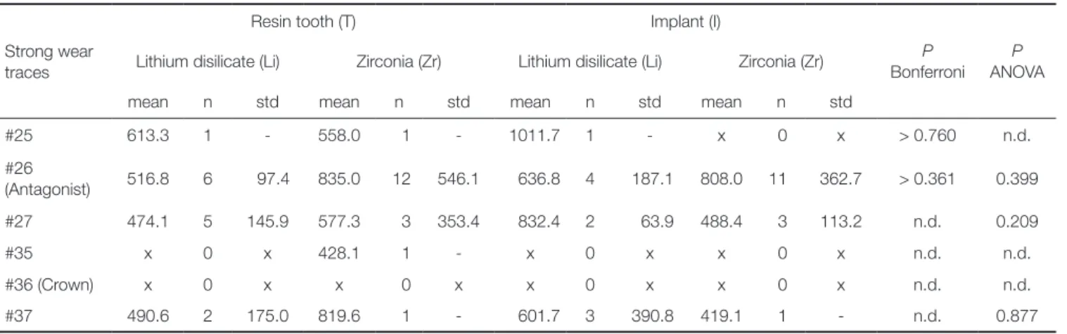
Strong wear traces were quantitatively evaluated. The highest number of strong wear traces was found for the antagonists in the crown groups. Mean depth of strong wear traces of the antagonists (tooth #26) varied between 516.8+/-97.4 µm (T-Li, n = 6), 835.0+/-546.1 µm (T-Zr, n = 12), 636.8+/-187.1 µm (I-Li, n = 4), and 808.0+/-362.7 µm (I-Zr, n = 11). No statistical differences were found between the individual systems (ANOVA: P = .399, Bonferrroni: P >
.361). Antagonists against resin tooth-supported Li crowns showed a tendency for lower values in comparison to implant-supported Li crowns.
Due to low numbers of strong wear traces for teeth
#25, #27, #35, and #37 statistical differences were not determined. Neither Li nor Zr crowns showed strong wear traces(see Table 2 and Table 3).
Table 3. Strong wear traces
Strong wear traces
Resin tooth (T) Implant (I)
Lithium disilicate (Li) Zirconia (Zr) Lithium disilicate (Li) Zirconia (Zr) P Bonferroni
P ANOVA
mean n std mean n std mean n std mean n std
#25 613.3 1 - 558.0 1 - 1011.7 1 - x 0 x > 0.760 n.d.
#26
(Antagonist) 516.8 6 97.4 835.0 12 546.1 636.8 4 187.1 808.0 11 362.7 > 0.361 0.399
#27 474.1 5 145.9 577.3 3 353.4 832.4 2 63.9 488.4 3 113.2 n.d. 0.209
#35 x 0 x 428.1 1 - x 0 x x 0 x n.d. n.d.
#36 (Crown) x 0 x x 0 x x 0 x x 0 x n.d. n.d.
#37 490.6 2 175.0 819.6 1 - 601.7 3 390.8 419.1 1 - n.d. 0.877
mean [µm], number (n), standard deviation (std); no strong traces: x; n.d.: not determined due to a low number of cases
Fig. 1. Overview of total number of wear traces on crown 36, antagonist 26, and adjacent teeth (25, 27, 35, 37) in the different groups.
Exemplary SEM pictures of worn crown and antagonist surfaces are given in Fig. 2. Reference tooth #36 and its tooth antagonist #26 showed typical wear traces without any cracks or other damages. T-Li and I-Li groups displayed wear traces on both tooth and crown, but no noticeable cracks or damages. For the T-Zr and I-Zr groups, wear trac- es on the antagonist were found. The zirconia crowns showed slightly worn areas of the glaze layer but no further damage or crack. On the antagonistic surface of T-Zr, a small enamel chipping was found.
DISCUSSION
The hypothesis that crown material (Li or Zr) and type of abutment (rigid implant, resin tooth with artificial periodon- tium) have an influence on the wear performance (traces, depth, and superficial damages) of crowns, antagonistic and adjacent teeth was accepted. The amount of wear differed between the Zr and Li crowns and partly between rigid implant or resilient resin tooth, and was influenced by the size of the root surfaces of the adjacent teeth.
While Zr crowns showed wear only in the glaze layer, resulting in the exposure of the underlying zirconia, Li crowns both for the resin tooth and implant situation pro- vided a high number (n = 30 - 31) of wear traces. The num- ber of wear traces on the antagonistic tooth 26 were com- parably high (n = 27 - 29) for both ceramic materials and abutment situations, but Zr crown groups showed a 2 - 3 - times higher number of strong traces than Li crown groups.
Furthermore, antagonists against resin tooth-supported Li crowns showed lower wear depth values in comparison to implant-supported Li crowns. Generally, adjacent tooth #37 and its antagonistic tooth #27 showed a higher number of strong wear traces than teeth #35 and #25. A tendency to a higher total number of wear traces was found for teeth #35 and #27 of group I-Li, and for tooth #37 of group T-Zr.
Differences between Zr, Li, and the human tooth refer- ence may be attributed to different material properties such as hardness (Li: HV 600, Zr: HV 1200 - 1300, enamel: HV 300 - 400) and flexural strength (Li: < 500 MPa, Zr: 1200 MPa, enamel 300 - 450 MPa). Thus, in contrast to Zr, Li showed wear, as it was reported in many previous stud- ies.7,28,29 Li crowns with wear may be flattened and compen- sate for chewing forces, resulting in lower loads on adjacent teeth. This effect is enhanced by the intrusion and the later- al movement of teeth with artificial periodontium. In con- trast, implant-supported Li crowns have no resilience, resulting in lower force absorbing capacity. As a conse- quence, adjacent teeth have to bear these additional chewing forces, resulting in increased wear. The larger the root sur- face, the smaller the expected intrusion of the adjacent teeth. Therefore, tooth #37 is supposed to show less resil- ience than tooth #35, and consequently the higher number of strong wear traces was found.
Damping effects of implant-supported crowns differ between materials,30 showing the lowest shock absorbing capacity and the highest load transfer for high-strength ceramics like zirconia. As Zr does not abrade itself, a higher force impact on the antagonist may result in increased wear, as it is reflected in a high number of strong antagonistic wear traces, both for resin tooth and implant groups. In the resin tooth group, intrusion of the crowned tooth caused a higher number of wear traces on the adjacent teeth, as the forces were not absorbed by the zirconia crown. In this situ- ation, the size of the root surface may play an important role: the larger the surface, the less the intrusion, resulting in increased wear of adjacent molars compared to premo- lars. Thus, the significantly higher number of wear traces on tooth #37 in group T-Zr may be explained. In contrast, in group I-Zr, mainly the antagonistic tooth is worn, as the highly wear-resistant Zr in combination with the rigid implant support takes the load off the adjacent teeth.
The results of all groups suggest that the size of the root surface of the adjacent teeth influences their wear behavior. It is assumed that molar teeth with larger root sur- faces show less resilience upon chewing forces than premo- lars, resulting in different absorption processes; molars are able to absorb forces via resilience only to a limited extent, and therefore the enamel layer in the area around the con- tact points absorbs comparably more chewing forces. This leads to a higher number of strong wear traces. In this con- text, it has to be considered that a comparison of the total number of wear traces between the different types of teeth is not significant because molars have a larger occlusal sur- face and consequently a higher number of contact points than premolars.
Fig. 2. Exemplary SEM pictures of worn crown surfaces and corresponding antagonistic enamel wear facets: (A) Occlusal surface of Li crown (30× magnification), (B) Wear facet of tooth antagonist of Li crown (100× magnifi- cation), (C) Occlusal surface of Zr crown (30× magnifica- tion), (D) Wear facet of tooth antagonist of Zr crown (100× magnification).
A B
C D
As the experimental set up of this pilot study did not allow for quantification of tooth intrusion, further studies that investigate the amount and effects of intrusion and consequent load distribution in a complex jaw model are necessary. Furthermore, it cannot be predicted if these observations correspond to the in vivo situation without restrictions since the in vitro simulation in the chewing simu- lator did not reproduce the three-dimensional structures of the temporomandibular joint. Any possibly occurring com- pressions of the temporomandibular joint may influence the loading situation of adjacent teeth. Furthermore, differ- ent wear behavior of ceramic crown materials compared to the natural teeth might have an influence on occlusion and jaw position or even result in craniomandibular dysfunc- tions.
Stronger wear traces for antagonists of ceramic crowns than for the reference tooth group were found. Antagonistic wear of Zr was generally higher than that of Li. These results highlight the necessity of a correct occlusal setup and regular recall sessions to control wear behavior and occlusal relation, or make adjustments if required. Long- time clinical studies are needed for a more significant pre- diction of the wear behavior of ceramic crowns, especially zirconia, with natural antagonistic and adjacent teeth.
Currently, there is only limited clinical evidence of enamel wear against zirconia, and adjacent teeth are usually not considered. First in vivo results indicated similar or more antagonistic enamel wear of Zr,1,11,18-21 which corresponded to previous in vitro results.4,6,15,16 The results indicated the necessity of considering the occlusal setup as well as the antagonistic and adjacent situation in clinical studies.
When comparing implant and resin tooth groups, the rigid positioning of the implants in comparison to the mobile positioning of the resin teeth, associated with the different properties of the abutment materials (modulus of elasticity: resin 2-3 GPa, titanium 110 GPa) or the different preparation geometries, had only limited influence on the wear performance. However, it can be assumed that the geometry and the abutment material can influence the force application and transmission and thus the abrasion behav- ior. Effects on the adjacent teeth and their antagonists may not be excluded. A limiting factor of the study design is that the artificial periodontal mobility simulates the clinical situa- tion only in rudiments and lacks tactile sensitivity and no proprioceptive motion feedback due to missing periodontal mechanoreceptors. Similarly, resin may not replace the com- plex bone structure of cortical and cancellous bone, result- ing in different damping effects.
Except for small chipping on the antagonistic surface of group T-Zr, no failures or damages were observed by SEM analysis. Chipping as major reason for clinical failure of veneered restorations 31-33 was not found in the present study as only monolithic crowns were applied. Based on identical initial surface states (polishing, glazing) of both materials, in the course of the wear simulation, Zr crowns only showed slight worn areas of the glaze layer with expo- sure of the underlying smooth surface, while Li revealed
typical rough wear facets. Wear of glaze and exposure of the underlying ceramic was also shown in previous studies on wear of different ceramics.8,34,35 For Li, removal of the glaze layer or glassy matrix and exposure of crystalline phases has superposed the original surface state. Because of its microstructure and inferior mechanical properties, lithi- um disilicate is more prone to microploughing, microcrack- ing and microcutting. The roughened surface also provoked wear on the antagonistic teeth, which is reflected in a flat- tening and a higher number of strong wear traces compared to the tooth reference.
As glaze layers are usually worn within the first months or years of clinical service, the surface treatment of the exposed ceramic may be of high importance. Smooth sur- faces are considered to be important for the long-term suc- cess of the restoration and reduced wear of the antagonist.
However, the hardness of zirconia might be a challenge for polishing. Several studies have dealt with the effect of pol- ishing procedures on surface properties of zirconia.36-40 Most studies agree that appropriate polishing instruments and procedures are effective in reducing surface roughness.
36,37,40 However, severely ground surfaces may not be fully restored by polishing.39 Partly, surface phase changes by pol- ishing were reported.37,38 Therefore, careful polishing is rec- ommended to achieve smooth surfaces and to keep phase changes low. Even if glaze layers are gradually worn in con- tact areas, they are clinically important for aesthetic charac- terization of crowns and may seal superficial pores and cracks in the ceramic surface. The glaze may also protect the zirconia surface from aging effects and low temperature degradation in the moist oral cavity. Furthermore, the glaze layer may be necessary for some favourable “fitting wear”,8 as it has to be considered that human teeth are also exposed to continuous height changes by wear under clinical condi- tions, which were reported to range between 30 and 40 µm per year.27
Potential effects of the humid oral environment on ceram- ic wear and aging were considered by thermocycling with water. Cyclic loading in water combined with temperature changes (5°C/ 55°C) might induce subcritical crack propa- gation and low temperature degradation (LTD) of zirconia, which may influence mechanical properties and wear.41-43
For a clinically relevant wear simulation and evaluation, human antagonistic and adjacent teeth were used. Although teeth with comparable size and shape were selected, individ- ual differences in morphology, differences in surface state and roughness, thickness and hardness of the enamel layer among other mechanical factors might account for a broad- er distribution of results. Optimal contact situations were achieved by a tooth-to-tooth situation with only one antago- nist per tooth. Nevertheless, different number and distribu- tion of contact points due to differences in the tooth mor- phology might have influenced loading and consequent wear. However, these variations in human teeth reflect the in vivo situation more realistically than artificial teeth with standardized geometry and composition. As the highest variations in the enamel structure as well as predamage are
expected primarily in the superficial enamel layer, evaluation of the wear depth allowed for better comparison between the groups than wear volume or area. Due to the lack of standardization in wear evaluation methods, comparison to other studies is difficult and determination of general refer- ence values for (strong) wear is not possible. Therefore, after evaluation of all wear data and comparison to the tooth reference group in this study, we defined a threshold value for wear (350 µm) that allowed a differentiation between the total number of wear facets and strong wear.
Assuming annual wear rates of about 30 - 35 µm,27 wear depths below 350 µm (after 10 - 15 years of simulation) were graded “normal”. Strong wear facets (> 350 µm) were further analyzed, and although their number varied among the groups, the values were in a realistic range (antagonists of crowns: about 500 - 800 µm). Especially for groups with a similar total number of wear facets, the number of strong wear facets allowed to clearly identify differences in the severity of wear (e.g. tooth #26: T-Li versus T-Zr, I-Li ver- sus I-Zr). As there were high variations in wear data due to individual tooth differences, this global differentiation of wear under consideration of all tooth representatives of one group is more significant than comparing individual wear values of every single tooth.
CONCLUSION
Wear performance of monolithic ceramic molar crowns was evaluated in a clinically relevant model with human antago- nistic and adjacent teeth. Wear depths and the number of wear traces differed between Zr and Li crowns, showing worn glaze with exposure of the underlying smooth surface for Zr, and deep wear facets for Li.
The crown material in combination with the type of abutment (implant, resin tooth) influenced the number of wear traces and the amount of strong wear (> 350 µm) of antagonistic and adjacent teeth. Antagonists against Zr showed about twice the amount of strong wear traces than Li irrespective of the abutment situation. Adjacent molar teeth of crowns and their antagonists revealed a higher amount of strong wear traces than adjacent premolar teeth due to differences in the size of the root surfaces that influ- ence their ability of intrusion. The results highlight the necessity of a correct occlusal setup and regular recall ses- sions to control wear behavior and occlusal relation.
Based on the results of this pilot study, the principal suitability of the applied in vitro jaw model for wear testing was shown. A limited comparative evaluation of the wear situation was enabled. Further development of the experi- mental set-up (e.g. measurement of tooth intrusion and load distribution) is recommended.
ORCID
Martin Rosentritt https://orcid.org/0000-0002-5227-288X Stephanie Krifka https://orcid.org/0000-0003-1454-2073 Verena Preis https://orcid.org/0000-0003-2625-7835
REFERENCES
1. Pathan MS, Kheur MG, Patankar AH, Kheur SM. Assessment of antagonist enamel wear and clinical performance of full- contour monolithic zirconia crowns: One-year results of a prospective study. J Prosthodont 2019;28:e411-6.
2. Rabel K, Spies BC, Pieralli S, Vach K, Kohal RJ. The clinical performance of all-ceramic implant-supported single crowns:
A systematic review and meta-analysis. Clin Oral Implants Res 2018;29:196-223.
3. Rauch A, Reich S, Dalchau L, Schierz O. Clinical survival of chair-side generated monolithic lithium disilicate crowns:10- year results. Clin Oral Investig 2018;22:1763-9.
4. Ludovichetti FS, Trindade FZ, Werner A, Kleverlaan CJ, Fonseca RG. Wear resistance and abrasiveness of CAD-CAM monolithic materials. J Prosthet Dent 2018;120:318.e1-8.
5. Hayashi S, Homma S, Takanashi T, Hirano T, Yoshinari M, Yajima Y. Wear properties of esthetic dental materials against translucent zirconia. Dent Mater J 2019;38:250-6.
6. Chong BJ, Thangavel AK, Rolton SB, Guazzato M, Klineberg IJ. Clinical and laboratory surface finishing procedures for zir- conia on opposing human enamel wear: A laboratory study. J Mech Behav Biomed Mater 2015;50:93-103.
7. Preis V, Grumser K, Schneider-Feyrer S, Behr M, Rosentritt M. Cycle-dependent in vitro wear performance of dental ce- ramics after clinical surface treatments. J Mech Behav Biomed Mater 2016;53:49-58.
8. Preis V, Behr M, Handel G, Schneider-Feyrer S, Hahnel S, Rosentritt M. Wear performance of dental ceramics after grinding and polishing treatments. J Mech Behav Biomed Mater 2012;10:13-22.
9. D’Arcangelo C, Vanini L, Rondoni GD, Vadini M, De Angelis F. Wear evaluation of prosthetic materials opposing them- selves. Oper Dent 2018;43:38-50.
10. D’Arcangelo C, Vanini L, Rondoni GD, De Angelis F. Wear properties of dental ceramics and porcelains compared with human enamel. J Prosthet Dent 2016;115:350-5.
11. Gou M, Chen H, Kang J, Wang H. Antagonist enamel wear of tooth-supported monolithic zirconia posterior crowns in vivo: A systematic review. J Prosthet Dent 2019;121:598-603.
12. Wiegand A, Credé A, Tschammler C, Attin T, Tauböck TT.
Enamel wear by antagonistic restorative materials under ero- sive conditions. Clin Oral Investig 2017;21:2689-93.
13. Fathy SM, Swain MV. In-vitro wear of natural tooth surface opposed with zirconia reinforced lithium silicate glass ceramic after accelerated ageing. Dent Mater 2018;34:551-9.
14. Zheng J, Zeng Y, Wen J, Zheng L, Zhou Z. Impact wear be- havior of human tooth enamel under simulated chewing con- ditions. J Mech Behav Biomed Mater 2016;62:119-27.
15. Nakashima J, Taira Y, Sawase T. In vitro wear of four ceramic materials and human enamel on enamel antagonist. Eur J Oral Sci 2016;124:295-300.
16. Zandparsa R, El Huni RM, Hirayama H, Johnson MI. Effect of different dental ceramic systems on the wear of human enamel: An in vitro study. J Prosthet Dent 2016;115:230-7.
17. Lee A, Swain M, He L, Lyons K. Wear behavior of human enamel against lithium disilicate glass ceramic and type III
gold. J Prosthet Dent 2014;112:1399-405.
18. Hartkamp O, Lohbauer U, Reich S. Antagonist wear by pol- ished zirconia crowns. Int J Comput Dent 2017;20:263-74.
19. Esquivel-Upshaw JF, Kim MJ, Hsu SM, Abdulhameed N, Jenkins R, Neal D, Ren F, Clark AE. Randomized clinical study of wear of enamel antagonists against polished mono- lithic zirconia crowns. J Dent 2018;68:19-27.
20. Yang SW, Kim JE, Shin Y, Shim JS, Kim JH. Enamel wear and aging of translucent zirconias: In vitro and clinical stud- ies. J Prosthet Dent 2019;121:417-25.
21. Lohbauer U, Reich S. Antagonist wear of monolithic zirconia crowns after 2 years. Clin Oral Investig 2017;21:1165-72.
22. Rashid H, Sheikh Z, Misbahuddin S, Kazmi MR, Qureshi S, Uddin MZ. Advancements in all-ceramics for dental restora- tions and their effect on the wear of opposing dentition. Eur J Dent 2016;10:583-8.
23. Rosentritt M, Behr M, Scharnagl P, Handel G, Kolbeck C.
Influence of resilient support of abutment teeth on fracture resistance of all-ceramic fixed partial dentures: an in vitro study. Int J Prosthodont 2011;24:465-8.
24. Scharnagl P, Behr M, Rosentritt M, Leibrock A, Handel G.
Simulation of physiological tooth mobility in in-vitro stress examination of dental restorations in the masticator. J Dent Res 1998;77:1260.
25. Rosentritt M, Behr M, van der Zel JM, Feilzer AJ. Approach for valuating the influence of laboratory simulation. Dent Mater 2009;25:348-52.
26. Rosentritt M, Siavikis G, Behr M, Kolbeck C, Handel G.
Approach for valuating the significance of laboratory simula- tion. J Dent 2008;36:1048-53.
27. Lambrechts P, Braem M, Vuylsteke-Wauters M, Vanherle G.
Quantitative in vivo wear of human enamel. J Dent Res 1989;
68:1752-4.
28. Preis V, Weiser F, Handel G, Rosentritt M. Wear performance of monolithic dental ceramics with different surface treat- ments. Quintessence Int 2013;44:393-405.
29. Lawson NC, Janyavula S, Syklawer S, McLaren EA, Burgess JO. Wear of enamel opposing zirconia and lithium disilicate after adjustment, polishing and glazing. J Dent 2014;42:1586- 91.
30. Rosentritt M, Schneider-Feyrer S, Behr M, Preis V. In vitro shock absorption tests on implant-supported crowns: Influence of crown materials and luting agents. Int J Oral Maxillofac Implants 2018;33:116-22.
31. Tsumita M, Kokubo Y, Ohkubo C, Sakurai S, Fukushima S.
Clinical evaluation of posterior all-ceramic FPDs (Cercon): a prospective clinical pilot study. J Prosthodont Res 2010;54:
102-5.
32. Teichmann M, Wienert AL, Rückbeil M, Weber V, Wolfart S, Edelhoff D. Ten-year survival and chipping rates and clinical quality grading of zirconia-based fixed dental prostheses. Clin Oral Investig 2018;22:2905-15.
33. Schmitter M, Mueller D, Rues S. Chipping behaviour of all- ceramic crowns with zirconia framework and CAD/CAM manufactured veneer. J Dent 2012;40:154-62.
34. Rosentritt M, Preis V, Behr M, Hahnel S, Handel G, Kolbeck C. Two-body wear of dental porcelain and substructure oxide
ceramics. Clin Oral Investig 2012;16:935-43.
35. Preis V, Behr M, Kolbeck C, Hahnel S, Handel G, Rosentritt M. Wear performance of substructure ceramics and veneer- ing porcelains. Dent Mater 2011;27:796-804.
36. Preis V, Grumser K, Schneider-Feyrer S, Behr M, Rosentritt M. The effectiveness of polishing kits: influence on surface roughness of zirconia. Int J Prosthodont 2015;28:149-51.
37. Preis V, Schmalzbauer M, Bougeard D, Schneider-Feyrer S, Rosentritt M. Surface properties of monolithic zirconia after dental adjustment treatments and in vitro wear simulation. J Dent 2015;43:133-9.
38. Bartolo D, Cassar G, Al-Haj Husain N, Özcan M, Camilleri J.
Effect of polishing procedures and hydrothermal aging on wear characteristics and phase transformation of zirconium dioxide. J Prosthet Dent 2017;117:545-51.
39. Al-Haj Husain N, Camilleri J, Özcan M. Effect of polishing instruments and polishing regimens on surface topography and phase transformation of monolithic zirconia: An evalua- tion with XPS and XRD analysis. J Mech Behav Biomed Mater 2016;64:104-12.
40. Al-Haj Husain N, Özcan M. A study on topographical prop- erties and surface wettability of monolithic zirconia after use of diverse polishing instruments with different surface coat- ings. J Prosthodont 2018;27:429-42.
41. Alghazzawi TF, Lemons J, Liu PR, Essig ME, Bartolucci AA, Janowski GM. Influence of low-temperature environmental exposure on the mechanical properties and structural stability of dental zirconia. J Prosthodont 2012;21:363-9.
42. Salazar Marocho SM, Studart AR, Bottino MA, Bona AD.
Mechanical strength and subcritical crack growth under wet cyclic loading of glass-infiltrated dental ceramics. Dent Mater 2010;26:483-90.
43. Zhang Y, Song JK, Lawn BR. Deep-penetrating conical cracks in brittle layers from hydraulic cyclic contact. J Biomed Mater Res B Appl Biomater 2005;73:186-93.