Received on November 11, 2009. Accepted on November 11, 2009.
CC This is an open access article distributed under the terms of the Creative Commons Attribution Non-Commercial License (http://creativecommons.org/licenses/by-nc/3.0) which permits unrestricted non-commercial use, distribu- tion, and reproduction in any medium, provided the original work is properly cited.
*Corresponding Author. Tel: 82-63-275-1515; Fax: 82-63-250-4215; E-mail: [email protected] Keywords: Cancer, Regulatory T cells, Suppressor T cells, MicroRNA, Immunotherapy
The Role of Regulatory T Cells in Cancer
Tai-You Ha*
Department of Immunology, Chonbuk National University Medical School, Chonju, Chonbuk, Korea
There has been an explosion of literature focusing on the role of regulatory T (Treg) cells in cancer immunity. It is becoming increasingly clear that Treg cells play an active and significant role in the progression of cancer, and have an important role in suppressing tumor-specific immunity. Thus, there is a clear rationale for developing clinical strategies to diminish their regulatory influences, with the ultimate goal of augmenting antitimor immunity. Therefore, manipulation of Treg cells rep- resent new strategies for cancer treatment. In this Review, I will summarize and review the explosive recent studies dem- onstrating that Treg cells are increased in patients with ma- lignancies and restoration of antitumor immunity in mice and humans by depletion or reduction of Treg cells. In addition, I will discuss both the prognostic value of Treg cells in tumor progression in tumor-bearing hosts and the rationale for strategies for therapeutic vaccination and immunother- apeutic targeting of Treg cells with drugs and microRNA.
[Immune Network 2009;9(6):209-235]
INTRODUCTION
It is well established and generally accepted that numerous innate and adaptive immune effector cells and molecules par- ticipate in the recognition and destruction of cancer cells, a process that is known as cancer immunosurveillance. Howev- er, cancer cells avoids such immunosurveillance through the outgrowth of poorly immunogenic tumor -cell variant (immu- noselection, a process that is also known as immunoediting) and through immunosubversion (1,2) The immune system in- teracts with tumors throughout their development, and the consequences of this interaction have substantial implications for cancer therapy. Although some host responses may inhibit
tumor growth and progression, the immune system can also promote cancer by provoking chronic inflammation and, in turn, elaborating factors that drive tumor growth, survival, and angiogenesis. Failure of immunity can predispose a per- son to tumor development (3). T cell-mediated immuno- suppression has been observed for decades without clar- ification as to which factors was responsible for this observation. Recently, a number of publications showed that progressive tumor growth evokes generation of immuno- suppressor cells and suppressor T cells (4,5). The identi- fication of regulatory T (Treg) cells represents a milestone in the field of immuology and provides an explanation for T-cell-mediated immunosuppression (6-11). Although Treg cells were originally identified for their ability to prevent or- gan-specific autoimmune disease in mice, emerging evidence suggests that Treg cells play a pivotal role in tumor immunol- ogy and contribute to tumor growth and progression, thereby having an important impact on the outcome of cancer pa- tients (3,12-17). Likewise, tumor-induced immune suppre- ssion could be responsible for the failure of many promising cell-base vaccine attempts (18) and other immune-based therapies against cancer (2,3,15,19-21). Tumor-induced im- mune suppression is caused by numerous mechanisms, many of which involve the accumulation of immune-suppressive in- filtrates in the tumor microenvironment. One of the most po- tent and well-studied suppressive phenotypes found in the tu- mor microenvironment is the Treg cells. Increasing evidence shows that Treg cell may also play an important role in im- mune evasion mechanisms employed by cancer (17,18,22-24).
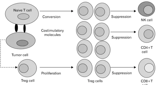
Tumors may differentiate, expand, recruit, and activate Treg (tumor Treg) cells via multiple mechanisms and potently ab- rogate antitumor immunity (25,26). Fig. 1 shows a schematic
Figure 1. Tumor-mediated genera- tion of regulatory T (Treg) cells and the effect on the tumor micro- environment. Tumor cells induce the generation of Treg cells through both cell contact-dependent and cell contact-independent mechani- sms. Soluble protein such as TGFβ produced by tumor cells promote the proliferation of Treg cells and induce the conversion of naive CD4+CD25- T cells into Treg cells.
Tumor cells also express costimula- tory molecules such as CD80/
CD86 or CD70 and interact with naive T cells to convert these naive T cells into Treg cells. The in- creased numbers of Treg cells inhibit the NK cells, CD4+ T cells, CD8+T cells and the other cells and contribute to the progression of tumors.
diagram of tumor-mediated generation of Treg cells in tumor microenvironment. Tumor Treg cells may promote local tu- mor growth at tumor site, and may be relevant to the pro- gression of systemic disease in the peripheral blood or lym- phoid organs (25,27,28). Thus, tumor Treg cells may be a ma- jor obstacle for immunoterapy of cancer (25,27,29,30).
Elucidating the mechanisms by which these cells traffic and accumulate in the tumor microenvironment could provide at- tractive therapeutic targets with which to combat tumor-in- duced immune suppression (11,31,32). An emerging interest is the potential role of Treg cells in cancer development and progression because they have been shown to suppress anti- tumor immunity (11,33). Thus, the critical role of Treg cells in tumor immunotherapy has been gradually elucidated (28,29,34,35). Treg cells (defined as CD4+CD25+FoxP3+
T cells) have been shown to be inversely related to the out- come of several human malignancies (26,36-41). Further- more, depletion of Treg cells can enhance rejection of endog- enous immune mediated tumor and improve tumor anti- gen-specific immunity, which highlights the impact of Treg cells in antitumor immunity (28,42,43) Depletion of Treg cells has also been shown to improve the efficacy of tumor im- munotherapy, including vaccination and CTLA4 blockade (28-30,44).
In this review, I will discuss the role of human Treg cell in cancer. With this in mind, I briefly outline the evidence describing the role of Treg cell in impairing antitumor im- mune responses. And, I will review the recent studies demon- strating that Treg cells are increased in patients with
malignancies. I will also review the effects of depletion or reduction of Treg cells on tumor progression and alterations of Treg homeostasis by cancer treatment. In addition, I will discuss both the prognostic value of Treg cells in tumor pro- gression or survival of tumor-bearing animals and humans and the rationale for clinical use of Treg cells. It will empha- size the difficulties, as well as the opportunities, encountered in transitioning from bench to bedsides. I will confine this discussion to CD4+CD25+ or CD4+CD25+FoxP3+ Treg cells that develop in both the thymus and periphery and rep- resent the major Treg cell populations that act critical for im- mune homeostasis (45). FoxP3−iTreg cells such as Tγ1 and Th3 cell will not be discussed here.
Treg CELLS
There has been an explosion of literature focusing on the role of these cells in several settings including cancer immunity, autoimmunity, transplantation tolerance allergic responses and microbial immunity (reviewed in 9,12-14,46-48). Treg cells can be described as a T-cell population that functionally suppresses an immune response by influencing the activity of another cell type (49,50). Treg cells were initially described by Gershon et al (6,7) in the early 1970s and were called suppressor T cells. Thereafter, a series of papers concerning thymic suppressor cell which could inhibit cell-mediated im- munity in many different in vivo models (51-55). Unfortunate- ly, despite the importance of these studies there was ex- tensive skepticism in the immunological field about existence
of these cell, and suppressor T cells left the center stage of immunology for decades. However, a rejuvenation of the Treg field was launched through the work of Sakaguchi et al (10,11) who in 1995 reported that a small group of T cells with particular cell surface phenotype (CD4+ cells which co-express the IL-2 receptor-α chain, CD25) maintain self-tol- erance and that breakdown of this tolerance could lead to autoimmune disease and CD4+CD25+ T cells were named Treg cells. Now the terms “suppressor T cells” and “regulato- ry T cells” are used interchangeably, but the term “regulatory T cells” is preferred by most researchers. Work in the field of Treg cell immunology was greatly enhanced in 2003 by the discovery and characterization of the Treg-specific gene, FoxP3 (45,56-59). So far the most specific marker for naturally occurring CD4+ Treg cells, at least in mice, is FoxP3, a mem- ber of the forkhead family of DNA-binding transcription factor (45,56,58,59). More recent studies have shown that FoxP3 is not only a key intracellular marker but is also a crucial devel- opment and functional factor for CD4+CD25+ Treg cells (45,59-61). Until now, there are at least three recognized sub- sets of CD4+ Treg cells involved in the negative regulation of immune response, which include CD4+CD25+ FoxP3+, type 1 Treg cells (Tγ1) secreting IL-10 and Th3 cells secret- ing TGFβ (62-66). Among these, classical Treg cells are CD4
+CD25+FoxP3+ T cells and depletion of CD4+CD25+
FoxP3 cells has attracted much attention in recent years (28,29,58,67,68). Treg cells, according to their sources, broad- ly belong to two classes, which are naturally occurring thy- mic-derived Treg cells and adaptive or inducible Treg cells.
Naturally occurring Treg (nTreg) cells are generated in the thymus during development of T lymphocytes (62,63,69).
These Treg cells, constituting 5-10% total lymphocytes, then enter peripheral circulation and are widely distributed in pe- ripheral reservoir lymph nodes and spleens (63,64,70). On the contrary, adaptive or inducible Treg (iTreg) cells are de- veloped in the periphery from naive T cells or nTreg cells under the influence of various inductive signals, most im- portantly TGFβ or IL-10 (54,62,63,69,71). Treg cells are pri- marily characterized by CD4+FoxP3+ or CD4+CD25+
FoxP3 T cells and FoxP3 has been considered as a master regulatory transcription factor for Treg cells (45,56,58,59). It has recently been reported that FoxP3+ Treg cells could also be generated outside the thymus under a variety of conditions (54,59,72). It has become evident that FoxP3+ Treg cells are one of the main barrier to implementation of cancer im- munotherapies (12,21). Tumor tissue promote the conversion
of naive T cells into FoxP3 Treg cells and accumulation of Treg cells in tumor site, thereby impairing the development of effector responses (33,60,61). Thus, FoxP3+ Treg cells ha- va a key function in obsructing tumor immunosurveillance (33,57,73). Although nTreg cells develop in the thymus, sev- eral reports have demonstrated that peripheral CD4+CD25+
T cells can be converted into adaptive FoxP3+Treg cells (74,75). Among the other major surface markers, glucocorti- coid-induced TNF-receptors-related (GITR) protein and cyto- toxic lymphocyte-associated angigen-4 (CTLA-4) are the most important with respect to development and function of Treg cells (45,47,48,69). Intrestingly, it has been reported that the large number of different cell types that are purported to be directly targeted by FoxP3+ Treg cells are CD4+, CD8+ T cell, dendritic cells, B cells, marophages, osteoblasts, mast cells, NK cells, and NKT cells (45).
INCREASED PREVALENCE OF Treg CELLS IN CANCER BEARING MICE
Recently, in murine models, a vast numbers of murine studies have shown that prevalence of Treg cells is increased in tu- mor bearing mice and the depletion or inhibition of Treg cells can enhance antitumor immunity (76-78). In context of can- cer, Treg-cell frequencies and function are important because increased number might favor tumor development or growth and influence the course of the disease. It has been also shown that CD8+ T cell immunity against a tumor or self-an- tigen is hindered by Treg cells and Treg cells inhibit adoptive immunotherapy in pme-l TCR transgenic mice (77). In a mouse pancreas cancer model, tumor actively promotes the accrual of Treg through several mechanisms involving activa- tion of nTreg cells as well as conversion of non-Treg into Treg cells (78). Both antitumor effector T cells and Treg cells capable of abrogating the antitumor reactivity of the effector T cells are primed in the same tumor-draining lymph nodes during tumor progression and the Treg cells generated in tu- mor-draining lymph nodes possess the same functional prop- erties as the Treg cells that arise naturally in the thymus but recognize tumor-associate antigen (79). One study reported that the numbers of FoxP3+CD4+nTreg cells are sig- nificantly increased in draining but not non-draining, lymph nodes and spleen of tumor-bearing mice (80). In this study, furthermore, a small number of nTreg cells could be also found at the tumor sites. In a transgenic mouse model of prostate dysplasia, functional studies confirmed a role for
CD4+CD25+ Treg cells in suppressing T cell proliferation as well as regulating the growth of transplanted prostate tu- mor cells (81). Additionally, these studies showed that an- ti-CD25 antibody treatment reduces, but does not prevent, tu- mor growth in a transgenic mouse model of prostate dyspla- sia, These transgenic mouse developed reproducible prostate specific tumors that can progress to mouse prostatic intra- epithelial neoplasia similar to human prostatic intraepithelial neoplasia (81). Interestingly, in the mouse tumor model, no difference was observed in the proportion of CD4+CD25+
/CD4+ in peripheral blood lymphocytes between colon-car- cinoma-bearing BALB/c mice and normal mice, while there was a significant increase in the proportion in spleen lympho- cytes in tumor bearing mice as compared with normal mice (82). Moreover, the proportion increased in accordance with the increase in the tumor size, suggesting that the CD4+
CD25+/CD4+ proportion in spleen lymphocytes might be a sensitive index to evaluate the Treg cell in tumor mouse models. In one study, the transforming growth factor-β (TGF β) secreting murine pancreas adenocarcinoma (Pan02) cell line was injected into syngeneic C57BL/6 mice. and the prev- alence of Treg cells in the tumor draining lymph nodes (TDLNs) and spleen was measured weekly. Compared with control mice, the prevalence of CD4+CD25+ Treg cells in TDLNs and in tumor spleen increased with tumor growth, providing evidence that Pan02 tumor promotes the preva- lence of Treg cells (32).
A study showed, using murine colorectal tumor, that tumor immunity stimulated in the absence of regulatory cells is not restricted to tumors of colorectal origin, but is effective against tumors of different histological types such as B cell lymphoma and a renal cell carcinoma (83). The data suggest that the generation of cross-reactive tumor immunity is a spe- cific manifestation of CD25 Treg cell depletion and the gen- eration of CD4+ T cells capable of mediating tumor rejection is another important feature of tumor immunity induced in the absence of CD25+ cells. Interestingly, Ghiringhelli et al investigated the mechanisms of immune tolerance raised by tumors by comparing immunogenic and tolerogenic tumor cell clones isolated from a rat colon carcinoma (84). When injected into syngeneic hosts, the immunogenic REGb cells yield tumors that are rejected, while the tolerogenic PROb cells yield progressive tumors and inhibit the regression of REGb tumors (84). They found that PROb tumor volume is correlated with an expansion of CD4+CD25+ Treg cells in lymphoid tissues. While total T cells from PROb tumor-bear-
ing rats yield no apparent antitumor immune response, deple- tion of CD25+ T cells restores this reactivity. A single admin- istration of cyclophosphamide depletes CD4+CD25+ Treg cells in PROb tumor-bearing animals, delays the growth of PROb tumors, and cures rats bearing established PROb tumor when followed by an imunotherapy which has no curative effect when administered alone. These data strongly suggest that role of Treg cells in tumor-induced immune tolerance and the interest of Treg cell depletion to sensitize established tumors to immunotherapy. Interestingly, furthermore, a recent study demonstrated the role of Treg cells in tumors of the central nervous system (85). The authors implanted syngeneic GL26 tumor cells in the brain of C57BL/6 mice, and tumor-in- filtrating lymphocytes (TILs) was analyzed and found that CD4+CD25+ Treg cells play an important role in suppress- ing the immune response to tumors of central nervous sys- tem, suggesting that these Treg cells may therefore represent a potentially novel target for immunotherapy of malignant glioma. Schabowsky et al showed that Treg cells are actively recruited and induced by tumors to block innate and adaptive immune priming, effector function and memory response which can inhibit the efficacy of therapeutic cancer vaccines (18). Combination of CTLA-4 blockade and depletion of CD25
+ Treg cells results in maximal tumor rejection (86). The synergism in the effects indicates that CD25+ Treg cells and CTLA-4 signaling represent two alternative pathways for sup- pression of autoreactive T cell immunity, suggesting that si- multaneous intervention with both regulatory mechanisms is therefore a promising concept for the induction of therapeutic antitumor immunity.
It has been reported that cyclooxygenase (COX)-2 and its product prostaglandin (PG)E2 underlie an immunosuppressive network that is important in the pathogenesis of non-small cell lung cancer (86). Tumor-derived COX-2/PGE2 induced expression of the Treg cell-specific transcription factor, FoxP3, and increased Treg cell activity. In vivo, COX-2 in- hibition reduced Treg cell frequency and activity, attenuated FoxP3 expression in TILs and decreased tumor burden, pro- viding evidence that inhibition of COX-2/PGE2 suppresses Treg cell activity and enhances antitumor responses (87).
Concomitant tumor immunity describes the immune re- sponse in a host bearing a progressive tumor that rejects an inoculum of the same tumor at distant site (34,88). Progres- sion of poorly immunogenic B16 melanoma did not sponta- neously elicit concomitant immunity (88). However, deple- tion of CD4+ T cells in tumor-bearing mice resulted in CD8
+ T cell-mediated rejection of challenge tumors. Concomi- tant immunity was also elicited by treatment of cyclophos- mamide. nTreg cell efficiently suppressed concomitant im- munity mediated by previously activated CD8+ T cells (88).
These results shows that Treg cells are the major regulator of concomitant tumor immunity against weakly immunogenic tumor. In a series of elegant studies, most recently, Lin et al found that CD8+ T cells, from mice that lost concomitant tumor immunity, possessed potent antitumor properties and strongly expressed effector molecules (34). Furthermore, in- terestingly, effector/memory Treg cells increased as the pri- mary tumor progressed. They also reported that these effec- tor/memory Treg cells inhibit concomitant tumor immunity in vivo. Their data suggest that effector/memory Treg cells are responsible for the loss of concomitant tumor immunity asso- ciated with tumor progression.
Valzasina et al reported that tumor injection in thymectom- ized and CD25-depleted mice shows that CD4+CD25+
T-cell expansion occurs even in the absence of thymus (74).
These newly generated cells are bonna fide Treg cells in terms of FoxP3 expression. In addition, they analyzed Treg cell number and function in tumor-bearing mice as well as their recovery after peripheral and central depletion by means of anti-CD25 monoclonal antibody (mAb) and thymectomy, and described CD4+CD25-T-cell conversion into Treg cells as the main mechanism of Treg cell replenishment and ex- pansion in tumor-bearing mice.
Recently, there is a large number of evidence illustrating the increase in the number of Treg cells in the tumor setting, but very little work has been done to address the functionality of these cells. Despite the numerous studies showing that Treg cell from tumor-bearing animals and cancer patients are functional, no studies have directly compared Treg cells from tumor-bearing and nontumor-bearing animals. A recent study directly compared Treg cells in the spleen and the tumor in- filtrate at both the molecular and functional levels (89). This study provides evidence that tumor-infiltrating Treg cells are functionally impaired, due to a loss of ability to respond to TCR stimulation and that murine Treg cells in the tumor mi- croenvironment display both enhanced proliferation and re- duced functionality. This enhanced proliferation leads to an intratumoral accumulation of Treg cells with a unique pheno- type: CD4+CD25+FoxP3+GITRhighCD27llowCD621L−. The data further demonstrate that the accumulation of Treg cells in the tumor is caused by multiple factors, including increased proliferation, decrease apoptosis, and altered expression of
chemokines, chemokine receptor, and cell-surface markers.
The trafficking of T cells is mediated in part by chemokines and the expression of their receptors. Higher expression of the chemikine receptor CCR4 was reported on Treg cells.
Likewise, CCR4 associated chemokines and macrophage-deri- ved chemokine (CCL22) have been correlated with higher fre- quencies of Treg cells in gastric cancer (90). In ovarian can- cer, tumor-derived CCL22 was shown to recruit Treg cells in vivo and predicts a negative prognosis (91). Most recently, Mailloux et al examined Treg cell levels and changes in che- mokine secretion in a murine model of Lewis lung carcinoma (LLC) and demonstrated increased number of CD4+CD25+
FoxP+ Treg cells in the lungs of C57BL/6 mice bearing a metastatic LLC (31).
It has been recently reported that in a syngeneic murine glioma model a time-dependent accumulation of CD4+
FoxP3 Treg cell in brain tumor (92). Further analysis revealed a time-dependent upregulation of CD25, CTLA-4, GITR on in- tratumoral CD4+FoxP3+ Treg cells during tumor growth.
Moreover, treatment with anti-CD25 mAbs significantly re- duced the number of these highly suppressive Treg cells within the growing tumor and provoked a CD4 and CD8 T cell dependent destruction of the glioma cells. Combining Treg cell depletion with administration of blocking CTLA-4 mAbs further boosted glioma-specific CD4+ and CD8+ ef- fector T cells as well as antiglioma IgG2a antibody titers re- sulting in complete tumor eradication (92). These data illus- trate that intratumoral accumulation and activation of Treg cells act as a dominant immune escape mechanisms for glio- ma and underline the importance of controlling tumor-in- filtrating Treg cells in glioma immunotherapy. Most recently, Liu et al have beautifully investigated the suppressor function of both tumor and naive Treg cells in vitro and in vivo and reported that Treg cells from mice bearing a breast tumor were elevated (tumor Treg cells) and that tumor Treg cells potently abrogate tumor-specific CD8+ T cell response in tu- mor-draining lymph nodes, thereby suppressing antitumor immunity at the early stage of the immune response induced by adoptively transferred tumor-primed CD4+ T cells (17).
RESTORATION OF ANTITUMOR IMMUNITY IN MICE BY DEPLETION OR REDUCTION OF Treg CELLS As describe before, accumulating data have shown that Treg cells play an important role in tumor immunology. Increasing evidence for their role in fostering immune privilege in both
mouse and human tumors has fueled interest in attempts to interfere with their number or function for therapeutic benefit (14,28). There is a large body of data in the literature demon- strating restoration of antitumor immunity by depletion or re- duction of Treg cells. An earlier study demonstrated that elim- ination of CD25+CD4 T cells can abrogate immunological unresponsiveness to syngeneic tumor in vivo and in vitro, leading to spontaneous development of tumor specific effec- tor cells as well as tumor-nonspecific ones (93). Using the weakly immunogenic MCA205 sarcoma and the poorly im- munogenic B16/BL/D5 melanoma, an early study demon- strated that although the antitumor immunity enhanced by the depletion of CD4+CD25+ Treg cells is insufficient to erad- icate tumors, it augments the sensitization of immune T cells in the draining lymph nodes, thus, facilitating adoptive im- munotherapy (94). Prasad et al showed that depletion of Treg cells before vaccination considerably increased the ability of vaccinated mice to survive tumor challenge, and allowed the development of long-lasting tumor protective immunity (95).
This study suggests that it is possible to generate effective im- munotherapeutics against weakly immunogenic tumors by combining treatment with dendritic cells (DC) loaded with apoptotic tumor cells and depletion of Treg cells. Van Meirvenne et al (96) described mRNA electroporation as an efficient gene delivery method to introduce tumor-antigen in- to murine immature DC. They showed in this report that a significant improvement in cytotoxic T lymphocytes (CTL) re- sponse is obtained both in the primary and in the memory effector phases when Treg cells are depleted in vivo prior to immunization, providing the convincing evidence for the merit of Treg cell depletion prior to immunization with mRNA-electroporated DC. Onizuka et al investigated the ef- fect of in vivo administration of anti-CD25 mAB on the growth of eight tumors, including five leukemias, a myeloma, and two fibrosarcoma derived from four different inbred mouse strains that grew progressively in syngeneic mice (97).
They found that a single injection of less than 0.125 mg of anti-CD25 mAb caused a reduction in the number of CD4+
CD25+cells and regression of six of the eight tumors, sug- gesting effective tumor rejection responses resulted from de- pletion of Treg cells. A study also showed that elimination of CD4+ T cells by anti-CD4 antibody caused regression of a methylcholantrene-induced sarcoma growing in syngeneic A/J mice (98). A CD4+ T-cell clone, designated T595B1, was also established to elucidate the characteristics of CD4+ sup- pressor T cells (98). Interestingly, one report has shown using
a murine model of melanoma that treatment of mice with an- ti-CD25 mAB facilitates long-term CD4+ T cell-mediated tu- mor immunity through depletion of CD25+ Treg cells (99).
Furthermore, depletion of CD25+ Treg cells allows the host mice to induce both CD4+ and CD8+ antitumoral responses following challenge of colon cancer cells (100). Simultaneous depletion of CD25+ and CD8+ cells with mAb revealed that tumor-specific CD4+ T cells were able to reject colon cancer cells, a MHC class II-negative tumor (100). Depletion of Treg cell alone or in combination wih a whole tumor cell vaccine promotes a tumor-specific immune response (76). Thus, strat- egies incorporating Treg cell depletion might improve the ef- ficacy of cancer vaccines. In addition, the suppression of anti- tumor immunity by Treg cells occurs predominantly at the tu- mor site, and that intratumor depletion of CD4+ cells, even at a late stage of tumor development, can be an effective treatment for well-established cancers (101). IL-2-induced an- titumor immunity is enhanced by Treg-cell depletion and is due to expansion of the tumor-infiltrating cytotoxic CD8+ T cell population. More recently, using B16/BL6, a trans- plantable murine melanoma model, Quezada et al demon- strated dichotomy between the effects of Treg cell depletion on tumor rejection dependent on whether depletion occurs before (prophylactic) or after (therapeutic) tumor engraftment (102). In these studies, failure to promote rejection with ther- apeutic depletion (i.e., depletion after tumor establishment, which is more relevant to the clinical seting) was not related to lack of Treg cell depletion, to elimination of effector T cells, or to a failure to enhance systemic antitumor T cell re- sponses, but correlates with failure of effector cells to infiltrate the tumor and increase the intratumor ratio of effector T cells/Treg cell, suggesting an alternative, clinically applicable strategy for the treatment of established tumor. In fact, care- fully timed depletion of Treg cells has been shown to en- hance tumor immunity.
In a series of elegant experiments, Wrzesinski et al re- ported that removal of Treg cells by nonmyeloablative total body irradiation of tumor-bearing mice improved the function of adoptively transferred cells for new antitumor immuno- therapies (103). Surprisingly, they also demonstrated that su- perior antitumor efficacy is achieved by further increasing the intensity of lymphodepletion to a level that required hema- topoietic stem cells. A recent study also showed that removal of CD25+ Treg cells enhanced antitimor immunity against lo- cal growth of B cell lymphoma and that induction or ex- pansion of Treg cells could be one mechanism by which the
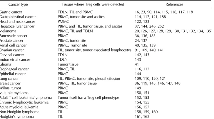
Table I. Increased prevalence of CD4+CD25+ regulatory T cells in patients with various cancers
Cancer type Tissues where Treg cells were detected References
Gastric cancer TDLN, TIL and PBMC 16, 23, 90, 114, 115, 116, 117, 118
Gastrointestinal cancer PBMC, tumor site and ascites 114, 117, 121, 188
Head and neck cancer PMMC 122, 123
Hepatocellular cancer PBMC and TIL, tumor tissue, and ascites 37, 144, 246, 252
Melanoma PBMC, TIL and TDLN 20, 126, 127, 128, 129, 130, 131, 132, 134, 135
Pancreatic cancer PBMC 36, 136, 185
Prostate cancer PBMC, tumor site 24, 137
Renal cell cancer PBMC, Tumor site 40, 135, 139
Ovarian cancer TIL, tumor site, tumor associated lymphocytes 91, 109, 140, 141
Cervical cancer TDLN 142, 143
Endometrial cancer TDLN 143
Glioma Tumor tissue 41
Esophageal cancer PBMC, TIL 116, 117
Epithelial cancer PBMC 144
Lung cancer TIL, PBMC, tumor site, pleural effusion 109, 110, 120, 121
Breast cancer PBMC, TIL, tumor tissue 36, 119, 145, 146, 147, 148
Wilms' tumor PBMC 149
Multiple myeloma PBMC 150, 151
Adult T cell leukemia/lymphoma Tumor itself has a Treg cell phenotype 152, 153
Chronic lymphocytic leukemia PBMC 154, 155
Acute myeloid leukemia PBMC 156, 157
Non-Hodgkin lymphoma TIL 158, 159, 160
Hodgkin's lymphoma TIL 161, 162
TDLN, tumor-draining lymph nodes; PBMC, peripheral blood mononuclear cells; TIL, tumor-infiltrating lymphocytes.
growing tumor evades immune surveillance in mice (104).
Treatment of mice with low-dose of cyclophosphamide or an- ti-25 antibody to deplete Treg cells unmasked latent T- cell antitumor activity and tumor-located ligation of CD3 and CD28 can activate both innate (NKT cells) and adaptive (CD4
+ and CD8+ T cells) responses to create a tumor-destructive environment to control tumor growth, but modulation of Treg cells is necessary to unmask local adaptive antitumor re- sponses (105). A recent study showed that inducible and acti- vated CD4+CD25+ Treg cells in the regional draining lymph nodes in mice suppress host local immunity during the growth of benzo[a]pyrene-induced forestomach carcinoma (106) and deletion of Treg cells can promote host local im- munity to suppress tumor growth (106).
In a variety of disease models, the chemokine receptors thus far identified as allowing Treg cells to migrate to sites of disease more efficiently than effector cells include CCR4, CCR5, CCR6, CCR7 and CCR8 (91,107). In one most recent study, Tan et al hypothesized that CCR5-dependent chemo- taxis is important for Treg cell migration into pancreatic ad- enocarinoma and that disruption of this signaling would result
in decrease migration of Treg cell into the tumor, thereby slowing tumor growth (107). In addition, they showed that in human and murine pancreatic adenocarcinoma, Treg cells found within the tumor microenvironment express CCR5 and demonstrated that CCR5 ligand, such as CCL5, are produced by the tumor cells themselves. Subsequently, they showed in the murine model that reduced CCL5 expression by the tumor results in decreased Treg cell migration to the tumor and slowed tumor growth. This study suggests that Treg cell mi- gration into tumor microenvironment is mediated by the CCL5-CCR5 axis, and that blockade of this pathway could rep- resent a novel immunomodulatory strategy for the treatment of cancer.
It has been recently reported that reduction of Treg cells can be exploited to provoke effective tumor immunity. Two cardinal features of FoxP3+ Treg cells are that they con- stitutively express CTLA-4 which only happens after activation in other T cell subsets, and that FoxP3 controls the expression of CTLA-4 in Treg cells. CTLA-4 is a potent negarive regulator of T cell immune resposnes, as illustrated by CTLA-4 knock- out mice (45,56,57). A recent study showed that a specific
deficiency of CTLA-4 in Treg cells results in spontaneous de- velopment of systemic lymphoproliferation, fatal T cell-medi- ated autoimmune disease, and hyperproduction of im- munoglobulin E in mice, and it also produces potent tumor immunity (108). Interestingly, Treg-specific CTLA-4 deficiency impairs in vivo and in vitro suppressive function of Treg cells, strongly suggesting that CTLA-4 is a key molecular target for controlling Treg cell-suppressive function in both physio- logical and pathological immune responses including tumor immunity, autoimmuniy and allergy (108).
INCREASED PREVALENCE OF Treg CELLS IN PATIENTS WITH MALIGNANCIES
T cells with regulatory function were reported in patients with cancer even in the early 1990s (12-14). However, these re- ports were not followed until the identification of CD4+
CD25+ Treg cells in 1995 (37). The first paper indicating the presence of CD4+CD25+ Treg cells in human tumor sam- ples emerged in 2001 when Woo et al reported when ob- served increased percentages of CD4+CD25+ cells from pa- tients with lung or ovarian cancer (109,110). Since then, there has been an explosion of literature demonstrating the pres- ence of CD4+CD25+ Treg cells in patients with a variety of cancers (see Table I). Treg cells play an important role in the prevention of antitumor immune responses (15,21, 64,93,111-113). The number of Treg cells in peripheral blood increases in many human cancers. The presence of the Treg cells is also likely to hinder the development of antitumor im- mune responses following the delivery of an immunother- apeutic agent. For this reason, methods of abrogating the ac- tivity of Treg cells may be critical for the successful im- munotherapeutic treatment of cancer (89). As such, modu- lation of Treg cell functions in cancer has been studied using various approaches, with encouraging preclinical and clinical findings. A clear understanding of how immune function is modulated by tumor cell is required to devise strategies to enhance the efficacy of such therapeutic intervention.
Treg cells in solid tumors
It has been shown that patients with gastrointestinal malig- nancies had a higher proportion of CD4+CD25+ T cells in peripheral blood (114,115). Among patients with gastric carci- noma, those with higher percentages of CD4+CD25+ T cells had a poorer prognosis than did those with lower per- centages. Interestingly, CD4+CD25+ T cells also were pres-
ent in greater proportions in ascites from patients who had advanced-stage disease with peritoneal dissemination (114, 115). A recent study showed that the percentages of CD4+
CD25high T cells in peripheral blood mononuclear cells (PBMCs) from patients with gastric and esophageal cancer were significantly higher than those for healthy donors (116).
Interestingly, after patients received curative resections of gas- tric cancer, the increased proportions of Treg cells were sig- nificantly reduced, and the levels were almost equal to those in normal healthy donors. A study also showed that the prev- alence of peripheral blood CD4+CD25+ Treg cells in both gastric and esophageal cancer patients was significantly high- er than that in healthy donors. The population of Treg cells in the TILs of gastric cancer patients with advanced disease was significantly higher than that in TILs of patients with ear- ly-stage disease (117). Most recently, it has been shown that prevalence of Treg cells in the peripheral blood of gastro- intestinal cancer patients is significantly higher than that in healthy volunteers, even in the early stages of the disease (23). Moreover, since Treg cell levels are significantly reduced after curative resection, it is possible that tumor cells may have induced and expanded the Treg cell pool (23). Current- ly, Shen et al have characterized CD4+CD25CD127low/− as the surface marker of Treg cells in gastric cancer (16). They found that the frequency of CD4+CD25+CD127low/− Treg cells in the peripheral blood of gastric cancer patients was significantly higher than that in healthy controls. Increased Treg cells were also present in the tumor environment, such as those found in the ascites fluid, tumor tissue or adjacent lymph nodes. They proposed that CD4+CD25CD127low/−
can be used as a selective biomarker to enrich human Treg cells and also to perform functional in vitro assay in gastric cancer. Currently, Xu et al also have investigated the preva- lence of Treg cells in gastrointestinal cancer patients treated with chemotherapy and demonstrated that not only was the prevalence of Treg cells in the peripheral blood of gastro- intestinal cancer patients significantly higher than that in healthy donors, but it also increased in parallel with tumor progression (118). In addition, among patients with advanced disease, 3 weeks after chemotherapy, those who higher per- centage of Treg cells had a poorer prognosis. Recently, Mizukami et al investigated the frequency of CD4+CD25+
FoxP3+ Treg cells in TILs, tumor-draining regional lymph nodes, and PBMCs of patients of gastric cancer, and evaluated the relationship between the presence of CCL17- or CCL22-producing cells and found that CC17 and CCl22 within
the tumor are related to the increased population of FoxP3+
Treg cells, with such an observation occurring in early gastric cancer (90). Most recently, some studies demonstrated that CCL22 chemokines derived from a tumor induce the migra- tion of Treg cells through CCR4, which is chemokine receptor for CCL22, and impair antitumor immunity in primary breast cancer and lung cancer (119,120). In patients with gastro- intestinal cancer, as described previously, it has been shown that patients with gastrointestinal malignancies had a higher proportion of CD4+CD25+ T cells in peripheral blood (114,117). Wieczorek et al also found that Treg cell numbers are significantly increased in the peripheral blood of patients with IL-2-treated melanoma and in formalin-fixed tissue from patients with lung and colon carcinoma (121). In addition, they also demonstrated that Treg cell numbers are pre- dictively elevated in the peripheral blood of patients with var- ious solid tumors. Patients with squamous cell carcinoma of the head and neck have increased number of CD4+CD25+
FoxP3+ Treg cells in their peripheral circulation compared with normal controls and have a depressed antitumor im- mnity (122,123). In addition, surprisingly, higher Treg fre- quency and levels of suppression were observed in patients with no evident disease than in untreated patients with active disease (122). Of interest, a recent study showed that patients with hepatocellular carcinoma also have increased numbers of CD4+CD25+ Treg cells in their peripheral blood, sug- gesting the increase in frequency of Treg cells might play a role in modulation of the immune response against hep- atocellular carcinoma and could be important in design of im- munotherapeutic approaches (37). Moreover, Treg cells are associated with hepatocellular carcinoma invasiveness, and intratumoral balance of regulatory and cytotoxic T cells is a promising independent predictor for recurrence and survival in hepatocellular carcinoma (124). It has been also demon- strated that primary hepatic cancers develop in liver that is immunosuppressed by a marked infiltration of CD4+CD25+
FoxP3+ Treg cells. A high prevalence of Treg cell infiltrating hepatocellular carcinoma cells is thought to be an unfavorable prognostic indicator (125).
One line of study showed that measurement of heavy (H)- and light chain(L)-ferritin by ELISA revealed that H-but not L-ferritin was elevated in the circulation of melanoma patients and that the ratio of H-ferritin:L-ferritin correlated with the levels of Treg cells. In addition, this study revealed a marked increase in the number of CD4+CD25+CD69- T cells in such patients (126). Several studies showed that CD4+
CD25high FoxP3+ Treg cells are overrepresented in human metastatic lymph nodes with a 2-fold increased frequency compared with both tumor-free lymph nodes (127-130) and that advanced melanoma is associated with increased num- bers of circulating Treg cells and DCs and suggested that mel- anoma induces immunosuppressive DCs and Treg cells in the systemic circulation of the patients (131). Vence et al also showed the presence of tumor antigen-specific CD4+ Treg cells in the blood of patients with metastatic melanoma (132).
These Treg cells recognize a broad range of tumor antigens, including gp100 and TRP1 (melanoma tissue differentiation antigens), NY-ESo-1 (cancer/testis antigen) and survivin (inhibitor of apoptosis protein family antigen). They suppress auatologous CD4+CD25- T cell responses in a cell-contact dependent manner. Such tumor antigen-specific Treg cells were not detected in healthy individuals. Thus, these tumor antigen-specific Treg cells might represent another target for immunotherapy of metastatic melanoma.
Interestingly, signal transducers and activation of tran- scription (STAT)5 and STAT3 oppose one another in regu- lation of the reciprocal development of CD4+CD25+FoxP3
+ Treg cells and a reduction in STAT3 is associated with up- regulation of Treg cells, and STAT5 activation promotes Treg cell differentiation or function (133,134). The effects of IFNα on STAT signaling in relation to tumor Treg cells were not documented in humans beyond the observation that IFNα2b downregulates STAT3 (133,134). Wang et al investigated the effect of high-dose IFNα2b on regional lymph node meta- stases of human melanoma (134). They found that IFNα2b upregulates STAT5 and downregulates STAT3, in conjunction with upregulation of Treg cells in melanoma tissues, suggest- ing the effects of IFNα may be potentiated through interfer- ence with the response of Treg cells and/or STAT5. A recent study showed that administration of high dose IL-2 in patients with metastatic melanoma and renal cell cancer increased the frequency of circulating CD4+CD25+FoxP3+ Treg cells (135). These studies suggest that selective inhibition of IL-2-mediated enhancement of Treg cells may improve the therapeutic effectiveness of IL-2 administration. Nicholaou et al most recently showed in patients with melanoma that al- though strong antibody responses were mounted, the gen- eration of delayed-type hypersensitivity response was sig- nificantly impaired and patients with advanced melanoma had a significantly higher proportion of circulating CD4+
CD25+FoxP3+ Treg cells compared with those with mini- mal residual disease (20). The prevalence of Treg cells in the
peripheral blood of pancreas cancer patients is increased when compared with normal individuals. Similarly, Treg cells are present in TILs and regional lymph nodes infiltrated by tumor. In addition, a recent study showed that the prevalence of Treg cells was significantly increased in the ductal ad- enocarcinomas compared with that in the stroma of nonneo- plastic inflammation and the increased prevalence of Treg cells was significantly correlated with certain clinicopathologic factors (136). They also showed that a better prognosis was observed in patients with a low prevalence of Treg cells and that in pancreatic ductal carcinoma, a high prevalence of Treg cells seems to be a marker of poor prognosis.
In patients with prostate cancer, the prevalence of CD4+
CD25high Treg cells inside the prostate was significantly higher in the tumor compared with benign tissue from the sam pros- tate (137). In this study, furthermore, the frequency of CD4+
CD25high T cells in peripheral blood was significantly higher in prostate cancer patients compared with normal donors.
Moreover, CD4+CD25+ T cells from blood and super- natants from cultured prostate tumor tissue samples exhibited immunosuppressive function in vitro. These findings indicate that Treg cells are an important cellular component of ear- ly-stage prostate tumors, and thus new therapeutic strategies aimed at inhibition or depletion of Treg cells may improve prostate cancer immunotherapy. Interestingly, a more recent study showed that although levels of CD4+CD25highFoxP3+
Treg cells in the peripheral blood of patients with prostate cancer were not significantly higher than those in healthy do- nors, Treg cells in patients with prostate cancer had sig- nificantly greater suppressive functionality than Treg cells from heathy donors (24). Additionally, there was a direct cor- relation between the serum levels of PGE2 and Treg cell func- tionality in patients with localized prostate cancers. Most re- cently, it has been reported that more than four hundred prostate cancer patients have elevated numbers of circulating and tumor infiltrating Treg cells and Treg cells increase tumor growth in vivo and these Treg cells potently inhibit tu- mor-specific T cells (138). In patients with renal cancer, inter- estingly, administration of high dose IL-2 in patients with re- nal cell cancer increased the frequency of circulating CD4+
CD25+Foxp3+ Treg cells (135). These studies suggest that selective inhibition of IL-2-mediated enhancement of Treg cells may improve the therapeutic effectiveness of IL-2 administration. More recently, a study showed that it is the FoxP3- subset of CD4+CD25+ T cells and not the FoxP3+
subset that correlated with worse pathologic feature of renal
cell carcinoma and cancer-specific survival (40). Most re- cently, Jensen et al also reported that intratumoral FoxP3+
Treg cells significantly increased during IL-2-based im- munotherapy, and high numbers of on-treatment FoxP3+
cells were correlated with poor prognosis in patients with metastatic renal cell carcinoma (139).
In patients with ovarian cancer, tumor-associated T cells from patients with late-stage ovarian cancer contain increased proportion of CD4+CD25 T cells and may be contributing to T-cell immune suppression (109). In addition, a study also showed that there is an increasing of the proportion of CD4+
CD25+ Treg cells in PBMC, TIL and tumor associated lym- phocytes of the patients with ovarian carcinoma (140). In a large scale study of 104 patients with ovarian carcinoma, CD4
+CD25+FoxP3+ Treg cells were shown to accumulate in tumors, in which they suppressed tumor-specific T cell im- munity (91). In this study, most notably, the numbers of Treg cells present within tumor biopsy specimens of different pa- tients was highly inversely correlated with patient survival.
Tumor cells and microenvironmental macrophage produce the chemokine CCL22, which mediates trafficking of Treg cells to the tumor. These studies provide evidence that this specific recruitment of Treg cells represents a mechanism by which tumor may foster immune privilege and blocking Treg cell migration or function may help to defeat human cancer (91). Most recently, one line of evidence showed that disease specific survival was not influenced by the presence of FoxP3
+ Treg cells in ovarian tissue, but median disease specific survival of patients with a high CD8+/FoxP3+ ratio in ovar- ian-derived tumor tissue was twice as high as for patients with a low CD8+/FoxP3+ ratio (141). Possibly, the positive prognostic effect of the of FoxP3+ cells in their samples can be attributed to the staining of not only suppressive, but also activated T cells expressing FoxP3. In patients with cervical carcinoma, all TDLN contained Treg cells whose frequency and suppressive function was not influenced by the neo- adjuvant therapy (142,143). These studies showed that neo- adjuvant therapy produces an enhancing effect on the im- mune competency of TDLNs from patients with cervical carcinoma. In endometrial cancer patients, similarly, Treg cells were observed in TDLN (143).
Of great interest, in malignancies of intrinsic central nerv- ous system, Heimberger et al have recently shown that Treg cells were not present in normal brain tissue and were very rarely found in low-grade gliomas and oligodendrogliomas (41). They observed significant differences in the prevalence
of Treg cells between astrocytic and oligodendorglial tumors, between different pathologic types of tumors. In addition, they identified Treg cells most frequently in glioblastoma mul- tiforme but very rarely in low-grade astrocytomas. Further- more, they also observed that the presence of Treg cells with- in glioblastoma multiformes did not alter the median survival in patients from whom the tumors were obtained. These re- sults strongly suggest that Treg infiltration differed signifi- cantly in the tumors according to lineage, pathology, and grade, and that Treg cells seemed to have the highest predi- lection for tumors of the astrocytic lineage and specifically in the high-grade gliomas, such as glioblastoma multifmorme.
The authors noted that the presence of Treg cells in glio- blastome multiformes seemed to be prognostically neutral. A study also showed that the prevalence of peripheral blood CD4+CD25+ Treg cells in esophageal cancer patients was significantly higher than that in healthy donors (117). In addi- tion, Kono et al showed that the percentages of CD4+
CD25high T cells in PBMCs from patients with esophageal can- cer were significantly higher than those for healthy donors (116). One study provided evidence of an increased pool of CD4+CD25+ Treg cells in the peripheral blood of cancer patients, including lung cancer, breast cancer, colorectal can- cer and gastiric cancer patients with potent immunosuppre- ssive features (144). In patients with lung cancer Woo et al showed that lung tumors contain large numbers of CD4+
CD25+ cells and they have constitutive high-level expression of CTLA-4 (110). Furthermore, the CD4+CD25+ T cells me- diate potent inhibition of autologous T cell proliferation, sug- gesting they could contribute to the progression of lung cancer. Wieczorek et al found that Treg cell numbers are sig- nificantly increased in the peripheral blood of patients with IL-2-treated melanoma and in formalin-fixed tissue from pa- tients with lung and colon carcinoma (121). In addition, they also demonstrated that Treg cell numbers are predictively ele- vated in the peripheral blood of patients with various solid tumors. Most recently, Qin et al showed that pleural fluid from lung cancer patients was chemotactic for Treg cells, and intrapleural administration of the chemokine CCL22 of pa- tients produced a marked progressive influx of Treg cells into pleural space, suggesting that CCL22 directly induce Treg cell infiltration into the pleural space in patients with malignant effusion (120). Tumor-associated T cells from patients with early-stage non-small cell lung cancer and late-stage ovarian cancer contain increased proportion of CD4+CD25 T cells and may be contributing to T-cell immune suppression (109).
In patients with breast cancer, the prevalence of Treg cells in the peripheral blood of breast and pancreas cancer patients is increased when compared with normal individuals.
Similarly, Treg cells are present in TILs and TDLNs infiltrated by tumor. These cells secret IL-10 and TGFβ and prevent activated CD4+CD25- and CD8+ from proliferating (36).
Quantification of FoxP3+ Treg cells in breast cancer is val- uable for assessing disease prognosis and progression, and Treg cells are an important therapeutic target for breast cancer. FoxP3+ Treg cells represent a novel marker for iden- tifying late-relapse patients (145). Most recently, immunohis- tochemical analysis of FoxP3 in primary breast tumors showed that a high number of tumor-infiltrating Treg cells within lymphoid infiltrates surrounding the tumor was pre- dictive of relapse and death (119). These Treg cells could be selectively recruited through CCR4. A most recent study also showed that Treg cell are involved in tumor onset and pro- gression in human primary breast cancer, possibly con- tributing to poor prognosis of patients with breast cancer (146). A recent study elegantly showed that breast cancer pa- tients have a greater percentage of circulating Treg cells along with serum levels of TGFβ than normal subjects, and that HER2/neu peptide (E75) vaccination increased circulating ac- tivated CD4+ T cells post-vaccination (147). However, Treg cells were significantly reduced after vaccination. Further- more, activated Treg cells increased. Importantly, post-vacci- nation decreases in Treg cells were temporally associated with increased E75 vaccine-specific CD8+ T cells and corre- sponding HER/neu+ tumor cytotoxicity. These studies sug- gest that successful cancer vaccination strategies may require the alteration of complex immune interaction. Most recently, a study demonstrated that FoxP3 expression in breast cancer was associated with worse overall survival probability and the risk increased with increasing FoxP3 immunostaining inten- sity. FoxP3 was also a strong prognostic factor for distant metastases-free survival. The probability of 10-year survival in node-negative subgroup was 100% for FoxP3-negative and 82% for FoxP3-positive patients. Authors proposed that FoxP3 expression as a new independent prognostic factor in breast carcinoma, which might help to improve the selection of pa- tients for appropriate therapy (148).
Treg cells in hematologic malignancies
The Wilms' tumor gene exerts an oncogenic function in vari- ous types of leukemias (149). It is also overexpressed in sev- eral solid tumors, and therefore, it has been considered as
an attractive targer for cancer immunotherapy (149). Recently, a study demonstrated that a human HLA-restricted CD4+
FoxP3+ Treg population has been detected in acute myeloid leukemia patients, suggesting that Wilms' tumor may be a novel target for leukemia-specific CD4+ Treg cells (149).
Beyer et al demonstrated a significantly increased frequency of CD4+CD25high FoxP3+ Treg cell with strong inhibitory function in the patients with monoclonal gammopathy of un- determined significance (MGUS) or multiple myeloma (150).
Prabhala et al also observed a significant increase in CD4+
CD25+ T cells in patients with MGUS and in patients with multiple myeloma compared with healthy donors (151).
However, Treg cells are significantly decreased in patients with MGUS and multiple myeloma compared with healthy donors. Moreover, even when they are added in higher pro- portions, Treg cells in patients with MGUS and multiple mye- loma are unable to suppress anti-CD3-mediated T-cell pro- liferation (151). This decrease number and function of Treg cells may account, at least in part, for the nonspecific increase in CD4+CD25+T cells, thereby contributing to dysfunctional T-cell responses. In patients with adult T cell leuke- mia/.lymphoma (ATLL), FoxP3 expression was observed in ATLL cells (151,153). In patients with chronic lymphocytic leukemia, increased frequencies of CD4+CD25high Treg cells have been recently described as an additional mechanism re- ducing immunity in many patients with cancer (154). More recently, Beyer et al assessed 73 patients with chronic lym- phocytic leukemia and demonstrated significantly increased frequencies of CD4+CD25highFoxP3+ Treg cells, with high- est frequencies in untreated or progressing patients presenting with extended disease (155). Most surprisingly, in the ma- jority of patients with chronic lymphocytic leukemia treated with fludarabine-containing therapy regimens the inhibitory function of Treg cells was decreased or even abrogated (155).
In addition, frequencies of Treg cells were significantly de- creased after therapy with fludarabine. The authors have pro- posed that in light of similar findings for cyclophosphamide the combination of fludarabine and cyclophosphamide might be further exploited in strategies reducing immunosuppre- ssion prior to cancer immunotherapy (155). In patients with acute myeloid leukemia, the frequency of CD4+CD25high Treg cells in peripheral blood is significantly higher when compared with healthy individuals. Notably, Treg cells in pa- tients with acute myeloid leukemia presented significantly higher apoptosis and proliferation than that of health in- dividuals (156). Most recently, it has been reported that Treg
cell accumulating in the peripheral circulation of acute mye- loid leukemia patients mediate vigorous suppression via con- tact-dependent and contact-independent mechanisms (157).
Patients with lower Treg cell frequency at diagnosis have a better response to induction chemotherapy. Unexpectedly, patients who achieved complete remission still had elevated frequency of Treg cells, which mediated high levels of sup- pressor activity, suggesting that Treg cell are resistant to con- ventional chemotherapy (157). Accumulating data have pro- vided that the presence of a high number of Treg cells in the solid tumors has been correlated with a poor prognosis of patients (25,38,141). However, most of these studies have been performed on carcinomas, with the role of Treg cells in hematologic malignancies such as non-Hodgkin lymphoma being less established. Contrary to these observations, Carre- ras et al reported that follicular lymphoma patients with high- er Treg cell numbers in their tumors had a better response to therapy and improved overall survival (158). In this series of follicular lymphoma patients, Follicular Lymphoma Interna- tional Prognostic Index (FLIPI) effectively segregated the pa- tients into prognostically relevant groups. Interestingly, the prognostic value of the number of Treg cells was indepen- dent of the FLIPI score, suggesting that this parameter reflects a different biologic aspect of the tumors. These studies sug- gest that the role of Treg cells in the pathogenesis of these B-cell lymphomas may be different than carcinomas. Most non-Hodgkin lymphomas are of B-cell origin, but the tumor tissue can be variably infiltrated with T cells. A recent study showed that a subset of CD4+CD25+FoxP3+with high lev- el of CTLA-4 are identified in biopsy specimens of B-cell non-Hodgkin lymphoma and these cells suppressed the pro- liferation and cytokine (IFN-γ and IL-4) production of in- filtrating CD4+CD25- T cells in response to phytohemag- glutinin stimulation (159). Using a human model of B-cell non-Hodgkin's lymphoma, Yang et al showed that intra- tumoral Treg cells inhibit the proliferation and granule pro- duction of activated autologous infiltrating CD8+ T cells (160). Moreover, their results showed that degranulation and subsequent cytotoxic acivity of infiltrating CD8+ T cells ex- posed to lymphoma B cell is completely attenuated by the presence of intratumoral Treg cells. In patients with Hodg- kin's lymphoma, Hodgkin lymphoma-infiltrating lymphocytes are highly enriched for Treg cells, which induce a profoundly immunosuppressive environment for the ineffective immune clearance of Hodgkin-Reed Sternberg cells (161). Alvaro et al examined the expression of granzyme B and TIA-1 (mar-
kers for cytotoxic T lymphocyte) and FoxP3 independently by immunochemistry in tissue microarray of classic Hodgkin's lymphoma patients and correlated with patient outcome (162). They found that low infiltration of FoxP3+ cells in conjunction with high infiltration of TIA-1+ cells in classic Hodgkin's lymphoma and this result may represent biological markers predicting an unfavorable outcome. In line with a previous study (161), the presence of FoxP3+ Treg cells was detected in the reactive background of their classic Hodgkin's lymphoma patients.
EFFECTS OF DEPLETION OR REDUCTION OF Treg CELLS ON HUMAN TUMOR PROGRESSION
A recent study showed that TILs isolated from patients with hepatocellular carcinoma are compromised and contain a subpopulation of suppressive CD4+CD25+FoxP3+ Treg cells and that functional deletion of tumor-infiltrating Treg cells could enhance tumor-specific immunotherapy (163).
Human CD4+CD25+ Treg cells has been isolated from hu- man peripheral blood, thymus, lymph nodes and cord blood.
In general, the characteristics of this T cell subset are strik- ingly similar between mouse and man (164). Depletion or blockade of Treg cells can also enhance immune protection form tumor-associated antigens that are expressed as self anti- gens (165). A most recent study showed that the relative in- crease in CD4+CD25+ Treg cells may be related to im- munosuppression and tumor progression in patients with gas- trointestinal cancer (118). In addition, chemotherapy in pa- tients with advanced disease showed that immunosuppression is enhanced early (about 1∼2 weeks) after chemotherapy, and increased Treg cells 3 weeks after chemotherapy corre- lated with a poor prognosis.
An early study showed that the IL-2 diphtheria toxin con- jugate DAB389IL-2 (denileukin diftitox, ONTAK) have shown clinical activity in a variety of diseases, including B-cell non- Hodgkin's lymphoma, cutaneous T-cell lymphoma, Hodgkin's disease, psoriasis, rheumatoid arthritis, and HIV infection (166). Moreover, the highest response rates were observed in cutaneous T-cell lymphoma (166). This drug, denileukin diftitox has been approved by the Food and Drug Admini- stration in the United States for treatment of CD25+cuta- neous T-cell leukemia and lymphoma (167,168). Dannull et al used denileukin diftitox to selectively eliminate CD25-ex- pressing Treg cells from cancer patients (169). This drug sig- nificantly reduced the number of Treg cells present in the pe-
ripheral blood of metastatic cell carcinoma patients and abro- gated Treg cell-mediated immunosuppressive activity in vivo. Moreover, they showed that this drug-mediated elimination of Treg cells followed by vaccination with RNA-transfected DCs significantly improved the stimulation of tumor-specific T cell responses in metastatic renal carcinoma patients when com- pared with vaccination alone. Their results may have im- plications in the design of immune-based strategies that may incorporate the Treg depletion strategy to achieve potent anti- tumor immunity with therapeutic impact. It has been ap- proved for treatment of patients with persistent or recurrent cutaneous T-cell lymphoma whose malignant cells express the CD25 component of the IL-2 receptor (166,168). A more recent study indicate that denileukin diftitox has activity in the setting of not only T-cell but also B-cell malignancies (167). A recent study showed that denileukin diftitoxin was a well-tolerated treatments in early- and advanced -stage cuta- neous T-cell lymphoma and was not associated with detri- mental immunologic effects on lymphocyte populations (168). Recently, there was a report demonstrating that the hu- man IL-2 recepotrs exist in three isoforms such as low, inter- mediate, and high affinity (170). A number of leukemias and lymphomas, including cutaneous T-cell lymphoma, chronic lymphocytic leukemia, and B-cell non-Hodgkin lymphoma, express a component of the receptors (166). Ex vivo studies have shown that denileukin diftitox interacts with the high-and intermediate-affinity IL-2 receptors on the cell sur- face and undergoes internalization (170). Subsequent clea- vages in the endosome releases the diphtheria toxin into the cytosol, which then inhibits cellular protein synthesis, result- ing in rapid cell death (29,166,169). The clinical profile and potential benefits of denileukin diftitox in the treatment of cu- taneous T-cell lymphoma were shown (170). More recently, a study provided the direct evidence that circulating CD4+
CD25highFoxP3+ Treg cells are depleted after multiple doses of immunotoxin, denileukin diftitox and indicated the poten- tial for combining Treg cell depletion with anticancer vaccines to enhance tumor antigen-specific immune response (29). In a series of elegant experiments, Litzinger et al recently re- ported that Treg cells in spleen, peripheral blood, and bone marrow of normal C57BL/6 mice were variously reduced after a single intraperitoneal injection of denileukin diftitox (171);
the reduction was evident within 24 hours and lasted approx- imately 10 days. Injection of denileukin diftitox 1 day before vaccination enhanced antigen-specific T cell responses above levels induced by vaccination alone. These studies demon-