Introduction
The incidence of cardiovascular disease (CVD) in women increases with age1-6). CVD occurrence is distinct in both men
and women, and onset begins about ten years later in women than men. Myocardial infarction is uncommon until women reach their sixth decade; and although women below the age of fifty rarely develop CVD, by age seventy their incidences
폐경 여성에서 폐경과 대사증후군 관련인자들의 상관관계
성균관대학교 의과대학 삼성서울병원 건강의학센터, 연세대학교 원주의과대학 내과학교실1, 연세대학교 원주의과대학 평생건강사업단2
고장현․이미영
1․남수민
1․성중경
1․정필문
1․노진규
1․신장열
1․신영구
1․정춘희
1,2Relationship between Menopausal Status and Metabolic Syndrome Components in Korean Women
Jang Hyun Koh, Mi Young Lee1, Soo Min Nam1, Joong Kyung Sung1, Pil Moon Jung2, Jin Kyu Noh1, Jang Yel Shin1, Young Goo Shin1, Choon Hee Chung1,2
Health Promotion Center, Samsung Seoul Hospital, Sungkyunkwan University School of Medicine;
Department of Internal Medicine, Yonsei University Wonju College of Medicine1; and Institute of Lifelong Health, Yonsei University Wonju College of Medicine2
Abstract
Background: Postmenopausal status is associated with a 60% increased risk for metabolic syndrome. It is thought to be associated with decreased estrogens and increased abdominal obesity in postmenopausal women with metabolic syndrome. The purpose of this study was to investigate the association between metabolic syndrome components and menopausal status.
Methods: A total of 1,926 women were studied and divided into three groups according to their menstrual stage (premenopausal, perimenopausal or postmenopausal). The presence of metabolic syndrome was assessed using the National Cholesterol Education Program's (NCEP) Adult Treatment Panel Ⅲ criteria.
Results: The prevalence of metabolic syndrome was 7.1% in premenopause, 9.8% in perimenopause, and 24.2% in postmenopause. The strong correlation was noted between the metabolic syndrome score and waist circumference in postmenopause (r = 0.56, P < 0.01) and perimenopause (r = 0.60, P < 0.01). Along the menopausal transition, the risk of metabolic syndrome increased with high triglyceride after the age-adjusted
(odds ratio (OR) 1.517 [95% confidence interval (CI) 1.014~2.269] in perimenopausal women and OR 1.573 [95% CI 1.025~2.414] in postmenopausal women). In addition, the prevalence of metabolic syndrome increased in accordance with elevated alanine aminotransferase (ALT) and gamma-glutamyl transpeptidase (GGT) levels.
Conclusion: Triglyceride and waist circumference were important metabolic syndrome components, though ALT and GGT may also be related for predicting metabolic syndrome during the transition to menopause.
(KOREAN DIABETES J 32:243-251, 2008)
Key Words: Menopause, Metabolic syndrome, Prevalence, Triglyceride, Women
접수일자: 2008년 05월 23일, 통과일자: 2008년 6월 21일, 책임저자: 정춘희, 연세대학교 원주의과대학 내과학교실
reach equally to that of men7). This suggests that estrogen deficiency contributes to the rise in CVD risk. The decrease in estrogen is also related to elevated low density lipoprotein (LDL) cholesterol levels1,3-6).
Postmenopausal status is associated with a 60% increased risk of metabolic syndrome8). Characterized by the association of different metabolic syndrome risk factors such as glucose intolerance, abdominal obesity, dyslipidemia, and hypertension, metabolic syndrome is an important determinant of cardiovascular morbidity and mortality9). The etiology is unknown, but is hypothesized to be caused by several factors.
Many believe that underlying pathophysiology of metabolic syndrome is related to increased visceral obesity and insulin resistance10-12). There are considerable age-dependent rise of both insulin resistance and visceral obesity, especially in women these factors are further altered by menopause.
In essence, each factor increases progressively after menopause.
However, there are few studies questioning the factors of metabolic syndrome during the transition to menopause4,13). This study shows our investigation into the relationship between metabolic syndrome components and menopausal status.
Patients and Methods
1. Study Population
We conducted a cross-sectional study. A total of 1,926 women, who had visited the health examination center in the Wonju Christian Hospital at Yonsei University Wonju College of Medicine between March 2005 and February 2006, were included in this study. They didn't take any hormonal medication like estrogen, progesterone, or mixtures in the past medical history. They were divided into three groups according to their menstrual stage. We defined the perimenopause as 3 to 11 months of amenorrhea or changes in menstrual regularity (either shortening or lengthening of time between menses) among women aged 45 to 55 years14) and the menopause is defined after 12 months of amenorrhea15). There were 1,274 premenopausal women with a normal menstrual history, 205 perimenopausal women and 447 postmenopausal women. We excluded women with pregnancy, 1 year or less post-parturition,
use of hormone replacement therapy, acute illness, oral contraceptive use, prior hysterectomy or oophorectomy.
2. Data Collection and Assays
The height and weight of each patient was examined with light clothing in the morning. Body mass index (BMI) was calculated as kg/m2 and blood pressure was measured with a mercury sphygmomanometer after the patient had been seated at rest for 10 minutes. Waist circumference was measured midway between the lateral lower rib margin and the anterior superior iliac crest according to World Health Organization (WHO) criteria.
All venous blood samples were drawn in the morning after 10 hours of overnight fasting. Total cholesterol, triglyceride, HDL cholesterol, glucose, ALT, aspartate aminotransferase (AST), GGT, and uric acid were measured using an autoanalyser (Hitachi, Tokyo, Japan). White blood cell counts were measured using an autoanalyser (Bayer, New York).
High sensitive C-reactive protein (hsCRP) levels were determined using the latex aggregation method (Roche, Indianapolis, IN).
3. Definition of Metabolic Syndrome
We adopted the NCEP Expert Panel on the Detection, Evaluation, and Treatment of High Blood Cholesterol in Adults (Adult Treatment Panel Ⅲ; ATP Ⅲ) as the criteria for metabolic syndrome in our study. But we adopted the Korean Society for the Study of Obesity (KSSO) criteria about waist circumference for women16). Criteria were as follows: central waist circumference ≥ 85 cm, triglyceride
≥ 1.7 mmol/L (150 mg/dL), HDL cholesterol < 1.29 mmol/L (50 mg/dL), blood pressure ≥ 130 mmHg (systolic) or ≥ 85 mmHg (diastolic), and fasting plasma glucose ≥ 5.6 mmol/L (100 mg/dL). The subjects were classified as having metabolic syndrome if they possessed three or more of the five components. The number of metabolic syndrome components determined the metabolic syndrome score.
4. Statistical Analysis
Results were expressed as the mean ± standard deviation, and differences were considered significant at a P value <
0.05. Multivariate ANOVA was used to compare the three groups, and correlations between variables were calculated
using the Spearman test. Logistic regression analyses were used to obtain an odds ratio for waist circumference, triglyceride, HDL cholesterol, blood pressure, and fasting plasma glucose. All analyses were performed by the program SPSS for Windows (version 12.0, SPSS Inc., Chicago, IL).
Results
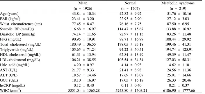
Table 1 shows the baseline clinical and biochemical characteristics of the women in the study. The average age was 43.8 years, and the patients classified with metabolic syndrome numbered 219 (11.3% overall prevalence).
1. Comparison of Anthropometric and Biochemical Characteristics Among the Groups
Of the 1,926 women, 66.2% were classified as premeno- pausal, 10.6% as perimenopausal, and 23.2% as postmeno- pausal. The prevalence of metabolic syndrome was 7.1% in premenopause, 9.8% in perimenopause, and 24.2% in postmenopause. Postmenopausal women were characterized by a higher body mass index, waist circumference, and blood pressure as compared to premenopausal women (P < 0.001).
Postmenopausal women also presented with higher fasting glucose, total cholesterol, triglyceride, uric acid, LDL cholesterol, white blood cell (WBC) count, and lower HDL cholesterol than premenopausal women (Table 2).
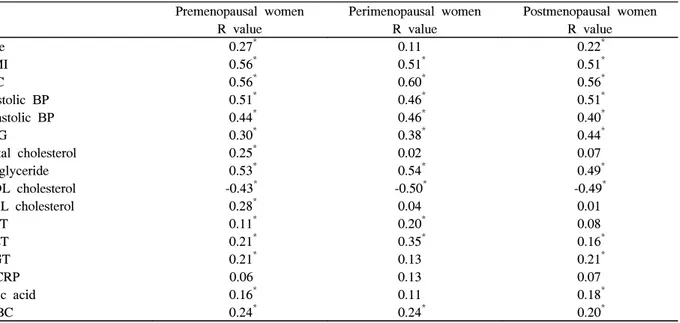
2. Relationships Between Metabolic Syndrome Score and Various Clinical and Biochemical Parameters
A strong positive correlation was noted between metabolic syndrome score and body mass index, waist circumference, systolic blood pressure, and triglyceride in premenopausal women (0.56, 0.56, 0.51, and 0.53). In perimenopausal women waist circumference, systolic blood pressure, and triglyceride were the strong positive correlation with the metabolic syndrome score (0.51, 0.60, and 0.54). In postmenopausal women, body mass index, waist circumference, and systolic blood pressure were the strong positive correlation with metabolic syndrome score (0.51, 0.56, and 0.51). A negative correlation was noted between the metabolic syndrome score and HDL cholesterol in premeno- pausal, perimenopausal, and postmenopausal women. Age was the positive correlation in premenopausal and postmeno- pausal women according to increase of metabolic syndrome
Table 1. Characteristics of subjects in the study samples
Mean
(n = 1926)
Normal (n = 1707)
Metabolic syndrome (n = 219)
Age (years) 43.84 ± 10.34 42.82 ± 9.92 51.76 ± 10.16
BMI (kg/m2) 23.41 ± 3.20 22.93 ± 2.90 27.12 ± 3.03
Waist circumference (cm) 77.45 ± 8.47 76.16 ± 7.75 87.50 ± 6.95
Systolic BP (mmHg) 116.68 ± 16.97 114.47 ± 15.67 133.88 ± 16.92
Diastolic BP (mmHg) 74.14 ± 11.65 72.97 ± 11.15 83.26 ± 11.48
FPG (mg/dL) 90.95 ± 19.91 88.71 ± 16.99 108.44 ± 29.92
Total cholesterol (mg/dL) 180.49 ± 36.55 178.05 ± 35.18 199.46 ± 41.31
Triglyceride (mg/dL) 105.65 ± 71.24 94.22 ± 50.51 194.74 ± 125.91
HDL-cholesterol (mg/dL) 61.31 ± 13.94 62.84 ± 13.49 49.39 ± 11.47
LDL-cholesterol (mg/dL) 106.21 ± 38.55 103.54 ± 34.34 127.03 ± 58.31
Uric acid (mg/dL) 4.20 ± 0.97 4.14 ± 0.93 4.62 ± 1.10
AST (U/L) 21.77 ± 9.33 21.41 ± 8.98 24.56 ± 11.36
ALT (U/L) 18.52 ± 14.46 17.69 ± 13.07 25.01 ± 14.66
GGT (U/L) 18.10 ± 16.97 17.05 ± 16.18 26.33 ± 20.46
hsCRP (mg/dL) 0.12 ± 0.40 0.11 ± 0.40 0.21 ± 0.37
WBC (/mm3) 5351.04 ± 1565.28 5243.80 ± 1503.21 6186.90 ± 1777.66 Data are expressed as means ± S.D. BMI, body mass index; BP, blood pressure; FPG, fasting plasma glucose; HDL, high density lipoprotein; LDL, low density lipoprotein; AST, aspartate aminotransferase; ALT, alanine aminotransferase; GGT, gamma-glutamyl transpeptidase; hsCRP, high sensitive C-reactive protein; WBC, white blood cell count.
score. Other parameters of metabolic syndrome, including ALT, GGT, uric acid, and WBC were also positively
correlated with metabolic syndrome score (Table 3).
Table 2. Anthropometric and biochemical characteristics in each group Premenopausal women
(n = 1274)
Perimenopausal women (n = 205)
Postmenopausal women (n = 447)
ANOVA P
Age 38.69 ± 6.58 46.51 ± 5.22 57.29 ± 8.05 < 0.001
BMI 22.91 ± 3.20 23.93 ± 3.12 24.57 ± 2.89 < 0.001
WC 75.72 ± 8.06 78.72 ± 8.60 81.77 ± 7.87 < 0.001
Systolic BP 113.08 ± 15.18 118.04 ± 15.60 126.31 ± 18.54 < 0.001
Diastolic BP 72.47 ± 10.97 75.32 ± 11.31 78.38 ± 12.54 < 0.001
FPG 89.53 ± 20.01 91.35 ± 18.33 94.84 ± 19.86 < 0.001
Total cholesterol 173.75 ± 29.79 187.03 ± 54.58 196.68 ± 38.20 < 0.001
Triglyceride 94.71 ± 62.83 116.17 ± 73.58 131.99 ± 84.12 < 0.001
HDL cholesterol 61.97 ± 13.87 61.51 ± 13.93 59.36 ± 14.00 0.003
LDL cholesterol 100.38 ± 34.41 110.71 ± 53.38 120.76 ± 37.59 < 0.001
AST 20.51 ± 7.00 22.47 ± 6.73 25.02 ± 14.15 < 0.001
ALT 17.04 ± 11.34 19.73 ± 11.85 22.18 ± 18.18 < 0.001
GGT 16.52 ± 15.60 19.68 ± 20.91 21.90 ± 18.06 < 0.001
hsCRP 0.10 ± 0.29 0.08 ± 0.12 0.20 ± 0.65 < 0.001
Uric acid 4.12 ± 0.89 4.22 ± 1.05 4.41 ± 1.08 < 0.001
WBC 5286.00 ± 1528.87 5429.51 ± 1609.70 5500.40 ± 1637.35 0.034
Met. SD (%) 7.1 9.8 24.2 < 0.001
Data are expressed as means ± S.D. BMI, body mass index; WC, waist circumference; BP, blood pressure; FPG, fasting plasma glucose; LDL, low density lipoprotein; HDL, high density lipoprotein; AST, aspartate aminotransferase; ALT, alanine aminotransferase; GGT, gamma-glutamyl transpeptidase; hsCRP, high sensitive C-reactive protein; WBC, white blood cell count; Met. SD, metabolic syndrome.
Table 3. Correlations between metabolic syndrome score and various parameters
Premenopausal women Perimenopausal women Postmenopausal women
R value R value R value
Age 0.27* 0.11 0.22*
BMI 0.56* 0.51* 0.51*
WC 0.56* 0.60* 0.56*
Systolic BP 0.51* 0.46* 0.51*
Diastolic BP 0.44* 0.46* 0.40*
FPG 0.30* 0.38* 0.44*
Total cholesterol 0.25* 0.02 0.07
Triglyceride 0.53* 0.54* 0.49*
HDL cholesterol -0.43* -0.50* -0.49*
LDL cholesterol 0.28* 0.04 0.01
AST 0.11* 0.20* 0.08
ALT 0.21* 0.35* 0.16*
GGT 0.21* 0.13 0.21*
hsCRP 0.06 0.13 0.07
Uric acid 0.16* 0.11 0.18*
WBC 0.24* 0.24* 0.20*
BMI, body mass index; WC, waist circumference; BP, blood pressure; FPG, fasting plasma glucose; HDL, high density lipoprotein; AST, aspartate aminotransferase; ALT, alanine aminotransferase; GGT, gamma-glutamyl transpeptidase;
hsCRP, high sensitive C-reactive protein; WBC, white blood cell count; Met. SD, metabolic syndrome. * P < 0.01.
3. Relative Risk Between Metabolic Syndrome and Its Components in Menopausal Transition
Based on a multiple regression analysis, a risk of metabolic syndrome was increased in postmenopausal women.
Triglyceride, blood pressure, and waist circumference
showed increased relative risk in perimenopausal women compared with premenopausal women (Table 4). However, when these components were adjusted by age, only triglyceride was significantly associated with menopausal transition (OR 1.517 [95% CI 1.014-2.269]; P = 0.042 in
Table 4. Risk for metabolic syndrome and its components
Premenopausal women Perimenopausal women Postmenopausal women OR (95% CI) for metabolic syndrome
Met. SD 1 1.405 (0.845~2.336) 4.142 (3.056~5.612)
Age-adjusted OR (95% CI) for metabolic syndrome
Met. SD 0.791 (0.464~1.346) 0.951 (0.571~1.583)
OR (95% CI) for components of metabolic syndrome
WC 1 1.803 (1.263~2.575) 3.763 (2.947~4.806)
Triglyceride 1 1.945 (1.331~2.844) 2.899 (2.216~3.791)
HDL-cholesterol 1 1.188 (0.822~1.718) 1.673 (1.298~2.158)
High BP 1 1.830 (1.307~2.561) 3.858 (3.049~4.881)
High FPG 1 1.262 (0.785~2.026) 2.803 (2.088~3.763)
Age-adjusted OR (95% CI) for components of metabolic syndrome
WC 1 1.076 (0.737~1.572) 1.046 (0.704~1.554)
Triglyceride 1 1.517 (1.014~2.269)* 1.573 (1.025~2.414)*
HDL-cholesterol 1 1.006 (0.682~1.484) 1.118 (0.751~1.664)
High BP 1 1.086 (0.758~1.555) 1.072 (0.736~1.561)
High FPG 1 0.737 (0.448~1.212) 0.710 (0.433~1.164)
Met. SD, metabolic syndrome; WC, waist circumference; HDL, high density lipoprotein; BP, blood pressure; FPG, fasting plasma glucose; OR, odds ratio; CI, confidence interval. * P < 0.05.
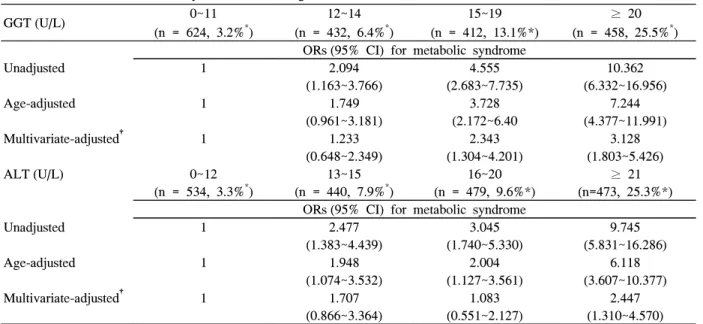
Table 5. Risk for metabolic syndrome according to GGT & ALT levels
GGT (U/L) 0~11
(n = 624, 3.2%*)
12~14 (n = 432, 6.4%*)
15~19 (n = 412, 13.1%*)
≥ 20 (n = 458, 25.5%*) ORs (95% CI) for metabolic syndrome
Unadjusted 1 2.094
(1.163~3.766)
4.555 (2.683~7.735)
10.362 (6.332~16.956)
Age-adjusted 1 1.749
(0.961~3.181)
3.728 (2.172~6.40
7.244 (4.377~11.991)
Multivariate-adjusted† 1 1.233
(0.648~2.349)
2.343 (1.304~4.201)
3.128 (1.803~5.426)
ALT (U/L) 0~12
(n = 534, 3.3%*)
13~15 (n = 440, 7.9%*)
16~20 (n = 479, 9.6%*)
≥ 21 (n=473, 25.3%*) ORs (95% CI) for metabolic syndrome
Unadjusted 1 2.477
(1.383~4.439)
3.045 (1.740~5.330)
9.745 (5.831~16.286)
Age-adjusted 1 1.948
(1.074~3.532)
2.004 (1.127~3.561)
6.118 (3.607~10.377)
Multivariate-adjusted† 1 1.707
(0.866~3.364)
1.083 (0.551~2.127)
2.447 (1.310~4.570) GGT, gamma-glutamyl transpeptidase; ALT, alanine aminotransferase; OR, odds ratio; CI, confidence interval. * percentage of women with the metabolic syndrome to each liver enzyme category. †after adjusting for age, body mass index, fasting plasma glucose, systolic blood pressure, and diastolic blood pressure.
premenopausal women, OR 1.573 [95% CI 1.025-2.414] P = 0.038 in postmenopausal women).
4. Relative Risk for Metabolic Syndrome According to Serum-glutamyl Transpeptidase and Alanine Aminotransferase Concentrations
Higher concentrations of GGT and ALT were risk factors for metabolic syndrome, although their levels were within normal range, and these relationships are independent of age (Table 5).
Discussion
In our study, the prevalence of metabolic syndrome increased throughout the menopausal transition. The relative risk of metabolic syndrome components increased in postmenopause. The prevalence of metabolic syndrome was 11.3% in all women, 7.1% in premenopausal women, 9.8% in perimenopausal women, and 24.2% in postmenopausal women. These results are similar to other studies8,12,17) even though we utilized NCEP ATP Ⅲ criteria (waist circumference ≥ 85 cm) to define metabolic syndrome.
Metabolic syndrome is a formidable risk factor for the development of type 2 diabetes, and the risk for diabetes is nearly fivefold in patients with metabolic syndrome18). Diabetes also increases the risk of coronary artery disease and stroke by two to four fold19,20). This study demonstrated an increase in fasting plasma glucose throughout the menopausal transition, and showed positive correlation between the metabolic syndrome score and elevated fasting glucose level in each group.
Metabolic syndrome is also a strong risk factor for CVD, and incidences in women increase with age1-6), followed by elevated total cholesterol, LDL cholesterol, and apolipoprotein B1,6). In this study total cholesterol, LDL cholesterol, and triglyceride increased in postmenopausal women and HDL cholesterol level decreased in postmenopausal women.
Triglyceride and HDL cholesterol were associated with metabolic syndrome score in each group. However the total cholesterol and LDL cholesterol correlated with metabolic syndrome score in premenopausal women and did not correlate in peri- and postmenopausal women. In premenopausal
women with metabolic syndrome, total cholesterol and LDL cholesterol relatively increased when compared with that of the other groups. Because of this difference, the total cholesterol and LDL cholesterol correlated with metabolic syndrome score only in premenopausal women.
The menopausal transition is associated with a preferential increase in abdominal adiposity, independent of age and total body adiposity21). Women with large amount of visceral fat have an excess of cardiovascular mortality and associated metabolic syndrome22). Obese postmenopausal women with metabolic syndrome are characterized by low lean mass and increased visceral fat23). The central obesity is inversely associated with estradiol level and may lead to menopause13). However, we did not investigate the correlation between serum estradiol level and metabolic syndrome because estradiol, follicular stimulating hormone, and luteinizing hormone were not routinely check in our health examination center.
Also, the combination of high waist circumference with elevated triglyceride appears to be the best indicators of cardiovascular risk in postmenopausal women24). Aging has been associated with increased total cholesterol, triglyceride and LDL cholesterol levels, and the rises of these lipids were particularly marked at the onset of menopause. In our study increased triglyceride and elevated waist circumference were strongly correlated with metabolic syndrome score in both peri- and postmenopausal groups. Especially, triglyceride was significantly associated with metabolic syndrome in both peri- and postmenopausal women after adjusting age.
hsCRP is associated with increased risk for CVD and diabetes. Several studies have drawn attention to elevated levels of hsCRP, a sensitive marker of inflammation, in subjects with metabolic syndrome or its components.
According to one study, hsCRP levels were significantly higher in those with metabolic syndrome, and waist circumference was the most important determinant of CRP levels in women25). In our study, hsCRP levels increased in postmenopausal women, but were not associated with metabolic syndrome score because hsCRP levels were not different between postmenopausal women with metabolic syndrome and without metabolic syndrome.
Some studies suggest serum uric acid as an independent
risk factor for CVD, especially in hypertensive and diabetic individuals26,27). According to a recent study, abdominal obesity is the main determinant of uric acid variance, and an increase in serum uric acid is associated with a higher incidence of metabolic syndrome28). In our data uric acid increased throughout menopausal transition and was correlated with metabolic syndrome components in postmenopausal women.
Several studies have demonstrated elevations of serum ALT and GGT in subjects with metabolic syndrome29-33). The elevated serum ALT and GGT are associated with increased oxidative stress and are related to CRP, a marker of systemic inflammation29). ALT and GGT are used as markers of hepatic insulin resistance31). Increased serum ALT levels are associated with waist circumference, fasting blood glucose, age, and white blood cell count in postmenopausal women with metabolic syndrome30). The raised GGT level is associated with hypertriglyceridemia and the presence of fatty liver32). In our study, ALT and GGT were increased throughout the menopausal transition and were related to metabolic syndrome in each group.
The menopause is the permanent cessation of menstruation due to loss of ovarian follicular function. In studying the effect of menopause, age is an important confounding factor.
In recent two studies, postmenopausal status is associated with an increased risk of the metabolic syndrome independent of age in Korean women34,35). In our study, while a risk of metabolic syndrome was not statistically significant in postmenopausal women after controlling for age, triglyceride was important factor of metabolic syndrome in peri- and postmenopausal women.
There are several limitations to this study. First, our study used a cross sectional design and then causality could not be determined. Second, we did not check sex hormones. Because the groups of menopausal transition were only classified by self-reported questionnaires, misclassification may have occurred. However, the reliability for age at menopause was about 80%. So, if possible, we tried to preempt such bias by including women who wrote out a documented gynecological history. Third, we did not exclude environmental factors such as exercise and diet, except alcohol intake.
In conclusion, triglyceride and waist circumference are
important metabolic syndrome factors. Even after adjusting age, serum ALT and GGT are useful for predicting metabolic syndrome in women.
요 약
연구배경: 폐경 여성에 있어서 심혈관계질환의 유병률은 증가하는 것으로 알려져 있으며, 이러한 증가는 폐경 전기 에서 폐경 후기로 변화하는 여성에서 나타나는 연속적인 호 르몬 변화에 의한 것으로 생각되고 있다. 특히 폐경 후기에 는 대사증후군의 위험도가 약 60% 정도 증가하는 것으로 알려져 있으며, 원인인자로서 저밀도지단백 콜레스테롤의 증가, 에스트로겐의 감소, 복부비만 등의 발생이 영향을 미 치는 것으로 알려져 있으나 아직 확실히 밝혀져 있지는 않 다. 따라서 본 연구의 목적은 폐경기를 지나가는 동안 각 단 계에서 폐경과 대사증후군과 관련된 주요인자들의 관계를 알아보고자 하였다.
방법: 2005년 3월부터 2006년 2월까지 연세대학교 원주 의과대학 원주기독병원 종합건강검진센터를 방문한 여성을 대상으로 하였다. 총 1,926명을 대상으로 검진 당시 시행한 설문지를 바탕으로 폐경 유무에 따라 폐경전기 (premeno- pausal status), 주폐경기 (perimenopausal status), 폐경후기 (postmenopausal status)의 3구분으로 분류하였다. 수축기 혈압, 이완기 혈압, 허리둘레, 체질량지수 등을 측정하였으 며, 공복 시에 혈당, ALT, GGT, 중성지방, 고밀도지단백 콜레스테롤 등을 측정하였다. 대사증후군의 진단기준으로는 NCEP ATP III의 기준을 보완하여 이용하였다.
결과: 대사증후군의 유병률은 폐경전기 7.1%, 주폐경기 9.8%, 폐경후기 24.2%이었다. 폐경전기에서 폐경후기로 갈 수록 허리둘레, 혈압, 체질량지수, ALT, GGT와 공복혈당 등은 통계적으로 유의하게 증가하였으며 (P < 0.001), 이중 허리둘레의 증가가 폐경후기에서 대사증후군 구성인자 수의 증가와 높은 상관관계를 나타냈다 (r = 0.56, P < 0.01). 폐 경전기에서 폐경후기로 갈수록 대사증후군 구성인자의 비교 위험률은 나이를 보정했을 경우 중성지방이 의미 있는 결과 를 보였다 (주폐경기, OR 1.517 [95% CI 1.014~2.269] 폐 경후기, OR 1.573 [95% CI 1.025~2.414]). 또한 여성전체 에서 ALT와 GGT의 상승에 따라 대사증후군 발생 위험률 도 증가하였다.
결론: 폐경 여성의 대사증후군 발생에 있어서 연령 이외에 중성지방과 허리둘레의 증가가 연관성이 있었고 ALT와 GGT 도 대사증후군의 발생을 예측하는 인자로서 사용할 수 있겠다.
References
1. Peters HW, Westendorp IC, Hak AE, Grobbee DE, Stehouwer CD, Hofman A, Witteman JC: Menopausal status and risk factors for cardiovascular disease. J Intern Med 246:521-8, 1999
2. Cabrera MA, Gebara OC, Diament J, Nussbacher A, Rosano G, Wajngarten M: Metabolic syndrome, abdominal obesity, and cardiovascular risk in elderly women. Int J Cardiol 114:224-9, 2007
3. Kuh D, Langenberg C, Hardy R, Kok H, Cooper R, Butterworth S, Wadsworth ME: Cardiovascular risk at age 53 years in relation to the menopause transition and use of hormone replacement therapy: a prospective British birth cohort study. Bjog 112:476-85, 2005
4. Matthews KA, Wing RR, Kuller LH, Meilahn EN, Plantinga P: Influence of the perimenopause on cardiovascular risk factors and symptoms of middle-aged healthy women. Arch Intern Med 154:2349-55, 1994
5. Matthews KA, Kuller LH, Sutton-Tyrrell K, Chang YF: Changes in cardiovascular risk factors during the perimenopause and postmenopause and carotid artery atherosclerosis in healthy women. Stroke 32:1104-11, 2001
6. Torng PL, Su TC, Sung FC, Chien KL, Huang SC, Chow SN, Lee YT: Effects of menopause on intraindividual changes in serum lipids, blood pressure, and body weight--the Chin-Shan Community Cardiovascular Cohort study. Atherosclerosis 161:409 -15, 2002
7. Carr MC: The emergence of the metabolic syndrome with menopause. J Clin Endocrinol Metab 88:2404-11, 2003
8. Park YW, Zhu S, Palaniappan L, Heshka S, Carnethon MR, Heymsfield SB: The metabolic syndrome: prevalence and associated risk factor findings in the US population from the Third National Health and Nutrition Examination Survey, 1988-1994.
Arch Intern Med 163:427-36, 2003
9. Isomaa B, Almgren P, Tuomi T, Forsen B, Lahti K,
Nissen M, Taskinen MR, Groop L: Cardiovascular morbidity and mortality associated with the metabolic syndrome. Diabetes Care 24:683-9, 2001
10. Despres JP: Abdominal obesity as important component of insulin-resistance syndrome. Nutrition 9:452-9, 1993
11. Eckel RH, Grundy SM, Zimmet PZ: The metabolic syndrome. Lancet 365:1415-28, 2005
12. Alberti KG, Zimmet P, Shaw J: Metabolic syndrome--a new world-wide definition. A Consensus Statement from the International Diabetes Federation.
Diabet Med 23:469-80, 2006
13. Mesch VR, Boero LE, Siseles NO, Royer M, Prada M, Sayegh F, Schreier L, Benencia HJ, Berg GA:
Metabolic syndrome throughout the menopausal transition: influence of age and menopausal status.
Climacteric 9:40-8, 2006
14. Brambilla DJ, McKinlay SM, Johannes CB: Defining the perimenopause for application in epidemiologic investigations. Am J Epidemiol 140:1091-5, 1994 15. Greendale GA, Lee NP, Arriola ER: The menopause.
Lancet 353:571-80, 1999
16. Lee SY, Park HS, Kim DJ, Han JH, Kim SM, Cho GJ, Kim DY, Kwon HS, Kim SR, Lee CB, Oh SJ, Park CY, Yoo HJ: Appropriate waist circumference cutoff points for central obesity in Korean adults.
Diabetes Res Clin Pract 75:72-80, 2007
17. Cameron AJ, Shaw JE, Zimmet PZ: The metabolic syndrome: prevalence in worldwide populations.
Endocrinol Metab Clin North Am 33:351-75, table of contents, 2004
18. Stern MP, Williams K, Gonzalez-Villalpando C, Hunt KJ, Haffner SM: Does the metabolic syndrome improve identification of individuals at risk of type 2 diabetes and/or cardiovascular disease? Diabetes Care 27:2676-81, 2004
19. Roper NA, Bilous RW, Kelly WF, Unwin NC, Connolly VM: Cause-specific mortality in a population with diabetes: South Tees Diabetes Mortality Study. Diabetes Care 25:43-8, 2002 20. Nakagami T: Hyperglycaemia and mortality from all
causes and from cardiovascular disease in five
populations of Asian origin. Diabetologia 47:385-94, 2004
21. Poehlman ET, Toth MJ, Gardner AW: Changes in energy balance and body composition at menopause:
a controlled longitudinal study. Ann Intern Med 123:673-5, 1995
22. Lapidus L, Bengtsson C, Larsson B, Pennert K, Rybo E, Sjostrom L: Distribution of adipose tissue and risk of cardiovascular disease and death: a 12 year follow up of participants in the population study of women in Gothenburg, Sweden. Br Med J (Clin Res Ed) 289:1257-61, 1984
23. You T, Ryan AS, Nicklas BJ: The metabolic syndrome in obese postmenopausal women: relationship to body composition, visceral fat, and inflammation. J Clin Endocrinol Metab 89:5517-22, 2004
24. Tanko LB, Bagger YZ, Qin G, Alexandersen P, Larsen PJ, Christiansen C: Enlarged waist combined with elevated triglycerides is a strong predictor of accelerated atherogenesis and related cardiovascular mortality in postmenopausal women. Circulation 111:1883-90, 2005
25. Florez H, Castillo-Florez S, Mendez A, Casanova -Romero P, Larreal-Urdaneta C, Lee D, Goldberg R:
C-reactive protein is elevated in obese patients with the metabolic syndrome. Diabetes Res Clin Pract 71:92-100, 2006
26. Bengtsson C, Lapidus L, Stendahl C, Waldenstrom J:
Hyperuricaemia and risk of cardiovascular disease and overall death. A 12-year follow-up of participants in the population study of women in Gothenburg, Sweden. Acta Med Scand 224:549-55, 1988
27. Lehto S, Niskanen L, Ronnemaa T, Laakso M: Serum uric acid is a strong predictor of stroke in patients with non-insulin-dependent diabetes mellitus. Stroke 29:635-9, 1998
28. Onat A, Uyarel H, Hergenc G, Karabulut A, Albayrak S, Sari I, Yazici M, Keles I: Serum uric acid is a determinant of metabolic syndrome in a population -based study. Am J Hypertens 19:1055-62, 2006
29. Yamada J, Tomiyama H, Yambe M, Koji Y, Motobe K, Shiina K, Yamamoto Y, Yamashina A: Elevated serum levels of alanine aminotransferase and gamma glutamyltransferase are markers of inflammation and oxidative stress independent of the metabolic syndrome. Atherosclerosis 189:198-205, 2006 30. Choi KM, Lee KW, Kim HY, Seo JA, Kim SG, Kim
NH, Choi DS, Baik SH: Association among serum ferritin, alanine aminotransferase levels, and metabolic syndrome in Korean postmenopausal women. Metabolism 54:1510-4, 2005
31. Marchesini G, Avagnina S, Barantani EG, Ciccarone AM, Corica F, Dall'Aglio E, Dalle Grave R, Morpurgo PS, Tomasi F, Vitacolonna E: Aminotransferase and gamma-glutamyltranspeptidase levels in obesity are associated with insulin resistance and the metabolic syndrome. J Endocrinol Invest 28:333-9, 2005 32. Sakugawa H, Nakayoshi T, Kobashigawa K, Nakasone
H, Kawakami Y, Yamashiro T, Maeshiro T, Tomimori K, Miyagi S, Kinjo F, Saito A: Metabolic syndrome is directly associated with gamma glutamyl transpeptidase elevation in Japanese women. World J Gastroenterol 10:1052-5, 2004
33. Kim DJ, Noh JH, Cho NH, Lee BW, Choi YH, Jung JH, Min YK, Lee MS, Lee MK, Kim KW: Serum gamma-glutamyltransferase within its normal concentration range is related to the presence of diabetes and cardiovascular risk factors. Diabet Med 22:1134-40, 2005
34. Cho GJ, Lee JH, Park HT, Shin JH, Hong SC, Kim T, Hur JY, Lee KW, Park YK, Kim SH:
Postmenopausal status according to years since menopause as an independent risk factor for the metabolic syndrome. Menopause 15:524-9, 2008 35. Kim HM, Park J, Ryu SY, Kim J: The effect of
menopause on the metabolic syndrome among Korean women: the Korean National Health and Nutrition Examination Survey, 2001. Diabetes Care 30:701-6, 2007