INTRODUCTION
Since the completion of the Human Genome Project, one of the biggest challenges in genetic research has been the iden
ti fication of inherited genetic variants that alter susceptibility to multifactorial and polygenic diseases such as cancer [1]. The polygenic model for cancer susceptibility presumes that the combined effects of variants in many genes, each conferring a
small to modest increase in cancer risk, cumulatively account for a substantial fraction of this heritable component of risk [2].
These genetic variants might function through interactions with different behavioral, environmental, or other external risk factors.
The weight of evidence indicates that cumulative, excessive exposure to estrogen across a woman’s life span contributes to and may be a causal factor for breast cancer [3]. Several endocrine related risk factors that are associated with an increased risk of breast cancer in postmenopausal women have been found consistently in many studies [47]. The mechanisms of carcinogenesis in the breast caused by estrogen include two different but complementary pathways.
In addition to the mitogenic properties of estrogen, the
Combined effect of CYP1B1, COMT, GSTP1, and MnSOD genotypes and risk of postmenopausal breast cancer
Jasmina-Ziva Cerne1, Maja Pohar-Perme2, Srdjan Novakovic3, Snjezana Frkovic-Grazio4, Vida Stegel3, Ksenija Gersak1
1Institute of Medical Genetics, Department of Obstetrics and Gynecology, University Medical Center Ljubljana, Slajmerjeva 3,
2Institute for Biostatistics and Medical Informatics, Faculty of Medicine, University of Ljubljana, Vrazov trg 2,
Departments of 3Molecular Diagnostics and 4Pathology, Institute of Oncology Ljubljana, Zaloska 2, 1000 Ljubljana, Slovenia
Received Dec. 14, 2010, Revised Feb. 10, 2011, Accepted Mar. 16, 2011 Correspondence to Ksenija Gersak
Institute of Medical Genetics, Department of Obstetrics and Gynecology, University Medical Center Ljubljana, Slajmerjeva 3, 1000 Ljubljana, Slovenia.
Tel: 386-1-522-51-81, Fax: 386-1-522-22-21, E-mail: [email protected]
Copyright © 2011. Asian Society of Gynecologic Oncology, Korean Society of Gynecologic Oncology and Colposcopy
Objective: Estrogen plays a key role in breast cancer development and functionally relevant genetic variants within the estrogen metabolic pathway are prime candidates for a possible association with breast cancer risk. We investigated the independent and the combined effects of commonly occurring polymorphisms in four genes encoding key proteins of estrogen metabolic pathway on their potential contribution to breast cancer risk.
Methods: We studied 530 breast cancer cases and 270 controls of the same age and ethnicity participating in a casecontrol study of postmenopausal women. Genotyping was conducted for CYP1B1 (rs1056836), COMT (rs4680), GSTP1 (rs1695), and MnSOD (rs4880) polymorphisms by polymerase chain reaction based restriction fragment length polymorphism and TaqMan allelic discrimination method. Adjusted ORs and 95% CIs were calculated using logistic regression.
Results: None of the 4 genetic variants examined contributed to breast cancer risk individually. When the combined effects of the risk genotypes were investigated, significant associations were observed among women with two highrisk genotypes in CYP1B1 and COMT (OR, 2.0; 95% CI, 1.1 to 3.5) and two highrisk genotypes in COMT and MnSOD (OR, 2.0; 95% CI, 1.0 to 3.8), compared to those with lowrisk genotypes.
Conclusion: Our results suggest that individual susceptibility to breast cancer incidence may be increased by combined effects of the highrisk genotypes in CYP1B1, COMT, and MnSOD estrogen metabolic genes.
Keywords: Breast neoplasms, Estrogen metabolism, Combined polymorphisms, Association study
metabolites of estrogen within the catechol estrogen (CE) pathway (semiquinones, quinones) damage DNA via the formation of superoxide radicals and depurinating DNA adducts [810]. Because estrogen metabolites have short halflives and are difficult to measure in vivo, assessing the associations of breast cancer risk with functional genetic variants that alter metabolite concentrations might be the most direct approach possible [11].
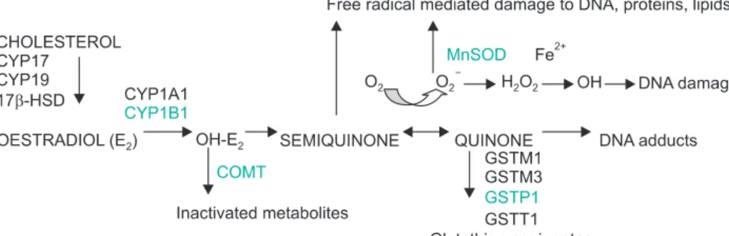
Candidate genes include CYP1A1 and CYP1B1, which encode phase I enzymes that lead to increased levels of estrogen metabolites and COMT, UGT1A1, SULT1A1, GSTPs, and SODs which are involved in phase II metabolism that leads to protective conjugation of estrogen metabolites or detoxify reactive oxygen species (ROS) formed in these reactions (Fig. 1) [12]. CYP1B1 appears to be the main CYP responsible for the extrahepatic 4hydroxylation [13]. The 4hydroxyestrogen (4CE) to 2hydroxyestrogen (2CE) concentration ratio has been reported to be 4:1 in a human breast cancer extract, thus a greater role in carcinogenesis has been suggested for 4CE [14]. Furthermore, quinones, the further oxidized metabolites of 4CE, may react with purine bases of DNA to form depurinating adducts that generates highly mutagenic apurinic sites. In contrast, quinones of 2CE produce less harmful, stable DNA adducts [9]. COMT enzyme is involved in methylating (and thereby inactivating) CEs. This is a quantitatively most active conjugation pathway for CEs, although they can also be conjugated by glucuronidation and sulfation [15]. Members of the GST family are thought to play a role in the conjugation of quinones and GSTP1 is the major GST expressed consistently in both normal and tumor breast tissue [16]. Finally, SODs catalyze the conversion of superoxide anion (O2) into hydrogen peroxide (H2O2) and molecular oxygen, and thus protect cells from the damage induced by free radicals emerging in estrogen metabolism.
As mitochondria consume over 90% of cell oxygen, mito
chondrial MnSOD is considered of particular importance for cellular defense against oxidative damage [17].
Several genetic polymorphisms within estrogen metabolic
genes have been shown to have functional effects on the catalytic properties of their corresponding enzymes. Altered activity of phase I and II enzymes may influence local hormone levels and cause variation in the extent of DNA damage [18,19].
These persontoperson differences may define subpopu
lations of women with higher lifetime exposure to hormone dependent growth promotion and/or to cellular damage from particular estrogen metabolites thus suggesting higher breast cancer risk. Therefore, the purpose of our study was to evaluate the independent and the combined effects of CYP1B1, COMT, GSTP1, and MnSOD genotypes on the development of breast cancer. Each of the selected genes is being highly expressed in breast tissue and involved in distinct estrogen metabolic subpathway.
MATERIALS AND METHODS 1. Study population
Postmenopausal women diagnosed with invasive primary breast cancer between January 1, 2006 and December 31, 2008 at the Institute of Oncology Ljubljana, who were 50
69 years old at the time of diagnosis and of Caucasian ethnic origin were eligible for inclusion in the study. The control group consisted of postmenopausal women randomly selected from the outpatient clinic records of the Department of Obstetrics and Gynecology, University Medical Center Ljubljana that were of the same age and ethnicity, and had no history of breast cancer.
2. Data collection
In addition to general information (socioeconomic status, weight, height), data on reproductive factors (age at menarche, number of pregnancies, age at first delivery, number of deli
veries, breastfeeding, age at menopause), family history of breast or ovarian cancer (firstdegree relatives), smoking and alcohol consumption were collected by means of a postal que stionnaire. Detailed questions were asked regarding drug
Fig. 1. A schematic presentation of enzymes with known gene polymorphisms involved in estrogen biosynthesis and metabolism.
Modified from [12] with permission from Elsevier.
intake, sex hormones in particular (oral contraceptive [OC]
use, hormone replacement therapy [HRT] use). A color chart displaying all preparations ever marketed in Slovenia was in
cluded in the questionnaire to aid recall. OC and HRT use for less than 1 year was considered no use. Women were assumed to be postmenopausal if they had no periods for at least 12 months before the reference date or had undergone a bila
teral oophorectomy.
Estrogen receptor (ER) status was assessed by immunohisto
chemistry, using monoclonal rabbit ER antibody, Clone SP1 (Neomarkers, Fremont, CA, USA). Tumors were categorized as ERpositive if nuclear staining was observed in at least 10% of nuclei.
Informed written consent was obtained from all women enrolled in the study. The study protocol was approved by the National Medical Ethics Committee of the Republic of Slovenia (No. 61/06/07).
3. Specimen collection and isolation of DNA
In case patients, DNA was extracted from formalinfixed paraffinembedded (FFPE) normal breast tissues using HP PCR Template Preparation Kit (Roche Diagnostics GmbH, Mannheim, Germany) following the manufacturer’s protocol.
The control group women were invited to provide blood sample; genomic DNA was extracted from whole blood using FlexiGene DNA Kit 250 (Qiagen GmbH, Hilden, Germany) following the manufacturer’s protocol.
4. Genotyping
Genotyping for the polymorphism c. 1294C>G (p. Leu432Val) in gene CYP1B1 was done using polymerase chain reaction (PCR)based restriction fragment length polymorphism (RFLP) method. Each PCR product was digested with restriction en
do nuclease Eco57I (Fermentas International Inc, Burlington, Canada) and DNA fragments were separated and visualized by electrophoresis on polyacrylamide gels. Genotyping for the polymorphisms c. 472G>A (p. Val108/158Met) in gene COMT, c. 313A>G (p. Ile105Val) in gene GSTP1 and c. 47T>
C (p. Val16Ala) in gene MnSOD was performed on 96well plates using the fluorogenic 5`nuclease assays on Light
Cycler 480 System (Roche Diagnostics GmbH). Each reaction mix contained genomic DNA, LightCycler 480 Probes Master (Roche Diagnostics GmbH) and Custom TaqMan SNP Geno
typing Assay (Applied Biosystems, Werterstadt, Germany). All genotyping protocols (PCR reaction conditions, primers and probes) will be provided upon request from the correspon
ding author. Positive control samples (homozygote for wild allele, heterozygote, homozygote for variant allele) and the negative control sample were included in each batch of sam
ples. Gels were scored by two different readers; discordant samples were repeated. Apart from CYP1B1 1294C>G, all poly
morphisms had no samples that failed to be genotyped. For CYP1B1 1294C>G, 1% of the samples failed. Samples that failed to be genotyped were scored as missing. Reliability was assessed by random selection of 5% of samples in which all genotypes were confirmed by sequencing using ABI PRISM 7000 sequence detection system (Applied Biosystems). Con
cordance was 100% for all genotypes.
5. Statistical analyses
We used the independent ttest to compare the values of the means between cases and controls. Differences in categorical characteristics between cases and controls were assessed using chisquare tests. Observed genotype frequencies were tested for deviation from HardyWeinberg equilibrium (HWE) with the chisquare goodnessoffit test.
Odds ratios (ORs) for breast cancer risk and the corresponding 95% confidence intervals (CI) were calculated using logistic regression analyses. The homozygous wildtype genotype, as determined by the presence of two putatively lowrisk alleles, served as a reference category, with the heterozygous genotype and homozygous variant genotype being collapsed into one category. The adjustment was made for age as a continuous variable. Additional factors (education level, body mass index, age at menarche, age at first full term pre
gnancy, parity, breastfeeding, OC use, age at menopause, HRT use, first degree family history of breast and/or ovarian cancer and smoking) are within the causal pathway between genetic factors and breast cancer, but could not be affecting genotypes and are thus not true confounders.
Therefore, we report the results without adjustment for these factors. A p<0.05 was considered statistically significant.
To account for multiple testing, we used the Westfall and Young [20] permutation method, which takes into account the interdependency of the variables tested. Our focus was different combinations of two and three risk genotypes, since their effect was the main hypothesis of the article. We used 10,000 simulation runs under the null hypothesis of no effect, both to correct each pvalue by itself and to calculate the probability that the significant results had occurred by chance.
The statistical analyses were done using SPSS ver. 18.0 (SPSS Inc., Chicago, IL, USA) and the R statistical software ver. 2.12 (R Development Core Team, Auckland, New Zealand).
RESULTS
Overall response rates were 82.5% (825/1,000) for cases and
73.2% (732/1,000) for controls. In the 3year period (2006
2008), we enrolled 1,493 postmenopausal women aged 50
69 years; of the 825 cases and 732 controls completing the questionnaire, complete data for all variables considered in the multivariate model were available for 78.4% (784/1,000) cases and 70.9% (709/1,000) controls. Only 38.1 % of the con
trol women agreed to provide a blood sample. The num ber of cases included in genotype analyses was therefore pro por
tionally decreased by random selection to gain the 2:1 ratio in casecontrol comparisons. The final analysis thus in cluded 800 postmenopausal women aged 5069 years: 530 were diag nosed with primary breast cancer and 270 were healthy volunteers (control group). The mean age for cases and con
trols was 60.45±5.84 and 60.10±5.85 years, re spectively, and did not differ significantly between the groups (p=0.432).
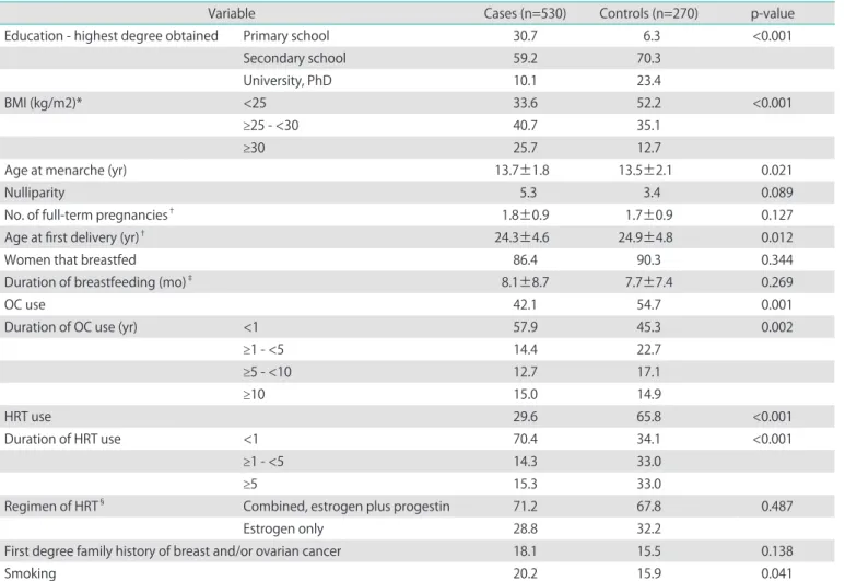
Selected characteristics of the study subjects are presented
in Table 1. Briefly, cases had significantly higher BMI at the time of diagnosis and were more likely to smoke. Control group women had a higher educational level, earlier age at menarche and were older at first delivery. Significantly more women in the control group were using both, OC and HRT. There was no significant difference between the groups in the percentage of nulliparity, number of full term pregnancies, percentage of women that breastfed, duration of breastfeeding, regimen of HRT and percentage of women having any first degree family history of breast or ovarian cancer.
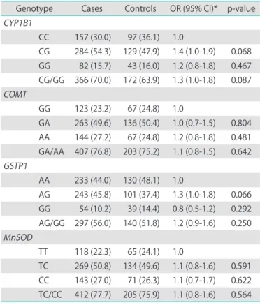
There was no deviation from the HWE except for GSTP1 (p=0.01). The chisquare tests for distribution revealed no significant difference between cases and controls in most of genotype frequencies except for GSTP1 (p=0.04). When adjusted for age, none of the 4 genetic variants studied was, by itself, statistically significantly associated with post
Table 1. Characteristics of study population
Variable Cases (n=530) Controls (n=270) p-value
Education - highest degree obtained Primary school 30.7 6.3 <0.001
Secondary school 59.2 70.3
University, PhD 10.1 23.4
BMI (kg/m2)* <25 33.6 52.2 <0.001
≥25 - <30 40.7 35.1
≥30 25.7 12.7
Age at menarche (yr) 13.7±1.8 13.5±2.1 0.021
Nulliparity 5.3 3.4 0.089
No. of full-term pregnancies† 1.8±0.9 1.7±0.9 0.127
Age at first delivery (yr)† 24.3±4.6 24.9±4.8 0.012
Women that breastfed 86.4 90.3 0.344
Duration of breastfeeding (mo)‡ 8.1±8.7 7.7±7.4 0.269
OC use 42.1 54.7 0.001
Duration of OC use (yr) <1 57.9 45.3 0.002
≥1 - <5 14.4 22.7
≥5 - <10 12.7 17.1
≥10 15.0 14.9
HRT use 29.6 65.8 <0.001
Duration of HRT use <1 70.4 34.1 <0.001
≥1 - <5 14.3 33.0
≥5 15.3 33.0
Regimen of HRT§ Combined, estrogen plus progestin 71.2 67.8 0.487
Estrogen only 28.8 32.2
First degree family history of breast and/or ovarian cancer 18.1 15.5 0.138
Smoking 20.2 15.9 0.041
Values are presented as percent (%) or mean±SD.
BMI, body mass index; OC, oral contraceptive; HRT, hormone replacement therapy.
*Calculated as weight in kilograms divided by height in meters squared at the age of the diagnosis. †Among women who had a full term pregnancy. ‡Among those who ever breastfed. §Among those who ever used HRT.
menopausal breast cancer risk (Table 2).
Additionally, we examined possible combined effects of CYP1B1 , COMT , GSTP1 , and MnSOD genotypes on breast cancer risk by calculating adjusted ORs for all of the combinations of two and three of the risk genotypes (Tables 3 and 4). The reference group consisted of individuals with the putatively most advantageous combinations of the genotypes, lowrisk genotypes, i.e., the presence of homozy
gous CC genotype for CYP1B1, GG genotype for COMT, AA genotype for GSTP1 and TT genotype for MnSOD.
When combinations of two putative atrisk genotypes were examined (Table 3), the concurrent presence of CYP1B1 (CG/
GG) and COMT (GA/AA) highrisk genotypes and COMT (GA/
AA) and MnSOD (TC/CC) highrisk genotypes posed a more than 2fold risk of breast cancer (OR, 2.0; 95% CI, 1.1 to 3.5) and (OR, 2.0; 95% CI, 1.0 to 3.8), respectively. In contrast, no statistically significant effects were seen for women simultaneously carrying the GSTP1 highrisk genotype in combination with any of the other three the risk genotypes.
When three of the putative atrisk genotypes were combined (Table 4), women with highrisk genotypes CYP1B1 (CG/GG), COMT (GA/AA), GSTP1 (AG/GG) and CYP1B1 (CG/
GG), COMT (GA/AA), MnSOD (TC/CC) were at a 2.7fold (95%
CI, 1.1 to 6.8) and 12.2fold (95% CI, 1.4 to 102.3) breast cancer risk, respectively, compared to those with lowrisk genotypes.
Similarly, clear combined effects were observed also in the other two combinations for the three atrisk genotypes, but the outcomes were not significant.
After the combinations of two and three risk genotypes were examined (Table 3 and 4), the results were further evaluated to account for multiple testing. While none of the single pvalues remained significant after the correction, the overall probability that the 8 observed pvalues were below 0.05 due
Table 2. Genetic variation and risk of postmenopausal breast cancer Genotype Cases Controls OR (95% CI)* p-value CYP1B1
CC 157 (30.0) 97 (36.1) 1.0
CG 284 (54.3) 129 (47.9) 1.4 (1.0-1.9) 0.068 GG 82 (15.7) 43 (16.0) 1.2 (0.8-1.8) 0.467 CG/GG 366 (70.0) 172 (63.9) 1.3 (1.0-1.8) 0.087 COMT
GG 123 (23.2) 67 (24.8) 1.0
GA 263 (49.6) 136 (50.4) 1.0 (0.7-1.5) 0.804 AA 144 (27.2) 67 (24.8) 1.2 (0.8-1.8) 0.481 GA/AA 407 (76.8) 203 (75.2) 1.1 (0.8-1.5) 0.642 GSTP1
AA 233 (44.0) 130 (48.1) 1.0
AG 243 (45.8) 101 (37.4) 1.3 (1.0-1.8) 0.066 GG 54 (10.2) 39 (14.4) 0.8 (0.5-1.2) 0.292 AG/GG 297 (56.0) 140 (51.8) 1.2 (0.9-1.6) 0.250 MnSOD
TT 118 (22.3) 65 (24.1) 1.0
TC 269 (50.8) 134 (49.6) 1.1 (0.8-1.6) 0.591 CC 143 (27.0) 71 (26.3) 1.1 (0.7-1.7) 0.622 TC/CC 412 (77.7) 205 (75.9) 1.1 (0.8-1.6) 0.564 Values are presented as number (%) or OR (95% CI).
OR, odds radio; CI, confidence interval.
*Adjusted for age as a continuous variable.
Table 3. Combined effects of two genotypes (CYP1B1, COMT, GSTP1, and MnSOD) and risk of breast cancer
Genotype Cases/controls OR (95% CI) * p-value CYP1B1 COMT
CC GG 27/26 1.0
CC GA/AA 130/71 1.8 (1.0-3.2) 0.071
CG/GG GG 94/40 2.3 (1.2-4.4) 0.014
CG/GG GA/AA 272/132 2.0 (1.1-3.5) 0.021
CYP1B1 GSTP1
CC AA 69/43 1.0
CC AG/GG 88/54 1.0 (0.6-1.7) 0.942
CG/GG AA 161/86 1.2 (0.7-1.8) 0.518
CG/GG AG/GG 205/86 1.5 (0.9-2.4) 0.088
CYP1B1 MnSOD
CC TT 33/22 1.0
CC TC/CC 124/75 1.1 (0.6-2.1) 0.733
CG/GG TT 84/43 1.3 (0.7-2.5) 0.413
CG/GG TC/CC 282/129 1.5 (0.8-2.6) 0.197
COMT GSTP1
GG AA 51/27 1.0
GG AG/GG 72/40 1.0 (0.5-1.8) 0.896
GA/AA AA 182/103 0.9 (0.6-1.6) 0.793
GA/AA AG/GG 225/100 1.2 (0.7-2.0) 0.515
COMT MnSOD
GG TT 19/19 1.0
GG TC/CC 104/48 2.2 (1.1-4.5) 0.037
GA/AA TT 99/46 2.1 (1.0-4.4) 0.041
GA/AA TC/CC 308/157 2.0 (1.0-3.8) 0.050
GSTP1 MnSOD
AA TT 44/28 1.0
AA TC/CC 189/102 1.2 (0.7-2.0) 0.541
AG/GG TT 74/37 1.3 (0.7-2.4) 0.435
AG/GG TC/CC 223/103 1.4 (0.8-2.4) 0.227
OR, odds ratio; CI, confidence interval.
*Adjusted for age as a continuous variable.
to chance only was 0.01 (1%).
We were restricted from evaluating the potential combined effects of all the four genotypes because none of the cases and only one of the controls carried all of the four putatively lowrisk genotypes simultaneously.
When stratified for ER status, 84.0% (n=445) of tumors were ERpositive and 16.0% (n=85) of tumors were ERnegative.
Genetic variants alone or combined effects of two genotypes did not affect differently breast cancer risk according to the ER
status (data not shown).
DISCUSSION
In this casecontrol study of postmenopausal Caucasian women, we investigated associations of functionally relevant genetic variants in four genes (CYP1B1, COMT, GSTP1, MnSOD) encoding key proteins of the estrogen metabolic pathway with breast cancer risk. Additionally, we evaluated the potential combined effects of these genotypes on the development of breast cancer.
We focused on polymorphic genes coding for enzymes that are relevant for the given exposure, are highly expressed in breast tissue, and act sequentially in the same metabolic pathway (Fig. 1 genes with polymorphisms included in our analysis are underlined). However, none of the 4 genetic variants in these genes contributed to breast cancer risk individually.
Several association studies in these candidate genes have been widely used to search for susceptibility alleles, but few definite associations have been established [21]. Such inconsistencies in results probably reflect the true variation in the underlying association between populations studied and the low penetrance of mutations in these multigenic pathways [22]. Although the effect of each individual single nucleotide polymorphism (SNP) was small and not significant, the genetic effect of combinations of functionally relevant SNPs may additively or synergistically contribute to increased breast cancer risk. Therefore, our a priori hypothesis specified that individual susceptibility to breast cancer may be increased by the combined effects of the risk genotypes in estrogen metabolic genes. This was confirmed by the results of our study since the concurrent presence of CYP1B1 and COMT highrisk genotypes and COMT and MnSOD high
risk genotypes posed a 2fold risk of breast cancer. Women with the three highrisk genotypes CYP1B1, COMT, GSTP1 and CYP1B1, COMT, MnSOD were at a 2.7fold and 12.2fold breast cancer risk, respectively. This is in agreement with the only genegene interaction study that investigated the same genegene combinations [23]. They observed only marginally increased breast cancer risk with the combination of highrisk genotypes in CYP1B1, COMT, and MnSOD genes in women with the BMI greater than 24 kg/m2 (OR, 1.4; 95% CI, 1.0 to 1.9) [23].
Several SNPs within CYP1B1 have been shown to have functional effects on the catalytic properties of the CYP1B1 enzyme, with the 4hydroxylase activity of the Val432 variant allele displaying a 3fold higher activity compared to Leu432 Table 4. Combined effects of three genotypes (CYP1B1, COMT,
GSTP1, and MnSOD) and risk of breast cancer
Genotype Cases/
controls OR (95% CI)* p-value CYP1B1 COMT GSTP1
CC GG AA 10/11 1.0
CCCC CG/GG
GGGA/AA GG
AG/GG
AAAA 116/62 2.1 (0.8-5.2) 0.114
CCCG/GG CG/GG
GA/AA GGGA/AA
AG/GG AG/GG
AA 246/135 2.0 (0.8-4.9) 0.118 CG/GG GA/AA AG/GG 151/61 2.7 (1.1-6.8) 0.029
CYP1B1 GSTP1 MnSOD
CC AA TT 14/10 1.0
CCCC CG/GG
AAAG/GG AA
TC/CC
TTTT 103/63 1.2 (0.5-2.8) 0.698
CCCG/GG CG/GG
AG/GG AAAG/GG
TC/CC TC/CC
TT 256/135 1.4 (0.6-3.2) 0.458 CG/GG AG/GG TC/CC 150/61 1.8 (0.8-4.2) 0.190
CYP1B1 COMT MnSOD
CC GG TT 1/6 1.0
CCCC CG/GG
GGGA/AA GG
TC/CC
TTTT 76/49 9.4 (1.1-80.4) 0.041
CCCG/GG CG/GG
GA/AA GGGA/AA
TC/CC TC/CC
TT 240/112 13.0 (1.5-109.0) 0.018 CG/GG GA/AA TC/CC 206/102 12.2 (1.4-102.3) 0.022
COMT GSTP1 MnSOD
GG AA TT 4/5 1.0
GGGG GA/AA
AAAG/GG AA
TC/CC
TTTT 102/59 2.2 (0.6-8.5) 0.256
GGGA/AA GA/AA
AG/GG AAAG/GG
TC/CC TC/CC
TT 258/129 2.5 (0.7-9.6) 0.173 GA/AA AG/GG TC/CC 166/77 2.7 (0.7-10.5) 0.143 OR, odds ratio; CI, confidence interval.
*Adjusted for age as a continuous variable.
[24]. On the other hand, the COMT Met158 variant allele has been hypothesized to produce an enzyme with a 3 to 4fold reduced functionality compared to the wildtype Val158 allele [25]. For GSTP1 gene, a point mutation results in a single amino acid change from isoleucine (Ile) to valine (Val) at codon 105.
This residue lies in close proximity to the hydrophobic binding site for electrophilic substrates, and the Val105 variant allele has been demonstrated to exhibit altered specific activity and affinity for electrophilic substrates [26]. A polymorphism at codon 16 of mitochondrial targeting sequence of the MnSOD gene leads to a substitution from Val to Ala. This substitution alters the secondary structure of the protein, which affects the localization and transport of the enzyme into the mitochondria, where it exerts its antioxidant action [27]. Therefore, it was first expected that the Val form was likely to be associated with increased risk of cancer. However, subsequent studies revealed a controversial picture [28]. If MnSOD is inhibited to enter the mitochondrial matrix, as is the case with Val form, O2 cannot be dismutated to H2O2, which causes cellular damage and consequently induces apoptosis.
Conversely, the Ala form of MnSOD efficiently dismutates O2
to H2O2, but the latter may react to yield other ROS, mostly hydroxyl radicals (·OH), which are highly detrimental to DNA.
In view of these findings, it is very likely that a combination of increased production of 4CE by Val432 form of CYP1B1 and concurrent decreased 4CE inactivation by Met158 form of COMT may result in elevated risk of breast cancer. Similarly, decreased 4CE inactivation by Met158 form of COMT leads to the production of O2, which are generated through the redox cycling between further oxidized metabolites, quinones and semiquinones. Therefore, while simultaneously carrying Ala16 form of MnSOD, excess production of H2O2 may result in the accumulation of DNA adducts and thus predispose to cancer.
On the other hand, rather unexpectedly, the GSTP1 Val105 variant allele did not prove to influence breast cancer risk when studied separately or when potential combined effects between the two risk genotypes were examined. It has been suggested that, depending on the chemical composition of the substrate, individuals with a given GSTP1 genotype may be at a differential risk for carcinogenesis [29]. Since GSTP1 is polymorphic with 2 singlenucleotide substitutions in the coding region (p. Ile105Val and p. Ala114Val), and both amino acid residues lie in close proximity to the substratebinding site, their concurrent determination will provide a clearer picture of the catalytic properties of the GSTP1 isozyme [29].
Furthermore, as the GST family of enzymes are known to have overlapping substrate specificities, another explanation for the discrepancy might be that the deficiency of GSTP1 isozyme was compensated by other isoforms (GSTM1, GSTM3,
GSTT1) [30]. A simultaneous determination of all relevant GST genotypes for a given exposure may therefore be a prerequisite for a reliable interpretation of the results.
Additionally, we observed a tendency of increased risk together with increased number of the putative highrisk genotypes. The ORs were more elevated in the women harboring three highrisk genotypes compared to two high
risk genotypes.
The results of the current study suggest that genetic variants without or with main effects, too small to be detected, may interact with others and confer an increased breast cancer risk. Since none of the single pvalue remained significant after the correction for multiple testing, we can not point on a single combination that we expect to have an effect in the population. However, as the overall probability that the 8 observed pvalues were below 0.05 due to chance was only 1%, we can claim at least some of the studied genotype combinations are associated with the increased risk of breast cancer. Genegene interactions among estrogen metabolic genes have been also investigated in other studies, with some studies [3034], but not all [3537], reporting associations between highrisk genotypes and breast cancer risk. However, with the exception of two studies reporting marginally significant increased risk of breast cancer associated with GSTP1, GSTM1, and GSTT1 risk genotypes and our study confirming the results of Kocabas et al. [23] indicating increased risk associated with CYP1B1, COMT, and MnSOD risk genotypes, no specific genegene combination has been observed in more than one study. Inconsistencies may be at least partly explained by the differences in the populations studied and in their exposure to the agents relevant to the development of breast cancer. Furthermore, for comparable statistical power larger study sizes are needed if investigating combined effects between different genotypes. The existing reports, including ours, were limited only to the interactions of SNPs within a single cancer pathway. Yet, there is also growing evidence regarding cross talk between the risk genotypes of different cancer pathways, including DNA repair, cell cycle, immune system and others [38]. The investigation of such genegene interactions presents new statistical challenges as the number of potential interactions between the SNPs can be substantial.
Since the current study was retrospective in design, the data was obtained on the basis of the postal questionnaire, and a low percentage of control group women agreed to provide a blood sample, we cannot rule out a possibility of introducing biased study results. The difference in how genetic data was collected from cases and controls (i.e., FFPE normal breast tissue samples in cases and blood samples in controls) might
have raised concerns about introducing bias toward a specific allele in any of the assays. However, studies have shown that, for most of the genotypes, the use of FFPE tissue samples is a valid alternative to peripheral blood, and vice versa [39,40].
Kweekel et al. [40] compared 11 genotypes in DNA isolated from blood and FFPE colorectal cancer tissue and found none of the individual genotypes that showed a discordance between FFPE cancer tissue and blood significantly different from 0.0%, except for GSTP1 (rs1695) with 95% CI 0.1 to 5.9.
Another study analyzed 5 genotypes selected from regions commonly known to have loss of heterozygosity in breast cancer from normal breast tissues adjacent to tumors and compared them with blood genotyping [41]. They found 100% concordance and concluded that the use of adjacent normal tissues provides accurate genotyping results with high specificity [41]. Therefore, by using FFPE normal (not cancerous) breast tissue samples, which are unlikely to harbour somatic alterations associated with carcinogenesis, this might not have affected our ability to identify germline genetic variants. We have no logical explanation regarding the lack of HWE for the GSTP1 (rs1695) in our study. The women in the control group were randomly enrolled from the outpatient clinic records of the Department of Obstetrics and Gynecology during their routine gynecologic exams. Genotyping was done in duplicates, using validated Custom TaqMan SNP Genotyping Assay and endpoint fluorescence scatter plots positioned the homozygotes for one allele, the heterozygotes and the homozygotes for the other allele clearly apart from each other. Furthermore, no other genetic variant appear next to this polymorphism arguing strongly against mismatch probe pairing. Additionally, GSTP1 genotyping reliability was assessed by sequencing 5% (n=40) of samples at random, and concordance was 100%. Therefore, we believe the lack of HWE for the GSTP1 is purely due to chance and, since it only affected the size of the groups that were compared, it might not have introduced any bias into our results. Given extensive data stratification in studying the combined effects of the three risk genotypes on breast cancer risk, the likelihood of a type I error (a falsepositive result) has to be considered. Our study population was of medium size and it is possible that some interactions were not significant due to insufficient power. The strengths of the study include homogenous study population and investigation of functionally relevant genetic variants in genes, each being highly expressed in breast tissue and involved in a distinct estrogen metabolic subpathway. Although the determination of a priori evidence of being functionally important in the disease process is quite challenging, selecting SNPs based on their functions clearly reduces the false positive report probability [42]. The
results are consistent with biologically plausible interactions and merit further investigation of the combined effects of polymorphisms in estrogen metabolic genes on breast cancer risk.
Our focus in this study was to find genegene interactions, which additively or synergistically contribute to breast cancer risk. Although our pool of SNPs was small, we have managed to show that the individual susceptibility to breast cancer incidence may be increased by combined effects of CYP1B1, COMT, GSTP1, and MnSOD highrisk genotypes, especially in certain combinations. With the exception of one study reporting marginally increased breast cancer risk, we are the first to report a large increase in breast cancer risk associated with these highrisk genotypes. Replication of our results is thus needed before any firm conclusion is drawn. These findings may provide new important data on the combined effects of putative genegene interactions in the etiology of human breast cancer. We believe that epistasis is likely to be a ubiquitous component of the genetic architecture of common diseases, such as breast cancer. This information could be used in clinical work to identify individuals at increased risk of breast cancer, and to develop preventive strategies.
CONFLICT OF INTEREST
No potential conflict of interest relevant to this article was reported.
ACKNOWLEDGMENT
We would like to thank Ms Mojca Pirc for revision of the English text.
REFERENCES
1. Hunter DJ, Riboli E, Haiman CA, Albanes D, Altshuler D, Chanock SJ, et al. A candidate gene approach to searching for lowpenetrance breast and prostate cancer genes. Nat Rev Cancer 2005;5:97785.
2. Pharoah PD, Dunning AM, Ponder BA, Easton DF. Associa
tion studies for finding cancersusceptibility genetic variants. Nat Rev Cancer 2004;4:85060.
3. Yager JD, Davidson NE. Estrogen carcinogenesis in breast cancer. N Engl J Med 2006;354:27082.
4. Key TJ, Appleby PN, Reeves GK, Roddam A, Dorgan JF, Longcope C, et al. Body mass index, serum sex hormones,
and breast cancer risk in postmenopausal women. J Natl Cancer Inst 2003;95:121826.
5. Key T, Appleby P, Barnes I, Reeves G; Endogenous Hor
mones and Breast Cancer Collaborative Group. Endo
genous sex hormones and breast cancer in postmeno
pausal women: reanalysis of nine prospective studies. J Natl Cancer Inst 2002;94:60616.
6. OnlandMoret NC, Kaaks R, van Noord PA, Rinaldi S, Key T, Grobbee DE, et al. Urinary endogenous sex hormone levels and the risk of postmenopausal breast cancer. Br J Cancer 2003;88:13949.
7. Missmer SA, Eliassen AH, Barbieri RL, Hankinson SE.
Endogenous estrogen, androgen, and progesterone con cen trations and breast cancer risk among postmeno
pausal women. J Natl Cancer Inst 2004;96:185665.
8. Yue W, Santen RJ, Wang JP, Li Y, Verderame MF, Bocchinfuso WP, et al. Genotoxic metabolites of estradiol in breast: potential mechanism of estradiol induced carcinogenesis. J Steroid Biochem Mol Biol 2003;86:477
86.
9. Cavalieri E, Frenkel K, Liehr JG, Rogan E, Roy D. Estrogens as endogenous genotoxic agents: DNA adducts and mutations. J Natl Cancer Inst Monogr 2000;(27):7593.
10. Yager JD. Endogenous estrogens as carcinogens through metabolic activation. J Natl Cancer Inst Monogr 2000;(27):
6773.
11. Ball P, Knuppen R. Catecholoestrogens (2and 4hydro
xyoestrogens): chemistry, biogenesis, metabolism, occurrence and physiological significance. Acta Endo
crinol Suppl (Copenh) 1980;232:1127.
12. Mitrunen K, Hirvonen A. Molecular epidemiology of sporadic breast cancer: the role of polymorphic genes involved in oestrogen biosynthesis and metabolism.
Mutat Res 2003;544:941.
13. Hayes CL, Spink DC, Spink BC, Cao JQ, Walker NJ, Sutter TR. 17 betaestradiol hydroxylation catalyzed by human cytochrome P450 1B1. Proc Natl Acad Sci U S A 1996;93:
977681.
14. Liehr JG. Is estradiol a genotoxic mutagenic carcinogen?
Endocr Rev 2000;21:4054.
15. Raftogianis R, Creveling C, Weinshilboum R, Weisz J.
Estrogen metabolism by conjugation. J Natl Cancer Inst Monogr 2000;(27):11324.
16. Forrester LM, Hayes JD, Millis R, Barnes D, Harris AL, Schlager JJ, et al. Expression of glutathione Stransferases and cytochrome P450 in normal and tumor breast tissue.
Carcinogenesis 1990;11:216370.
17. Fridovich I. Superoxide radical and superoxide dismutases.
Annu Rev Biochem 1995;64:97112.
18. Tworoger SS, Chubak J, Aiello EJ, Ulrich CM, Atkinson C, Potter JD, et al. Association of CYP17, CYP19, CYP1B1, and COMT polymorphisms with serum and urinary sex hormone concentrations in postmenopausal women.
Cancer Epidemiol Biomarkers Prev 2004;13:94101.
19. Dunning AM, Dowsett M, Healey CS, Tee L, Luben RN, Folkerd E, et al. Polymorphisms associated with circulating sex hormone levels in postmenopausal women. J Natl Cancer Inst 2004;96:93645.
20. Westfall PH, Young SS. Resamplingbased multiple testing. New York: John Wiley & Sons Inc.; 1993.
21. Pharoah PD, Tyrer J, Dunning AM, Easton DF, Ponder BA;
SEARCH Investigators. Association between common variation in 120 candidate genes and breast cancer risk.
PLoS Genet 2007;3:e42.
22. Colhoun HM, McKeigue PM, Davey Smith G. Problems of reporting genetic associations with complex outcomes.
Lancet 2003;361:86572.
23. Kocabas NA, Sardas S, Cholerton S, Daly AK, Elhan AH, Karakaya AE. Genetic polymorphism of manganese superoxide dismutase (MnSOD) and breast cancer susceptibility. Cell Biochem Funct 2005;23:736.
24. Li DN, Seidel A, Pritchard MP, Wolf CR, Friedberg T.
Polymorphisms in P450 CYP1B1 affect the conversion of estradiol to the potentially carcinogenic metabolite 4hydroxyestradiol. Pharmacogenetics 2000;10:34353.
25. Lachman HM, Papolos DF, Saito T, Yu YM, Szumlanski CL, Weinshilboum RM. Human catecholOmethyl
transferase pharmacogenetics: description of a func
tional polymorphism and its potential application to neuropsychiatric disorders. Pharmacogenetics 1996;6:24350.
26. Zimniak P, Nanduri B, Pikula S, BandorowiczPikula J, Singhal SS, Srivastava SK, et al. Naturally occurring human glutathione Stransferase GSTP11 isoforms with isoleucine and valine in position 104 differ in enzymic properties. Eur J Biochem 1994;224:8939.
27. Sutton A, Khoury H, PripBuus C, Cepanec C, Pessayre D, Degoul F. The Ala16Val genetic dimorphism modulates the import of human manganese superoxide dismutase into rat liver mitochondria. Pharmacogenetics 2003;13:14557.
28. Bag A, Bag N. Target sequence polymorphism of human manganese superoxide dismutase gene and its associ
ation with cancer risk: a review. Cancer Epidemiol Biomarkers Prev 2008;17:3298305.
29. Hu X, Xia H, Srivastava SK, Pal A, Awasthi YC, Zimniak P, et al. Catalytic efficiencies of allelic variants of human glutathione Stransferase P11 toward carcinogenic anti
diol epoxides of benzo[c]phenanthrene and benzo[g]
chrysene. Cancer Res 1998;58:53403.
30. Mitrunen K, Jourenkova N, Kataja V, Eskelinen M, Kosma VM, Benhamou S, et al. Glutathione Stransferase M1, M3, P1, and T1 genetic polymorphisms and susceptibility to breast cancer. Cancer Epidemiol Biomarkers Prev 2001;10:22936.
31. Helzlsouer KJ, Selmin O, Huang HY, Strickland PT, Hoffman S, Alberg AJ, et al. Association between glutathione Stransferase M1, P1, and T1 genetic polymorphisms and development of breast cancer. J Natl Cancer Inst 1998;90:5128.
32. Park SK, Yim DS, Yoon KS, Choi IM, Choi JY, Yoo KY, et al.
Combined effect of GSTM1, GSTT1, and COMT genotypes in individual breast cancer risk. Breast Cancer Res Treat 2004;88:5562.
33. Mitrunen K, Kataja V, Eskelinen M, Kosma VM, Kang D, Benhamou S, et al. Combined COMT and GST genotypes and hormone replacement therapy associated breast cancer risk. Pharmacogenetics 2002;12:6772.
34. Cheng TC, Chen ST, Huang CS, Fu YP, Yu JC, Cheng CW, et al. Breast cancer risk associated with genotype polymorphism of the catechol estrogenmetabolizing genes: a multigenic study on cancer susceptibility. Int J Cancer 2005;113:34553.
35. Reding KW, Weiss NS, Chen C, Li CI, Carlson CS, Wilkerson HW, et al. Genetic polymorphisms in the catechol estrogen metabolism pathway and breast cancer risk.
Cancer Epidemiol Biomarkers Prev 2009;18:14617.
36. Millikan R, Pittman G, Tse CK, Savitz DA, Newman B, Bell D. Glutathione Stransferases M1, T1, and P1 and breast cancer. Cancer Epidemiol Biomarkers Prev 2000;9:56773.
37. Curran JE, Weinstein SR, Griffiths LR. Polymorphisms of glutathione Stransferase genes (GSTM1, GSTP1 and GSTT1) and breast cancer susceptibility. Cancer Lett 2000;153:11320.
38. Onay VU, Briollais L, Knight JA, Shi E, Wang Y, Wells S, et al.
SNPSNP interactions in breast cancer susceptibility. BMC Cancer 2006;6:114.
39. Rae JM, Cordero KE, Scheys JO, Lippman ME, Flockhart DA, Johnson MD. Genotyping for polymorphic drug metabolizing enzymes from paraffinembedded and immunohistochemically stained tumor samples.
Pharmacogenetics 2003;13:5017.
40. Kweekel DM, Van der Straaten T, Koopman M, Meijer GA, Nortier JW, Cornelis JA, et al. Comparison of genetic polymorphisms in DNA isolated from blood and paraffin embedded colorectal cancer tissue [Internet]. [cited 2011 May 10]. Available from: https://www.openaccess.
leidenuniv.nl/bitstream/1887/13820/15/03.pdf.
41. Xie B, Freudenheim JL, Cummings SS, Singh B, He H, McCann SE, et al. Accurate genotyping from paraffin
embedded normal tissue adjacent to breast cancer.
Carcinogenesis 2006;27:30710.
42. Briollais L, Wang Y, Rajendram I, Onay V, Shi E, Knight J, et al. Methodological issues in detecting genegene interactions in breast cancer susceptibility: a population
based study in Ontario. BMC Med 2007;5:22.