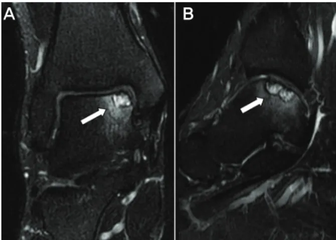
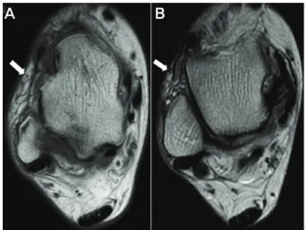
자기공명영상에서 나타난 만성 족근관절 외측 불안정성 동반 병변
전체 글
수치
관련 문서
Objective: The purpose of this study was to analyze recent trend in incidence of basal cell carcinoma and squamous cell carcinoma in patients from the Gwangju City
The purpose of this study was to identify the frequency and related factors of advanced airway management for patients with cardiac arrest by the
This retrospective chart review was conducted to determine the effectiveness of antipsychotics when given as antidepressant augmentation agents to inpatients
The purpose of this study was to analyze the impaction pattern of the impacted mandibular third molar and the relationship with the inferior alveolar nerve
The purpose of this study was to evaluate the curvature of Vertucci's type II mesial canals of mandibular molar using new method; The radius and angle
The purpose of this study was to investigate the relationship between vocabulary learning strategies and vocabulary ability of Korean middle school students
The purpose of the present study was to compare pulsatile type (TPLS) with rotary type (AK95) in order to reduce of dialysis time and to improve the
The purpose of this study was to determine the critical thinking disposition, professional autonomy, knowledge about home visiting nursing work, and
