- 19 -
∙Address for correspondence In-Tak Chu, MD
Department of Orthopedic Surgery, Seoul St. Mary’s Hospital, College of Medicine, The Catholic University, 505 Banpo-dong, Seocho-gu, Seoul, 137-040, Korea
Tel: +82-2-2258-2837 Fax: +82-2-535-9834 E-mail: [email protected]
* 본 논문의 요지는 2008년도 대한정형외과학회 추계학술대회에서 발표되었음.
대한족부족관절학회지: 제13권 제1호 2009
J Korean Foot Ankle Soc. Vol. 13. No. 1. pp.19-22, 2009
자기공명영상에서 나타난 만성 족근관절 외측 불안정성 동반 병변
가톨릭대학교 의과대학 서울성모병원 정형외과 이호진⋅주인탁⋅최광천
Associated Lesions of Magnetic Resonance Image in the Chronic Lateral Ankle Instability
Ho-Jin Lee, M.D., In-Tak Chu, M.D., Kwang-Cheon Choi, M.D.
Department of Orthopedic Surgery, Seoul St. Mary’s Hospital, College of Medicine, The Catholic University of Korea, Seoul
=Abstract=
Purpose: This retrospective study was designed to determine the type and frequency of associated lesions in patients with chronic lateral ankle instability who had modified Brostrom lateral ankle ligament reconstruction.
Materials and Methods: Between 2004 and 2007, 60 cases of 60 patients were enrolled in this study. A retrospective review of the magnetic resonance images of the affected ankle was conducted by two orthopedic surgeons who did not get any information about intraoperative findings and the lesions were admitted when two doctors were coincident.
Results: The overall incidence of associated lesions found in this study was about 83%. Peroneal tenosynovitis was the highest frequency (32%), followed by osteochondral lesion of talus (28%), anterolateral impingement (15%), Os subfibula (13%), Os trigonum (12%), ankle synovitis (12%), anterior tibiofibular ligament tear (15%), anterior bony spur (7%). Another findings were loose bodies (5%), flexor tendon tenosynovitis (5%), medial osteophyte (3%).
Conclusion: Identifying these associated lesions will be helpful in treating chronic lateral ankle instability especially when the surgeon have a plan to operate the instability. We suggest that the better results can be obtained when the associated lesions are corrected simultaneously.
Key Words: Ankle, Chronic lateral instability, Associated lesions
서 론
염좌는 족근관절의 가장 흔한 손상의 하나로 대부분 족
부와 족근관절에 내번력이 가해져서 발생한다. 이때 가장 다치기 쉬운 구조물이 족근관절 외측에 존재하는 전방 거비 인대와 종비인대이다3,6). 급성 염좌는 대부분 비수술적 방법 으로 활동력 변화, 물리치료, 보조기 등을 이용할 수 있지만, 20~40%에서는 보존적 요법에 실패하여 만성 족근관절 불 안정성이나 지속적인 통증이 발생하여 수술이 필요할 수 있
다9,12). 만성 족근관절 불안정성에서 6개월 이상의 보존적인
치료에도 호전이 없거나, 순발력이 떨어지거나, 기능적인 장애가 발생하거나, 동반 손상이 있어 해결되지 않으면 수 술을 고려한다12). 수술 방법은 일반적으로 해부학적인 재건 방법이 선호되고 있으나, 수술 후 결과에 대해서는 저자에 따라 다양하게 보고되고 있다1,7,8,11,12). 저자들은 만성 족근
이호진⋅주인탁⋅최광천
- 20 -
A B
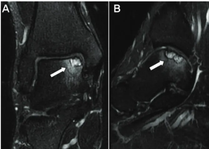
Figure 1. (A) Coronal T2-weighted image shows an osteochondral lesion along the medial aspect of the talar dome. (B) Sagittal T2- weighted image of same patient demonstrates increased signal within the talar dome.
관절 불안정성에 동반되는 족근관절의 병변이 수술 후 결과 에 영향을 미칠 수 있다고 판단이 되어 동반된 병변과 그 빈 도를 후향적으로 연구를 하고 문헌고찰과 함께 보고하는 바 이다.
대상 및 방법
2004년 1월부터 2007년 12월까지 본원에서 만성 족근 관절 불안정성으로 인대 재건술식을 받은 환자 60명의 60 예를 대상으로 하였다. 환자의 수술 전 자기공명영상을 후향적 연구를 하였다. 자기공명영상은 정형외과 전문의 2명이 검토하여 의견이 일치되는 판독소견을 병변으로 인 정하였다. 환자의 평균 연령은 33.4세(범위, 18~55세)이며 남자 44예, 여자 16예이었다. 수상 후 본원에 내원할 때까지 걸린 시간은 평균 16.7개월이었고 자기공명검사까지는 평균 17.7개월이었다.
결 과
동반 병변은 60예 중 50예(83%)에서 발견되었다. 비골건 병변이 19예(32%)로 가장 흔하였으며, 거골의 골연골 병변 이 17예(28%)에서 발견되었다(Fig. 1). 전외측 충돌증후군 이 9예(15%), 비골하 부골이 8예(13%), 족근 삼각골과 족근 관절 활액막염이 각각 7예(12%), 원위 전방 경비인대 손상 이 6예(10%), 족근관절 전방 골극이 4예(7%)로 관찰되었다.
그 외 병변으로는 관절 내 유리체와 굴곡건 건초염이 각각 3예(5%), 내측 골극이 2예(3%) 있었다. 기타의 소견으로 원위 경골의 골연골 병변이 1예가 있었다. 비골건 병변의 경우 19예 중 2예에서 단 비골건의 부분파열이 있었다. 두 명의 정형외과의사가 자기공명영상을 판독한 결과는 거골 의 골연골 병변, 비골하 부골, 족근 삼각골, 족근관절 전방 골극, 내측 골극과 같은 골과 연관된 병변에 관하여서는 평 균 95.3% 일치하였고, 족근관절 활액막염, 원위 전방 경비 인대손상 등의 진단은 평균 90.1% 이상 일치하는 소견을 보 였으나 비골건과 관련한 건초염과 전외측 충돌증후군의 진 단에서 평균 82.4%로 일치율이 가장 낮았다(Fig. 2, 3).
고 찰
만성 족근관절 불안정성과 동반된 병변은 최근의 보고에 의하면 드물지 않으며, 족관절의 내번 손상으로 외측 족근 관절 인대가 흔하게 손상되지만 다른 구조물도 내번 기전으 로 손상을 입을 수 있다6). 따라서 동반 손상의 가능성을 인
지하는 것은 임상적 결과에 영향을 미칠 수 있으므로 매우 중요하다. 저자들은 자기공명영상을 통하여 동반 병변의 빈도는 83%이고, 그 중 비골건 병변이 19예(32%)로 가장 흔하였으며 거골의 골연골 병변이 17예(28%)임을 알 수 있었다. Komenda와 Ferkel15)은 만성 불안정성 환자의 관 절경 소견에서 93%의 환자에서 관절내 병변이 있다고 하 였으며, 활액막염(70%), 소골(ossicle, 26%), 유리체(22%), 연골 연화증(22%), 거골 골연골 병변(17%) 등의 순이라고 하였다. DiGiovanni 등6)은 만성 족근관절 불안정성 환자의 수술장 소견으로 모든 예에서 동반 병변이 있으며, 비골건 건초염(77%), 전외측 충돌증후군(67%), 비골건 지지대 손상 (54%), 족근관절 활액막염(49%), 관절내 유리체(26%) 등 의 빈도 순으로 보고하였다. 이와 같이 연구 방법으로 인해 발견되는 동반 병변의 종류와 빈도의 차이는 존재하는 것을 알 수 있다. 관절경을 이용하는 방법은 관절 내 병변을 진단 함에 있어 수술장 소견이나 자기공명영상을 이용하는 방법 보다 더 우월하게 보고되었다. 실제로 Takao 등23)은 족근 관절의 손상 이후 2개월 이상 지속적인 증상이 있는 72명의 환자를 관절경을 이용하여 그 원인을 연구하였다. 관절경을 시행하기 전에 이학적 검사, 방사선 검사, 그리고 자기공명 영상검사의 결과와 관절경 시술 후 결과를 이중맹검법으로 비교하였는데 관절경 검사가 임상적, 방사선학적 검사로 진 단하지 못하는 원인을 찾는데 사용할 수 있다고 하였다. 따 라서 만성 불안정성의 수술시 관절경을 이용하여 족근관절 전체를 확인하여 관절 내 동반 병변을 진단하고 적절한 치 료를 하는 것도 좋은 방법으로 생각된다. 수술장 소견을 이 용하는 방법은 동반 병변의 빈도가 가장 높게 보고되었는데 병변의 직접관찰이 가능한 것은 장점일 수 있으나 관절경에
자기공명영상에서 나타난 만성 족근관절 외측 불안정성 동반 병변
- 21 -
A B
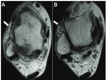
Figure 2. Peroneal tenosynovitis. MR imaging demonstrate significant fluid within the joined tendon sheath.
A B
Figure 3. Anterolateral impingement syndrome. MR imaging demon- strate (A) abnormal soft tissue mass in the anterolateral gutter as low signal on T1-weighted and (B) low to intermediate signal on T2- weighted images.
비하여 관절 내 병변을 접근하는데 있어 제한이 있을 수 있고 침습적인 면이 있다. 이에 비해 자기공명영상은 수술 전에 시행할 수 있으며 비침습적인 장점이 있다. 그러나 어떤 방법을 사용하거나 동반 병변의 빈도가 높다는 것에는 일치하는 소견을 보인다.
본 연구는 수술 소견에 정보가 없는 두 명의 정형외과 의 사가 공통된 의견을 나타내었을 때만 병변으로 인정한 것 으로 그 결과 거골의 골연골 병변, 비골하 부골, 골극과 같은 골과 연관된 병변에 관하여서는 평균 95.3%일치하였 으나 비골건과 관련한 건초염과 전외측 충돌증후군의 진단 에서 평균 82.4%로 일치율이 상대적으로 낮았다. 이는 족 근관절 주위 연부조직의 자기공명영상의 진단에 있어 각각 의 정형외과 의사에 따라 일정한 기준을 갖지 못한 것을 알 수 있고, 자기공명영상만으로 진단을 내리기가 어려운 경우 에는 반드시 정확한 이학적 검사를 바탕으로 진단을 내려야 함을 알 수 있다.
비골건 건초염은 자기공명영상에서의 비골건막(common peroneal tendon sheath)의 액체집적(fluid collection)이 존재하는 것으로 진단할 수 있는데14), 족근관절 주위에서 관상면, 시상면, 축면상에서 같은 액체집적이라 하더라고 그 정도가 다르게 보일 수 있고 정상에서도 액체집적이 있을 수 있으므로 주의해야 한다. 실제로 본 연구에서도 두 명의 정형외과 의사의 판독이 가장 일치하지 않았던 진단도 비골건 건초염이라는 것은 이 사실을 증명하여 준다. 그러 나 Kijowski 등13)은 비골건 건초염의 자기공명영상에서 비 골건막 주위에 최대 3 mm 이상의 액체집적이 있다면 그 특이도가 매우 높다고 주장하였다. 거골의 골연골 병변은 그 동반 빈도가 23~66%까지 보고되고 있는데2,5,15), 자기 공명영상에서 전위된 골연골편과 유리체를 발견하는데 정
확하다3,16,18,19). 그러나 병기(stage)결정에는 정확도가 81%
이며, 특히 연골하 부종과 동반된 단독 연골 병변은 정확한 병기를 결정하는데 제한이 있다고 하였다17). 따라서 거골의 골연골 병변의 유무와 그 빈도는 자기공명영상으로 알 수 있지만 그 정확한 병기를 결정하는 것은 어려울 것으로 생 각된다. 전외측 충돌 증후군은 자기공명영상에 족근관절의 외측구(lateral gutter)에 증식된 활액막과 외측 측부인대의 손상이 존재한다9,21). 외측구에 비정상적인 음영(signal)이 T1 이미지상 저음영, T2 이미지상 저-중간 음영으로 보인 다10). 일반적인 자기공명영상의 민감도와 특이도는 다양 하게 보고되며, 족근관절에 충분한 관절삼출액이 있어 관절 확장(distension)이 일어나면 더 정확한 진단이 가능하다21). 따라서 관절조영술을 이용하면 전외측 충돌증후군의 진단 에 도움을 줄 수 있으며 실제로 Robinson 등20)은 자기공명 영상 관절조영술로 정확도 97%, 민감도 96%, 특이도 100%
를 보고하였다.
동반 병변 중 예후가 좋지 않는 것으로 알려진 것은 전반 적으로 인대의 이완이 존재하는 경우나 족근관절의 내반변 형이 동반되어 있는 경우이다5). 실제로 Strauss 등22)의 연 구에서 재재건술을 시행한 20예에서 지속적인 통증의 유발 인자 또는 재발한 불안정성의 원인을 후족부 내반과 비골근 손상이라고 하였다. 따라서 만성 족근관절 불안정성을 수술 적으로 치료할 때는 전반적 인대의 이완, 후족부 내반, 비골 근의 상태을 판단해야 하며6), 그에 따른 적절한 치료가 인대 재건술시 동시에 이루어져야 좋은 결과를 기대할 수 있을 것이다.
이와 같이 수술 전 보존적 요법에 실패하여 인대 재건술 을 계획한다면 자기공명영상 검사를 실시하여 만성 족근
이호진⋅주인탁⋅최광천
- 22 - 관절 외측 불안정성과 동반될 수 있는 질환을 고려하여 함께 치료하는 것이 좋은 임상적 결과를 얻을 것으로 사료 된다. 그러나 자기공명영상의 소견만으로 임상적 진단을 내 려 수술을 시행하는 것보다 정확한 신체검사를 시행하여 환 자에게 꼭 필요한 동반 수술을 시행하는 것이 불필요한 수 술을 줄이고 좋은 결과를 기대할 수 있을 것으로 생각한다.
결 론
자기공명영상에 나타난 만성 족근관절 외측 불안정성과 연관된 동반 병변은 흔하게 존재할 수 있으며 수술시 동시 에 교정하지 않으면 족근관절 외측 인대 재건술 이후에 지속적인 통증과 장애를 유발할 수 있으므로 수술 전 신체 검사를 철저히 함과 동시에 자기공명영상 검사를 실시하여 동반 병변을 정확히 인지하고 그에 따른 수술 술식을 적용 하여 동시에 치료하는 것이 환자의 회복에 많은 도움이 될 것으로 사료된다.
REFERENCES
1. Bell SJ, Mologne TS, Sitler DF and Cox JS: Twenty-six-year results after Brostrom procedure for chronic lateral ankle instability. Am J Sports Med, 34: 975-978, 2006.
2. Cannon LB and Slater H: The role of ankle arthroscopy and surgical approach in the lateral ankle ligament repair. Foot Ankle Surg, 11: 1-4, 2005.
3. De Smet AA, Fisher DR, Burnstein MI, Graf BK and Lange RH: Value of MR imaging in staging osteochondral lesions of the talus (osteochondritis dissecans): results in 14 patients.
Am J Roentgenol, 154: 555-558, 1990.
4. DeMaio M, Paine R and Drez D: Chronic lateral ankle instability--inversion sprains: Part I. Orthopedics, 15: 87-96, 1992.
5. DiGiovanni BF, Fraga CJ, Cohen BE and Shereff MJ:
Associated injuries found in chronic lateral ankle instability.
Foot Ankle Int, 21: 809-815, 2000.
6. DiGiovanni BF, Partal G and Baumhauer JF: Acute ankle injury and chronic lateral instability in the athlete. Clin Sports Med, 23: 1-19, 2004.
7. Ferkel RD and Chams RN: Chronic lateral instability:
arthroscopic findings and long-term results. Foot Ankle Int, 28: 24-31, 2007.
8. Hamilton WG, Thompson FM and Snow SW: The modified Brostrom procedure for lateral ankle instability. Foot Ankle,
14: 1-7, 1993.
9. Jackson W and William M: Update on the treatment of chronic ankle instability and syndesmotic injuries. Curr Opin Orthop, 17: 97-102, 2006.
10. Jordan LK, Helms CA, Cooperman AE and Speer KP: Magnetic resonance imaging findings in anterolateral impingement of the ankle. Skeletal Radiol, 29: 34-39, 2000.
11. Karlsson J, Bergsten T, Lansinger O and Peterson L: Recon- struction of the lateral ligaments of the ankle for chronic lateral instability. J Bone Joint Surg, 70-A: 581-588, 1988.
12. Karlsson J and Lansinger O: Chronic lateral instability of the ankle in athletes. Sports Med, 16: 355-365, 1993.
13. Kijowski R, De Smet A and Mukharjee R: Magnetic resonance imaging findings in patients with peroneal tendinopathy and peroneal tenosynovitis. Skeletal Radiol, 36: 105-114, 2007.
14. Kirby AB, Beall DP, Murphy MP, Ly JQ and Fish JR: Magnetic resonance imaging findings of chronic lateral ankle instability.
Curr Probl Radiol, 34: 196-203, 2005.
15. Komenda GA and Ferkel RD: Arthroscopic findings associated with the unstable ankle. Foot Ankle Int, 20: 708-713, 1999.
16. Kramer J, Stiglbauer R, Engel A, Prayer, L and Imhof H: MR contrast arthrography (MRA) in osteochondrosis dissecans. J Comput Assist Tomogr, 16: 254-260, 1992.
17. Lee KB, Bai LB, Park JG and Yoon TR: A comparison of arthroscopic and MRI findings in staging of osteochondral lesions of the talus. Knee Surg Sports Traumatol Arthrosc, 16:
1047-1051, 2008.
18. Loredo R and Sanders TG: Imaging of osteochondral injuries.
Clin Sports Med, 20: 249-278, 2001.
19. Mintz DN, Tashjian GS, Connell DA, Deland JT, O'Malley M and Potter HG: Osteochondral lesions of the talus: a new magnetic resonance grading system with arthroscopic correlation. Arthroscopy, 19: 353-359, 2003.
20. Robinson P, White LM, Salonen DC, Daniels TR and Ogilvie- Harris D: Anterolateral ankle impingement: MR arthrographic assessment of the anterolateral recess. Radiology, 221: 186- 190, 2001.
21. Rubin DA, Tishkoff NW, Britton CA, Conti SF and Towers JD: Anterolateral soft-tissue impingement in the ankle:
diagnosis using MR imaging. AJR Am J Roentgenol, 169:
829-835, 1997.
22. Strauss JE, Forsberg JA and Lippert FG: Chronic lateral ankle instability and associated conditions: a rationale for treatment.
Foot Ankle Int, 28: 1041-1044, 2007.
23. Takao M, Uchio Y, Naito K, Fukazawa I and Ochi M: Arthro- scopic assessment for intra-articular disorders in residual ankle disability after sprain. Am J Sports Med, 33: 686-692, 2005.