298 www.kjlm.org
Isolation of a Klebsiella pneumoniae Isolate of Sequence Type 258 Producing KPC-2 Carbapenemase in Korea
Kyoung Ho Roh, M.D.1, Chang Kyu Lee, M.D.1, Jang Wook Sohn, M.D.2, Wonkeun Song, M.D.3, Dongeun Yong, M.D.4, and Kyungwon Lee, M.D.4
Departments of Laboratory Medicine1 and Internal Medicine2, Korea University College of Medicine, Seoul; Department of Laboratory Medicine3, Hallym University College of Medicine, Seoul; Department of Laboratory Medicine and Research Institute of Bacterial Resistance4,
Yonsei University College of Medicine, Seoul, Korea
Carbapenem-resistant Klebsiella pneumoniae isolates producing K. pneumoniae carbapenemases (KPC) were first reported in the USA in 2001, and since then, this infection has been reported in Europe, Israel, South America, and China. In Korea, the first KPC-2- producing K. pneumoniae sequence type (ST) 11 strain was detected in 2010. We report the case of a patient with a urinary tract in- fection caused by KPC-2-producing K. pneumoniae. This is the second report of a KPC-2-producing K. pneumoniae infection in Ko- rea, but the multilocus sequence type was ST258. The KPC-2-producing isolate was resistant to all tested β-lactams (including imi- penem and meropenem), amikacin, tobramycin, ciprofloxacin, levofloxacin, and trimethoprim-sulfamethoxazole, but was suscep- tible to gentamicin, colistin, polymyxin B, and tigecycline. The KPC-2-producing isolate was negative to phenotypic extended-spec- trum β-lactamase (ESBL) and AmpC detection tests and positive to modified Hodge test and carbapenemase inhibition test with aminophenylboronic acid.
Key Words: KPC-2, Klebsiella pneumoniae, ST258
Received: April 28, 2011 Manuscript No: KJLM-11-046 Revision received: June 13, 2011
Accepted: July 14, 2011
Corresponding author: Wonkeun Song, M.D.
Department of Laboratory Medicine, Kangnam Sacred Heart Hospital, 948-1 Daerim 1-dong, Youngdeungpo-gu, Seoul 150-950, Korea Tel: +82-2-829-5259, Fax: +82-2-847-2403, E-mail: [email protected] ISSN 1598-6535 © The Korean Society for Laboratory Medicine.
This is an Open Access article distributed under the terms of the Creative Commons Attribution Non-Commercial License (http://creativecommons.org/licenses/by-nc/3.0) which permits unrestricted non-commercial use, distribution, and reproduction in any medium, provided the original work is properly cited.
Korean J Lab Med 2011;31:298-301 http://dx.doi.org/10.3343/kjlm.2011.31.4.298
Case Report
Clinical Microbiology
KJLM
INTRODUCTION
The resistance of Klebsiella pneumoniae to carbapenems is mainly associated with acquired carbapenemases [1]. These carbapenemases can be Ambler class A (KPC, GES), class B (VIM, IMP), and class D (OXA-48) enzymes [2]. The most common class A carbapenemases in K. pneumoniae are the K. pneumoniae carbapenemases (KPCs) [3]. KPC-produc- ing K. pneumoniae strains were first reported in 2001 in the USA [4] and dissemination has been reported in Europe, Israel, South America, and China [5-9]. The first KPC-2- producing K. pneumoniae strain in Korea was isolated from
bronchial aspirates from a patient admitted to the intensive care unit (ICU) in 2010 [10]. KPC-producing K. pneumo
niae also produce VIM or CTX-M, making it difficult to se- lect appropriate antibiotics [11]. In addition, the mortality rate is significantly higher for patients with KPC-producing isolates than those with imipenem susceptible isolates [12].
In this report, we describe a case of infection with a KPC-2- producing K. pneumoniae isolate, sequence type (ST) 258 in Korea and various phenotypic methods for screening and confirmation.
CASE REPORT
A 70-year-old woman was admitted to the Plastic Surgery (PS) department on October 5, 2010, with a 24-h history of fever and dizziness. She had a known history of unstable angina and diabetes mellitus (2001). In November 2009, she was admitted to the PS department for a skin flap operation (February, 2010) to treat a third-degree burn to the sacral area. Two months ago, a sore, approximately 10×10 cm in size, developed at the sacral area and progressed to osteo- myelitis in the sacral bone. She had no recent travel history abroad. At admission, she was pale and febrile with a tem-
Roh KH, et al. • KPC-2-producing K. pneumoniae ST258
www.kjlm.org 299
http://dx.doi.org/10.3343/kjlm.2011.31.4.298
KJLM
perature of 38.2°C. An intermittent fever of 37.3-38.1°C last ed until hospital day (HD) 5. Her blood pressure was 110/80 mmHg, her pulse was 78/min, and her respiratory rate was 20/min. A laboratory investigation at the time of admission revealed a peripheral white blood cell (WBC) count of 8,730/μL (73.5% neutrophils), a hemoglobin level of 8.6 g/dL, and a platelet count of 261,000/μL. Routine blood chemistry results were AST/ALT of 7/11 U/L, alka- line phosphatase of 77 U/L, blood urea nitrogen/creatinine of 23.4/1.42 mg/dL, and total protein/albumin of 5.6/2.9 g/
dL. Erythrocyte sedimentation rate and C-reactive protein were both increased to 53 mm/hr and 220.03 mg/L, respec- tively. The urine was yellow and turbid and routine urinaly- sis revealed a positive WBC (3+), and positive protein (1+).
Microscopic examination of urine revealed >60 WBCs and yeast organisms in a high power field. A chest radiograph showed right pleural thickening and a little collapse of the right lower lung. An abdomen and pelvic computed tomog- raphy showed signs of cystitis and fluid collection in both the abdomen and pleural cavity.
Two sets of blood culture bottles and a urine sample were taken for microbiologic study. Aerobic and anaerobic blood cultures were all negative after 5 days of incubation. In the urine culture, Candida albicans (8×10⁴ CFU/mL) grew on a blood agar plate. On HD 32, the patient had a fever of 38.1°C. Two sets of blood culture bottles and a urine sample were collected again for culture study. The blood culture re- sults were negative. Multidrug-resistant K. pneumoniae (KPN 1010, >105 CFU/mL) was isolated from the urine. Vitek2 GN and AST-N044 (bioMérieux, Marcy l’Étoile, France) were used for species identification and antimicrobial sus- ceptibility test, respectively. With the exception of gentami-
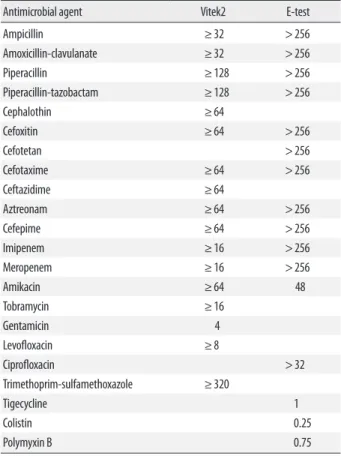
Table 1. MICs (μg/mL) of the KPC-2-producing Klebsiella pneumoniae isolate
Antimicrobial agent Vitek2 E-test
Ampicillin ≥32 >256
Amoxicillin-clavulanate ≥32 >256
Piperacillin ≥128 >256
Piperacillin-tazobactam ≥128 >256
Cephalothin ≥64
Cefoxitin ≥64 >256
Cefotetan >256
Cefotaxime ≥64 >256
Ceftazidime ≥64
Aztreonam ≥64 >256
Cefepime ≥64 >256
Imipenem ≥16 >256
Meropenem ≥16 >256
Amikacin ≥64 48
Tobramycin ≥16
Gentamicin 4
Levofloxacin ≥8
Ciprofloxacin >32
Trimethoprim-sulfamethoxazole ≥320
Tigecycline 1
Colistin 0.25
Polymyxin B 0.75
Abbreviations: MIC, minimum inhibitory concentration; KPC, K. pneumoniae carbap- enemase.
Fig. 1. Results obtained with a Modified Hodge test (A) and carbapenemase inhibition test (B) for carbapenem-resistant Klebsiella pneumoniae isolate (KPN 1010).
Abbreviations: MEM, meropenem; APB, aminophenylboronic acid; CLX, cloxacillin; EDTA, ethylenediaminetetraacetic acid; DPA, dipicolinic acid.
A B
KPN 1010
E. coli
ATCC 25922 KPN 1010
with DPA
with APB
with EDTA
with CLX
cin, all susceptibility results showed high-level minimum inhibitory concentration (MIC) values. MICs were also as- sessed by E-test (bioMérieux). Most antibiotics were resis- tant and consistent with the MIC of Vitek2. However, MICs
300 www.kjlm.org
Roh KH, et al. • KPC-2-producing K. pneumoniae ST258
http://dx.doi.org/10.3343/kjlm.2011.31.4.298
KJLM
of tigecycline and colistin were 1 and 0.25 μg/mL, respec- tively (Table 1). The modified Hodge test [13] demonstrated strong positivity (Fig. 1A) but AmpC and ESBL phenotypic tests [14] were negative. Carbapenemase inhibition tests were performed for discrimination of carbapenemases. Briefly, meropenem disks (Becton-Dickinson, Cockeysville, MD, USA) were supplemented with 10 μL of 4 different β-lacta- mase inhibitors: 60 mg/mL aminophenylboronic acid (APB;
Sigma St. Louis, MO, USA), 75 mg/mL cloxacillin (Sigma), 100 mg/mL dipicolinic acid (DPA; Sigma), and 0.2 M ethyl- enediaminetetraacetic acid (EDTA; Sigma). A 0.5 McFar- land inoculum was prepared and spread on Mueller-Hinton agar plates (Becton-Dickinson). Five disks were placed on each plate: meropenem, meropenem+APB, meropenem+
cloxacillin, meropenem+DPA, meropenem+EDTA. A pos- itive response was achieved when there was a greater than 5 mm increase of the inhibition zone diameter around disks containing β-lactamase inhibitors, as compared with the meropenem disk alone [15]. The positive result was seen only with APB (Fig. 1B).
PCR and DNA sequencing were performed with primers specific for the blaVIM, blaIMP, blaSIM, blaNDM, and bla KPC genes [16-18]. We found only the blaKPC-2 gene. Mul tilocus sequen ce typing (MLST) with 7 housekeeping genes (rpoB, gapA, mdh, pgi, phoE, infB, and tonB) was performed [19]. The MLST showed that the isolate belonged to the epidemic clone ST258.
Initially, the patient was treated with ertapenem (1 g once a day). After multidrug-resistant (MDR) K. pneumoniae was isolated from urine and the sacral sore wound, tigecycline (50 mg twice a day) was administrated for 14 days. How- ever, neither pyuria nor the wound subsided, and MDR K.
pneumoniae was repeatedly isolated. Lastly, the patient was treated with colistin (150 mg once a day), but clinical im- provement was not observed and kidney function was de- clining. She was managed in ICU, but expired on the 90th HD due to septic shock and multiorgan failure.
DISCUSSION
The most common mechanism of carbapenem-resistant K. pneumoniae in Korea is ESBL and/or AmpC β-lactamase plus porin loss [20]. Only a single case of KPC-2-producing strain has been described in Korea and the MLST type was ST11 [10]. This isolate showed an MDR pattern to various antibiotics, including colistin. The previous patient was treat ed with colistin, but the prognosis was also poor. This is the second report of a KPC-2-producing K. pneumoniae in Ko- rea. The isolate was not associated with travel and the MLST type was ST258. ST258 accounts for 70% of KPC-producing
K. pneumoniae in the USA [21]. Isolates of ST258 have also been identified in Europe and Israel, indicating an interna- tional spread of ST258 among KPC-producing K. pneumo
niae [6, 7].
Most KPC-producing K. pneumoniae have been associ- ated with other β-lactamase genes, such as the widespread ESBL gene blaCTX-M [22]. The previous KPC-2-producing K.
pneumoniae isolate in Korea also contained blaCTX-M-15 [10].
However, the KPN 1010 isolate contained neither ESBL nor AmpC gene in this study.
The KPN 1010 isolate was susceptible to gentamicin, co- listin, polymyxin B, and tigecycline, but the previous Korean isolate was nonsusceptible to gentamicin, colistin, polymyxin B, and tigecycline [10].
A modified Hodge test accurately detects KPC, but is not able to discriminate from other carbapenemases [23]. A car- bapenemase inhibition test, comprising a meropenem disk, and meropenem disks supplemented with APB (for detec- tion of class A carbapenemases), cloxacillin (for detection of AmpC β-lactamases plus porin loss), DPA or EDTA (for detection of class B metallo-carbapenemases) accurately distinguishes between several different mechanisms medi- ating reduced susceptibility to carbapenems in Enterobacte
riaceae [15]. The isolate was positive to APB and negative to cloxacillin, DPA, and EDTA, suggesting a class A carbapen- emase producing isolate.
Authors’ Disclosures of Potential Conflicts of Interest No potential conflict of interest relevant to this article was reported.
REFERENCES
1. Nordmann P and Poirel L. Emerging carbapenemases in Gram- negative aerobes. Clin Microbiol Infect 2002;8:321-31.
2. Queenan AM and Bush K. Carbapenemases: the versatile β-lacta- mases. Clin Microbiol Rev 2007;20:440-58.
3. Nordmann P, Cuzon G, Naas T. The real threat of Klebsiella pneu
moniae carbapenemase-producing bacteria. Lancet Infect Dis 2009;
9:228-36.
4. Yigit H, Queenan AM, Anderson GJ, Domenech-Sanchez A, Bid- dle JW, Steward CD, et al. Novel carbapenem-hydrolyzing β-lacta- mase, KPC-1, from a carbapenem-resistant strain of Klebsiella pneu
moniae. Antimicrob Agents Chemother 2001;45:1151-61.
5. Giakkoupi P, Pappa O, Polemis M, Vatopoulos AC, Miriagou V, Zioga A, et al. Emerging Klebsiella pneumoniae isolates coproduc- ing KPC-2 and VIM-1 carbapenemases. Antimicrob Agents Che- mother 2009;53:4048-50.
6. Navon-Venezia S, Leavitt A, Schwaber MJ, Rasheed JK, Srinivasan A, Patel JB, et al. First report on a hyperepidemic clone of KPC-3- producing Klebsiella pneumoniae in Israel genetically related to a strain causing outbreaks in the United States. Antimicrob Agents
Roh KH, et al. • KPC-2-producing K. pneumoniae ST258
www.kjlm.org 301
http://dx.doi.org/10.3343/kjlm.2011.31.4.298
KJLM
Chemother 2009;53:818-20.
7. Samuelsen Ø, Naseer U, Tofteland S, Skutlaberg DH, Onken A, Hjetland R, et al. Emergence of clonally related Klebsiella pneu
moniae isolates of sequence type 258 producing plasmid-mediated KPC carbapenemase in Norway and Sweden. J Antimicrob Che- mother 2009;63:654-8.
8. Villegas MV, Lolans K, Correa A, Suarez CJ, Lopez JA, Vallejo M, et al. First detection of the plasmid-mediated class A carbapene- mase KPC-2 in clinical isolates of Klebsiella pneumoniae from South America. Antimicrob Agents Chemother 2006;50:2880-2.
9. Wei ZQ, Du XX, Yu YS, Shen P, Chen YG, Li LJ. Plasmid-medi- ated KPC-2 in a Klebsiella pneumoniae isolate from China. Anti- microb Agents Chemother 2007;51:763-5.
10. Rhee JY, Park YK, Shin JY, Choi JY, Lee MY, Peck KR, et al. KPC- producing extreme drug-resistant Klebsiella pneumoniae isolate from a patient with diabetes mellitus and chronic renal failure on hemodialysis in South Korea. Antimicrob Agents Chemother 2010;
54:2278-9.
11. Pournaras S, Poulou A, Voulgari E, Vrioni G, Kristo I, Tsakris A.
Detection of the new metallo-β-lactamase VIM-19 along with KPC- 2, CMY-2 and CTX-M-15 in Klebsiella pneumoniae. J Antimicrob Chemother 2010;65:1604-7.
12. Marchaim D, Navon-Venezia S, Schwaber MJ, Carmeli Y. Isolation of imipenem-resistant Enterobacter species: emergence of KPC-2 carbapenemase, molecular characterization, epidemiology, and outcomes. Antimicrob Agents Chemother 2008;52:1413-8.
13. Lee K, Kim CK, Yong D, Jeong SH, Yum JH, Seo YH, et al. Impro- ved performance of the modified Hodge test with MacConkey agar for screening carbapenemase-producing Gram-negative bacilli. J Microbiol Methods 2010;83:149-52.
14. Song W, Jeong SH, Kim JS, Kim HS, Shin DH, Roh KH, et al. Use of boronic acid disk methods to detect the combined expression of plasmid-mediated AmpC β-lactamases and extended-spectrum β-lactamases in clinical isolates of Klebsiella spp., Salmonella spp., and Proteus mirabilis. Diagn Microbiol Infect Dis 2007;57:315-8.
15. Giske CG, Gezelius L, Samuelsen Ø, Warner M, Sundsfjord A, Wo- od ford N. A sensitive and specific phenotypic assay for detection
of metallo-β-lactamases and KPC in Klebsiella pneumoniae with the use of meropenem disks supplemented with aminophenylbo- ronic acid, dipicolinic acid and cloxacillin. Clin Microbiol Infect 2011;17:552-6.
16. Patzer JA, Walsh TR, Weeks J, Dzierzanowska D, Toleman MA.
Emergence and persistence of integron structures harbouring VIM genes in the Children’s Memorial Health Institute, Warsaw, Poland, 1998-2006. J Antimicrob Chemother 2009;63:269-73.
17. Smith Moland E, Hanson ND, Herrera VL, Black JA, Lockhart TJ, Hossain A, et al. Plasmid-mediated, carbapenem-hydrolysing β-lac- tamase, KPC-2, in Klebsiella pneumoniae isolates. J Antimicrob Chemother 2003;51:711-4.
18. Zarfel G, Hoenigl M, Leitner E, Salzer HJ, Feierl G, Masoud L, et al. Emergence of New Delhi metallo-β-lactamase, Austria. Emerg Infect Dis 2011;17:129-30.
19. Diancourt L, Passet V, Verhoef J, Grimont PA, Brisse S. Multilocus sequence typing of Klebsiella pneumoniae nosocomial isolates. J Clin Microbiol 2005;43:4178-82.
20. Park YJ, Yu JK, Park KG, Park YG, Lee S, Kim SY, et al. Prevalence and contributing factors of nonsusceptibility to imipenem or me- ropenem in extended-spectrum β-lactamase-producing Klebsiella pneumoniae and Escherichia coli. Diagn Microbiol Infect Dis 2011;
in press.
21. Kitchel B, Rasheed JK, Patel JB, Srinivasan A, Navon-Venezia S, Carmeli Y, et al. Molecular epidemiology of KPC-producing Kleb
siella pneumoniae isolates in the United States: clonal expansion of multilocus sequence type 258. Antimicrob Agents Chemother 2009;
53:3365-70.
22. Cai JC, Zhou HW, Zhang R, Chen GX. Emergence of Serratia mar
cescens, Klebsiella pneumoniae, and Escherichia coli Isolates possess- ing the plasmid-mediated carbapenem-hydrolyzing β-lactamase KPC-2 in intensive care units of a Chinese hospital. Antimicrob Agents Chemother 2008;52:2014-8.
23. Anderson KF, Lonsway DR, Rasheed JK, Biddle J, Jensen B, Mc- Dougal LK, et al. Evaluation of methods to identify the Klebsiella pneumoniae carbapenemase in Enterobacteriaceae. J Clin Micro- biol 2007;45:2723-5.