Molecular Cloning and Expression of Dihydroflavonol 4-reductase Gene in Tuber Organs of Purple-fleshed Potatoes
Won-jin Kang
1, Yong-hwa Lee
1, Hyun-soon Kim
1, Hyouk Joung
1and Jae-heung Jeon
1,*
1 Plant Cell Biotechnology Lab. Korea Research Institute of Bioscience and Biotechnology (KRIBB), Daejon, 305-333, South Korea
Received July 13, 2006; Accepted September 7, 2006
A full-length cDNA encoding dihydroflavonol 4-reductase (st-dfr) of potato was isolated by rapid amplification of cDNA ends, and their expression was investigated from purple-fleshed potato (Solanum tuberosum L. cv. Jashim). The st-dfr exists as a member of a small gene family and its transcripts was abundant in the order of tuber flesh, stem, leaf, and root. The expressions of st-dfr gene were light inducible and cultivar dependant. Transgenic potato plants harboring antisense st-dfr (AS-DFR) sequences were analyzed. The accumulation of mRNA was nearly completely inhibited as a result of introducing an AS-DFR gene under the control of the 35S CaMV promoter into the red tuber skin Solanum tuberosum L. cv. Desiree. The anthocyanin content of the tuber peels of the transgenic lines was dramatically decreased by up to 70%. The possible production of flavonols in the peels of AS-DFR transgenic potatoes was discussed.
Key words: Dihydroflavonol 4-reductase, Solanum tuberosum L. cv. Jashim, anthocyanin, antisense, transgenic potato
Flavonoids represent a large class of plant secondary metabolites, of which anthocyanins are the most conspicuous class, due to their wide range of colors. Anthocyanins, the largest subclass of flavonoids, are major pigments in flowers and fruits, where they serve as insect and animal attractants and therefore may play a role in pollination and seed dispersal.
Anthocyanins and other flavonoids can also be important as feeding deterrents and as protection against damage from UV irradiation.1,2) The flavonoids pathway has been studied extensively in many plants.3-5) Flavonoids also have significant biological activity when ingested by animals, and there is a great deal of interest in their potential health benefits, which include antioxidant activity, estrogenic and anti-cancer activity, and vasodilatory actions.6)
A number of genes that encode anthocyanin biosynthetic enzymes have been characterized and cloned from maize, snapdragon, and petunia. Dihydroflavonol 4-reductase (DFR, EC 1.1.1.219) mediates the first step of anthocyanin biosynthetic pathways, and the expression of this enzyme is the most pronounced in spatial and temporal patterns in corolla and inflorescence development.7,8) Dfr genes have been isolated from maize and snapdragon by transposon tagging. A snapdragon dfr clone was used to isolate a homologous gene from petunia. Petunia contains three different dfr genes (dfrA, dfrB, and dfrC), but only the dfrA gene is transcribed in floral tissue.9)
The red or purple coloration of potato skin is derived from anthocyanins. Potato cultivars can be classified according to the organ types (skin, flesh, and flower) in which anthocyanins accumulate and colors (white, yellow, red, pink, and purple). In recent years potato breeders have developed new cultivars, such as anthocyanin-containing purple and red-fleshed cultivars, for the purpose of using them as a functional food. Potatoes with pigmented flesh show antioxidant capacities 2-3 times greater than unpigmented varieties. However, only a few molecular studies on the anthocyanin pathway in potatoes have been reported.10-12) As one of the best-characterized natural product pathways in plants, flavonoid biosynthetic enzymes are attractive targets for metabolic engineering in providing enhanced feed crops, food sources, and medicinal agents.13-16)
In this experiment, we were successful in cloning the potato dfr gene from Solanum tuberosum L. cv. Jashim, which has purple skin and flesh color. Expression studies of potato dfr gene from purple-fleshed potato were carried out. We focused to make a transgenic potato plants harboring antisense st-dfr sequence (AS-DFR) to check the possible change of metabolic flux on flavonoids biosynthetic pathway.
Materials and Methods
Plant material and tissue culture
. S. tuberosum cv.Jashim, purple skin and flesh color, and cv. Desiree plants cultured in vitro were used for the expression studies and transformation, respectively. Plants were vegetatively propagated from nodal stem segments in tissue culture on MS medium17) containing 3% (w/v) sucrose at 23oC under 16 h light conditions.
*Corresponding author
Phone: +82-42-860-4492; Fax: +82-42-860-4599 E-mail: [email protected]
76 Won-jin Kang et al.
Leaves and stems were collected from in vitro grown plants.
For chemical analysis, plants were cultivated for 4 months in a greenhouse.
Cloning of
st-dfrcDNA
. We used a polymerase chain reaction (PCR) strategy to clone st-dfr cDNA. The tuber of Jashim cultivar grown in a field was used for extraction of total RNA. Total RNA was isolated from the purple flesh part of developing tubers according to the Phenol/SDS methods,18) and first-strand cDNA was synthesized with oligo dT-anchor primer using the 5'/3'-rapid amplification of a cDNA ends (RACE) Kit (Roche Molecular Biochemicals). Based on the amino acid sequences conserved among four dfr genes [gene products of tomato (S38474), petunia (P14720), Ipomoea batatas (BAA34637), Forsythia x intermedia (CAA70345)]clustered in the phylogenetic tree, a degenerate primer was designed. Using a degenerate primer DFR-DG and PCR anchor primer, we amplified a DNA fragment with expected size of about 900 bp from the Solanum tuberosum L. cv.
Jashim cDNA. The degenerate primer DFR-DG was based on the peptide sequences KMTGWMYF with the nucleotide sequence 5'-AA(A/G)ATGAC(A/C/T)GG(A/C)TGGATGTA (C/T)TT-3'(Fig. 2). The amplified fragment was cloned and the nucleotide sequence was determined. A 5'-terminal portion of cDNA was isolated by the 5'-rapid amplification of cDNA ends (RACE) method using a newly synthesized first-strand cDNA with a specific antisense primer, DFR-R1 (5'-GCACC ATCTTAGCCACATCGTAGATG-3'). Another specific antisense primer, DFR-R2 (5'-CCAACAACCAGTGGTGGTATGATG CT-3'), was used for the 5'-RACE using the 5'/3'-RACE System (Roche Molecular Biochemicals).
Southern analysis of
st-dfrin the Jashim cultivar
. To examine the copy number of the Jashim dfr gene, Southern blot analysis was performed on genomic DNA extracted from leaves of Jashim cultivar. Genomic DNA was isolated according to the protocol.19) After digestion with the appropriate restriction enzymes, the DNA was subjected to electrophoresis through a 0.7% agarose gel and transferred to a nylon membrane. The membranes were hybridized with probes labeled with digoxigenin (DIG) using a PCR DIG probe synthesis kit (Roche Molecular Biochemicals). Hybridization lasted for 16 h at 42oC in a hybridization buffer, a DIG Easy Hyb. The membranes were washed twice in 2× SSC, 0.1% (w/v) SDS at room temperature for 5 min each and then washed twice in 0.1× SSC, 0.1% SDS at 68oC for 15 min each. Target DNAs were detected by using a DIG luminescent detection kit as described in the manufacturer’s instructions (Roche Molecular Biochemicals).Construction of a
dfrplant antisense expression vector
. According to the cDNA sequence cloned from Jashim cultivar, a dfr coding region DNA fragment starting with the start codon and continuing through the stop codon was produced by PCR amplification using Pfu DNA polymerase (Stratagene, La Jolla, CA) using primers: 5'-CACGTGAAAA TGGCAAGTGAAGTTC-3' and 5'-CCATGGTTGCAGTACT AAATTTCACC-3', and the cDNA clone as a template. A newenzyme site of NcoI and PmlI was inserted into the end of these primers for cloning. The resulting 1172 bp fragments was subcloned in a Zeroblunt PCR cloning kit (Invitrogene).
After cutting out the NcoI and PmlI fragment from this cloning vector, the resulting dfr gene fragment was ligated into the NcoI and PmlI site of pCAMBIA- binary vector between the CaMV 35S promoter and the Nos 3' region. We confirmed the antisense oriented pCAMBIA-AS-DFR vector (pCAM-AS-DFR) by sequencing.
Transformation of potato and PCR analysis of transgenic lines.
pCAM-AS-DFR was transformed into Agrobacterium tumefaciens strain LBA4404 and introduced into Solanum tuberosum L. var. Desiree by leaf disc transformation method.20) Plants regenerated from transformed calli on 5 mg· l−1 of bialaphos containing plates were transferred to MS media and subcultured for two weeks before analysis. Transgenic lines with a high inhibition level of endogenous dfr gene were selected by Northern blot analysis, transferred to soil and then grown for chemical analysis. PCR from genomic DNA derived from leaf tissues was employed using well-characterized PCR primers. Antisense sequences were amplified using CaMV specific (CAMV-SP1, 5'-GATATCTCCACTGACGTAAGG GAT) and DFR specific (DFR-SP1, 5'-AAGATGACAGGAT GGATGTATTT) primers, which amplified the characteristic fragments from the antisense constructs. Since one of the introns of the dfr gene (unpublished data) is located in the middle of the DFR-SP1 primer sequence, only the transgene AS-DFR sequence can be amplified. The expected size of the amplified fragments was 768 bp.Northern analysis of AS-DFR mRNA.
The expression of dfr genes was investigated by Northern blot experiments using transgenic potato plants. To recognize the expression of the dfr genes, 25 mg of total RNA was separated by electrophoresis in a 1.2 % agarose gel containing 2.2 M formaldehyde and then transferred to a nylon membrane. The membranes were hybridized and detected by the same method as in Southern blot analysis.Analysis of anthocyanin and total UV-absorptive compounds
. Anthocyanin analysis was performed using a previously described protocol.21) Plant tissues were ground in liquidnitrogen and extracted with 80% (v/v) methanol, as described above.The methanol extracts were mixed with an equal volume of 0.5%(v/v) HCl, and then extracted with 2ml of chloroform. The aqueous/methanolphase was assayed spectrophotometrically at A530, with A657 subtracted,and the resulting absorbance value was normalized to the freshweight for each sample. Total UV-absorptive compounds were assayed using a previously described protocol,22) as follows.Plant tissues were ground in liquidnitrogen and extracted with 80% (v/v) methanol/1% (v/v) HCl. Theextracts were clarified by centrifugation at 14,000g for 10 min,followed by filtration through nylon centrifuge tube filters (Corning Inc., Corning, NY). Flavonoid compounds were assayed by measurement at A330 and absorbance values were normalized to the fresh weight ofeachsample.
Results and Discussion
Cloning and characterization of st-dfr. The full-length cDNA was achieved by final PCR using primers from the 5'- and 3'-untranslated regions and the cDNA pools as a template.
The deduced amino acid sequence from this DNA fragment showed significant identity with the dfr from tomato, and thus the putative corresponding gene was named st-dfr ofJashim cultivar (Genbank Accession Number, AF449422). The cDNA sequence is 1535 nucleotides long and consists of an open reading frame encoding a peptide of 382 amino acids (Fig. 1), with a calculated molecular mass of 42.7 kDa and an isoelectric point of 6.47. The BLAST analysis23) on the homology of the deduced amino acid sequence shows high homologies with Lycopersicon esculentum (S38474), Petunia x hybrida (P14720), Forsythia x intermedia (CAA70345), and Zea mays (CAA75996), 94.5, 87.9, 74, and 62%
identities, respectively (Fig. 1).
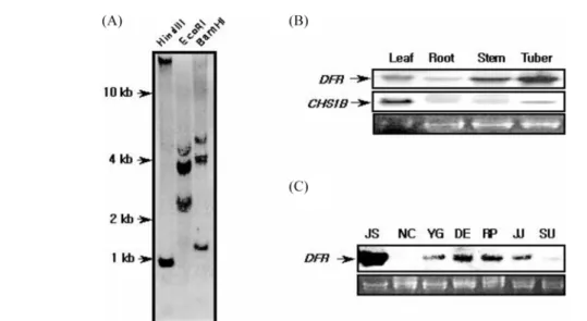
To examine the copy number of the st-dfr gene, Southern blot analyses were performed on genomic DNA extracted from leaves of Jashim cultivar. There are two internal BamHI and HindIII sites in st-dfr cDNA sequence. And we made a DIG probe using PCR DIG probe synthesis kit with two specific primers (DFR-F1, 5'-CCAGAGAACGAAGTAATT CAAC-3'; DFR-R1, 5'-GCACCATCTTAGCCACATCGTAG ATG-3') located between two HindIII sites. And there are three 105, 107 and 96 bp introns between two specific primers
(data not shown). As shown in Fig. 2A, digestions of genomic DNA with BamHI and HindIII generate hybridizing fragments of 1.36 kb and 0.99 kb, respectively. Moreover, in the absence of EcoRI site in st-dfr, there are at least three copies of st-dfr genes. Therefore, the genomic Southern result suggests that st-dfr exists as a member of a small gene family (Fig. 2A).
The results of the Northern blot analysis (Fig. 2B, C) showed that high expressions of RNA were observed in tuber and stem, while expressions in leaves and roots were very weak and the expression of potato chalcone synthase 1B gene was the highest in leaf tissues. The expressions of st-dfr gene were the highest in the microtuber of cv. Jashim, a purple-fleshed potato, and the lowest in the microtuber of cv. Norchip, a white-skin and -fleshed potato. The expression of st-dfr gene showed light inducible pattern in the stem and leaf of cv.
Jashim and cv. Desiree in high light condition. The expression of st-dfr gene was the highest level at 24 h after high light treatment and decreased gradually thereafter. About 72 h after the highest RNA expression, anthocyanin content reached the highest level (Fig. 3).
Introduction of the AS-DFR gene into potato.The st-dfr gene is strongly expressed in potato tuber flesh of Jashim cultivar (Fig. 2B). In order to increase the levels of flavonols in the potato tuber, we attempted to use an antisense technique with this dfr gene (Fig. 4). A schematic representation of the AS-DFR construct is given in Figure 4A. The binary vector used in these experiments was pCAMBIA3301, which includes Fig. 1. Alignment of predicted amino acid sequences of S. tuberosum cv. Jashim(st-dfr). The amino acid sequences conserved in more than two proteins are shaded in the figure. The amino acid sequences of st-dfr is aligned to those of DFRs of Lycopersicon esculentum (LE; S38474), Petunia x hybrida (PH; P14720), Forsythia x intermedia (FI; CAA70345), and Zea mays (ZM;
CAA75996). The residues with asterisks correspond to conserved region of degenerate primer designed for PCR cloning.
78 Won-jin Kang et al.
the CaMV and nos sequences within its T-DNA. Solanum tuberosum L. cv. Jashim dfr sequences were inserted between these sequences in an antisense orientation relative to the CaMV promoter. A bar gene is also located within its T-DNA.
The regenerated potato shoots were again selected by treatment with bialaphos (Fig. 4B). To verify the transformed lines selected from a 5 mg· l−1 bialaphos treatment, PCR analysis was carried out on genomic DNA extracted from control and antisense plants. CAMV-SP1 and DFR-SP1 primer pairs were used to detect the transgene sequences inserted into genomic DNA. An intron of the st-dfr gene is located in the middle of the DFR-SP1 primer sequence. Therefore, we were able to successfully amplify the transgene AS-DFR sequence, not an endogenous st-dfr gene. The generation of amplification fragments is shown in Fig. 4C. Seven independent transgenic lines were recovered. Highly inhibited transgenic plants were selected by Northern blot analysis from the PCR positive lines
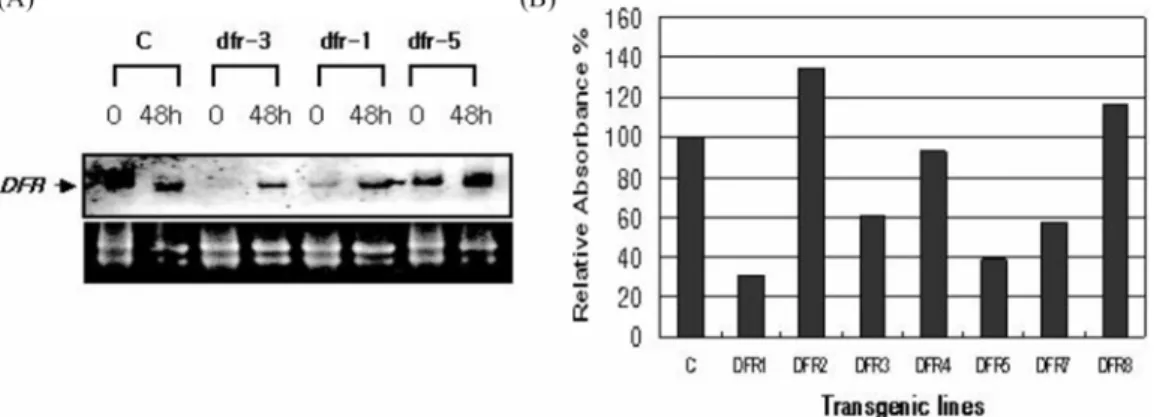
for the AS-DFR construct (Fig. 5A). An in vitro culture system also could be used to effectively screen the transformants (DFR-1 and DFR-3 lines) in which endogenous st-dfr levels were inhibited. After treatment with high white-light during tissue culture for 2 days, we were able to detect the inhibition of st-dfr transcripts and anthocyanin biosynthesis from the AS- DFR transgenic lines (Fig. 5). The transgenic DFR-1 and DFR- 3 lines expressing the AS-DFR gene at the highest level were further grown in a greenhouse for use in flavonol analysis. No obvious effects of tissue culture, microtuberization, or plant morphologic characteristics between control and transgenic plants were observed, although one of the antisense lines (DFR- 1 line) appeared to be a dark green phenotype of leaf and stem and showed growth inhibition during cultivation in the greenhouse. In the case of the transgenic lines, the brick-red skin color of the tuber changed from brick red to pale brick red (Fig. 6).
Fig. 2. Copy number prediction of st-dfr gene by genomic Southern blot analysis (A) and the expression profile of st-dfr genes and chalcone synthase 1B (st-chs1B) gene in different tissues and cultivars were investigated by Northern blot experiments (B, C). Solanum tuberosum L. cvs. Jashim (lane JS), Norchip (lane NC), Yukon Gold (lane YG), Desiree (lane DE), Red Pontiac (lane RP), Jaju (lane JJ) and Superior (lane SU).
Fig. 3. Induced expression of st-dfr genes and accumulation of anthocyanins in high light condition. (A) Northern blot and antho- cyanin content (B) of stem and leaf tissues were analyzed at different time intervals (0, 6, 12, 24, 48, 96, 168 hr) after high light treatment. (C) Northern blot analysis was carried out from different potato cultivars for detection of light inducibility (C; control cv.
Jashim, 24D; 24 hr dark, 24L; 24 hr light treatment. DC, D24D, D24L; same treatment but for cv. Desiree plant).
Antisense inhibition of DFR expression decreases anthocyanin levels. The wild type potato tuber of cv. Desiree has a brick-red skin in color (Fig. 6A). The brick-red color is restricted to the tuber skin in this variety. The tuber skin is composed of two layers of cells: a layer of single cells (the epidermis), and several layers of corky cells (the periderm).
The cells in the periderm layer may contain the red pigments that produce the red color. Only tuber peels were used for the detection of anthocyanin and flavonols content in the transgenic lines harvested from the greenhouse. As shown in Fig. 6, a dramatic reduction in anthocyanin content was observed for the transgenic line DFR-1 (Fig. 6B). TLC was carried out on KC 18F reverse phase plate with tert-Butanol : Acetic acid : Water (3 : 1 : 1) as the mobile phase. It clearly shows that the anthocyanin content is reduced almost completely in the DFR-1 line but that some amount of anthocyanins remains in the DFR-3 line. In the transgenic lines, levels of all anthocyanins appear to be reduced to a
similar extent (Fig. 6D). In the tuber peels of transgenic lines DFR-1 and DFR-3, the anthocyanin contents were decreased by 30% and 70%, respectively. Compared to anthocyanin accumulation patterns in tuber skin, no anthocyanin accumulation was observed in the leaf tissue of potato cv.
Desiree. These results were in agreement with other reports. A number of reports concerning changes in flower coloration have appeared. A number of strategies for introducing dfr
genes from maize or Gerbera into Petunia, the co-suppression of flavone synthase in tobacco, and the introduction of alfalfa chalcone reductase into Petunia have been reported.2,8,14,24) In another similar work,25) down-regulation of potato dfr gene and decrease of anthocyanidins in potato tubers using petunia
dfr gene were reported. Therefore, the manipulation of an anthocyanin pathway with this gene from new potato cultivars, such as anthocyanin-containing purple and red-fleshed cultivars, will result in decreased anthocyanin composition in tuber flesh also.
Fig. 4. Structure of AS-DFR constructs and the selection of transgenic lines. (A) RB, Right border of T-DNA; LB, left border of T-DNA, T, nos polyA site. Positions of the bar gene and AS-DFR construction within T-DNA are indicated. (B) Screening of trans- formants on a selection media containing 5 mg/L of bialaphos. Letter T means transgenic plant screened. (C) PCR analysis of trans- genic lines. PCR from genomic DNA derived from leaf tissues was employed using transgene specific primers. Lane M, DNA ladder;
PC, positive control from vector DNA; NC, negative control of non transformed tissue; T1~11, putative transgenic line numbers for PCR analysis.
Fig. 5. Expression of the DFR gene and a comparison of anthocyanin content from different AS-DFR transgenic lines. (A) Northern analysis was carried out for each of the transgenic potato lines. After about a 48-h white light treatment to induce the endog- enous DFR gene, total RNA from the leaves and stems of each transformant was isolated and analyzed. (B) After about a 48-h white light treatment, the relative anthocyanin content was determined from the leaves and stems of each transformant.
80 Won-jin Kang et al.
Potatoes have different flowers, tuber skin, and flesh colors, such as white, buff, cream, brick red, red, purple, depending on the anthocyanin pigmentation pattern. The pigmentation of a tuber can vary from red or pink, through purple, to blue. The anthocyanin content of tubers and shoots of the purple potato, Solanum tuberosum L. cv. Congo has been determined.
Glycosylated petunidin and malvidin were found to be the major anthocyanidins both in tubers and shoots.26) The major anthocyanin in red-fleshed potatoes was identified as pelagonidin-3-rutinoside-5-glucoside acylated with γ-coumaric acid.27) Petunidin and malvidin are derived from delphinidin and delphinidin is from dihydromyricetin (DHM) while pelargonidin is from dihydrokaempferol (DHK). This substrate specificity may explain the preferential accumulation pattern of flavonols. Therefore the DHK and DHM inhibited any further conversion to anthocyanins and may have accumulated as kaempferol and myricetin glycosides produced by endogenous flavonol synthase (data not shown). These preliminary results are consistent with the fact that the major anthocyanidins of tubers and shoots of the purple potato are glycosylated petunidin and malvidin and that the major anthocyanin in red- fleshed potatoes was identified as pelagonidin.26) Therefore, it maybe possible to produce potato plants containing elevated flavonol end products, such as kaempferol and myricetin, in tuber flesh from red or purple-fleshed potatoes using an antisense strategy. A strategy for producing health-promoting
crops is needed because food crops typically are not rich in flavonols. Flavonols such as quercetin, myricetin, and kaempferol have health-promoting activities. By using this same strategy, we think it is possible to produce transgenic fruit with increased fruit flavonol content.
Acknowledgments
This research was supported by a grant from Plant Diversity Research Center of 21st Century Frontier Research Program funded by Ministry of Science and Technology of Korean government.
References
1. Dooner, H. K., Robbins, T. P. and Jorgensen, R. A. (1991) Genetic and developmental control of anthocyanin biosyn- thesis. Annu. Rev. Genet.25, 173-199.
2. Holton, T. and Cornish, E. C. (1995) Genetics and biochem- istry of anthocyanin biosynthesis. Plant Cell7,1071-1083.
3. Koes, R. E., Quattrocchio, F. and Mol, J. N. M. (1994) The flavonoid biosynthetic pathway in plants: function and evo- lution. BioEssays16, 123-132.
4. Tanaka, Y., Tsuda, S. and Kusumi, T. (1998) Metabolic engineering to modify flower color. Plant Cell Physiol. 39, 1119-1126.
Fig. 6. Photographs of AS-DFR transgenic potatoes and TLC chromatogram. (A) Wild-type potatoes and transgenic potatoes (DFR3 line) were harvested after being grown in a green house. (B) TLC analysis of anthocyanin and total UV-absorptive com- pounds from wild type and the transgenic DFR1 and DFR3 lines. (C) Reduced red color of the tuber skin in the transgenic DFR1 and DFR3 lines and (D) Quantitation of anthocyanin levels in the leaf and tuber peels of AS-DFR lines. Each bar represents three inde- pendent experiments. Anthocyanin content. A530 minus A 657 was taken as a measure of anthocyanin content. The results were normal- ized using an arbitrary value of 10 for the tuber peels of wild-type line.
5. Winkel-Shirley, B. (2001) Flavonoid biosynthesis. A color- ful model for genetics, biochemistry, cell biology, and bio- technology. Plant Physiol.126,485-493.
6. Bohm, B. A. (1988) Introduction to flavonoids, Harcourt, Singapore.
7. Beld, M., Martin, C., Huits, P., Stuitje, A. R. and Gerats, A.
G. M. (1989) Flavonoid synthesis in Petunia hybrida: par- tial characterization of dihydroflavonol-4-reductase genes.
Plant Mol. Biol.13, 491-502.
8. Helariutta, Y., Elomaa, P., Kotilainen, M., Seppanen, P. and Teeri, T. H. (1993) Cloning of cDNA coding for dihydrofla- vonol-4-reductase (DFR) and characterization of dfr expres- sion in the corollas of Gerbera hybrida var. Regina (Compositae). Plant Mol. Biol.22, 183-193.
9. Gerats, A. G. M., de Vlaming, P., Doodeman, M., Al, B.
and Schram, A. W. (1982) Genetic control of the conver- sion of dihydroflavonols into flavonols and anthocyanins in flowers of Petunia hybrida. Planta155, 364-368.
10. Jeon, J. H., Kim, H. S., Choi, K. H., Joung, Y. H., Joung, H. and Byun, S. M. (1996) Cloning and characterization of one member of the chalcone synthase gene family from solanum tuberosum L. Biosci. Biotechnol. Biochem. 60, 1907-1910.
11. van Eldik, G. J., Reijen, W. H., Ruiter, R. K., van Herpen, M. M. A., Schrauwen, J. A. M. and Wullems, G. J. (1997) Regulation of flavonol biosynthesis during anther and pistil development, and during pollen tube growth in Solanum tuberosum. Plant J.11, 105-113.
12. De Jong, W. S., De Jong, D. M., De Jong, H., Kalazich, J.
and Bodies, M. (2003) An allele of dihydroflavonol 4- reductase associated with the ability to produce red antho- cyanin pigments in potato (Solanum tuberosum L.). Theor.
Appl. Genet.107, 1375-1383.
13. Dixon, R. A. and Steele, C. L. (1999) Flavonoids and isoflavonoids: a gold mine for metabolic engineering.
Trends Plant Sci.4,394-400.
14. Davies, K. M., Bloor, S. J., Spiller, G. B. and Deroles, S. C.
(1998) Production of yellow colour in flowers: redirection of flavonoid biosynthesis in Petunia.Plant J. 13, 259-266.
15. DellaPenna, D. (1999) Nutritional Genomics: Manipulating Plant Micronutrients to Improve Human Health. Science
285, 375-379.
16. Jung, W., Yu, O., Sze-Mei, C. L., O’Keefe, D. P., Odell, J.,
Fader, G. and McGonigle, B. (2000) Identification and expression of isoflavone synthase, the key enzyme for bio- synthesis of isoflavones in legumes. Nat Biotechnol. 18, 208-213.
17. Murashige, T. and Skoog, F. (1962) A revised medium for rapid growth and bioassays with tobacco tissue cultures.
Physiol. Plant. 15, 473-479.
18. Ausubel, F. M., Brent, R., Kingston, R. E. and Moore, D.
D. (1987) Seidman J. G., Smith J. A., and Struhl K., Cur- rent protocols in molecular biology, Greene Publishing Associates and Wiley-Interscience, John Wiley and Sons, New York.
19. Rogers, S. O. and Bendich, A. J. (1988) Extraction of DNA from plant tissues. Plant Molecular Biology Manual A6,1- 20. Choi, K. H., Yang, D. C., Jeon, J. H., Kim, H. S., Joung, Y.10.
H. and Joung, H. (1999) Expression of chitinase gene in Solanum tuberosum L. J. Plant Biotechnol.1, 85-90.
21. Bariola, P. A., MacIntosh, G. C. and Green, P. J. (1999) Regulation of S-like ribonuclease levels in Arabidopsis:
antisense inhibition of RNS1 or RNS2 elevates anthocyanin accumulation. Plant Physiol.119,331-342.
22. Li, J., Ou-Lee, T. M., Raba, R., Amundson, R. G. and Last, R. L. (1993)Arabidopsis flavonoid mutants are hypersensi- tive to UV-B irradiation. Plant Cell5,171-179.
23. Altschul, S. F., Gish, W., Miller, W., Myers, E. W. and Lip- man, D. J. (1990)Basic local alignment search tool. J. Mol.
Biol. 215, 403-410.
24. Meyer, P., Heidmann, I., Forkmann, G. and Saedler, H.
(1987) A new petunia flower colour generated by transfor- mation of a mutant with a maize gene. Nature 330, 677- 25. Stobiecki, M., Matysiac-Kata, I., Franski, R., Skala, J. and678.
Szopa, J. (2003) Monitoring changes in anthocyanin and steroid alkaloid glycoside content in lines of transgenic potato plants using liquid chromatography/mass spectrome- try. Phytochemistry62,959-969.
26. Fossen, T. and Andersen, O. M. (2000) Anthocyanins from tubers and shoots of the purple potato, Solanum tubero- sum. J. Horti. Sci. Biotech.75, 360-363.
27. Rodriguez-Saona, L. E., Giusti, M. M. and Wrolstad, R. E.
(1998) Anthocyanin pigment composition of red-fleshed potatoes. J. Food Sci.63, 458-465.