SURFACE ANALYSES OF TITANIUM SUBSTRATE MODIFIED BY ANODIZATION AND NANOSCALE Ca-P DEPOSITION
Joung-Min Lee, D.D.S., M.S.D., Chang-Whe Kim, D.D.S., M.S.D., Ph.D., Young-Jun Lim, D.D.S., M.S.D., Ph.D., Myung-Joo Kim, D.D.S., M.S., Ph.D.
Department of Prosthodontics, Graduate School, Seoul National University
Statement of problem.Nano-scale calcium-phosphate coating on the anodizing titanium sur- face using ion beam-assisted deposition (IBAD) has been recently introduced to improve the early osseointegration. However, not much is known about their surface characteristics that have influence on tissue-implant interaction.
Purpose. This study was aimed to investigate microtopography, surface roughness, surface composition, and wettability of the titanium surface modified by the anodic oxidation and cal- cium phosphate coating using IBAD.
Material and methods.Commercially pure titanium disks were used as substrates. The exper- iment was composed of four groups. Group MA surfaces represented machined surface. Group AN was anodized surface. Group CaP/AN was anodic oxidized and calcium phosphate coat- ed surfaces. Group SLA surfaces were sandblasted and acid etched surfaces. The prepared tita- nium discs were examined as follows. The surface morphology of the discs was examined using SEM. The surface roughness was measured by a confocal laser scanning microscope. Phase com- ponents were analyzed using thin-film x-ray diffraction. Wettability analyses were per- formed by contact angle measurement with distilled water, formamide, bromonaphtalene and surface free energy calculation.
Results.(1) The four groups showed specific microtopography respectively. Anodized and calcium phosphate coated specimens showed multiple micropores and tiny homogeneously dis- tributed crystalline particles. (2) The order of surface roughness values were, from the lowest to the highest, machined group, anodized group, anodized and calcium phosphate deposited group, and sandblasted and acid etched group. (3) Anodized and calcium phosphate deposit- ed group was found to have titanium and titanium anatase oxides and exhibited calcium phos- phorous crystalline structures. (4) Surface wettability was increased in the order of calcium phos- phate deposited group, machined group, anodized group, sandblasted and acid etched group.
Conclusion. After ion beam-assisted deposition on anodized titanium, the microporous struc- ture remained on the surface and many small calcium phosphorous crystals were formed on the porous surface. Nanoscale calcium phosphorous deposition induced roughness on the micro- porous surface but hydrophobicity was increased.
Key Words
Ion beam-assisted deposition, Calcium phosphate, Anodic oxidation, Surface characteristics, J Korean Acad Prosthodont : Volume 45, Number 6, 2007
T
itanium and titanium alloys are widely used for dental and orthopedic implants under load- bearing condition, because of their superior mechanical properties and excellent biocompat- ibility.1Titanium is a reactive metal that forms, spontaneously, in the air, water or any other electrolyte, a thin native oxide film, which is responsible for titanium biocompatibility. This oxide layer is responsible for the bone-bonding characteristics of titanium implants.2New research aims are to optimize bone bonding by modifying and improving this passive layer.3Among vari- ous methods, anodic oxidation (anodization) is reported to be a one interesting method to form thin rough and porous oxide film. The chemical composition, crystallinity, roughness, and topog- raphy of the implant surface are changed after the anodizing procedure.4Hydroxyapatite (HA) ceramic has been shown to be biocompatible, nontoxic, and capable of forming a biochemical bond with bone owing to its chemical similarlity to bond mineral, and there have been a number of studies concerning the properties of HA implants.5To produce HA coatings on implant materials, a plasma-spraying technique is commonly used. It has been demon- strated that this HA coating on cp Ti implants enhances rapid bone formation because of their improved osteoconductive properties compared with uncoated cp Ti implants.6However, problems such as the low adhesive strength of the coating substrate interface and high biodegradation or bioresorption governed by the chemical com- position, crystal structure, crystal and grain size, and microporosity have not been resolved.7,8
To overcome these problems, thin film coating methods using ion application have been recent- ly developed. Ion beam-assisted deposition (IBAD) has shown favorable effects. Jung et al.9
investigated that thin HA coating by IBAD method on the as-machined surfaces has shown results comparable to or more favorable than those obtained with a blasted implants. Park et al.10suggested the HA coating produced by the IBAD method was also very effective on the alu- minum oxide blasted surface, as demonstrated by significantly higher removal torque, bone-to- implant contact, and bone volume than the oth- er group.
Surface properties of an implant play a critical role in the biologic process as bone cells can rec- ognize and respond to surfaces. It has been shown that methods of implant surface preparation can significantly affect the resultant properties of the surface and subsequently the biologic respons- es and rates of cellular attachment that occur at the surface.11,12Recent efforts have shown that the success or failure of dental implants can be relat- ed not only to the chemical properties of the implant surface, but also to its micromorpho- logic nature.13,14In addition, cell adhesion to and spreading on a biomaterial are, among other factors, dependent on the surface wettability of the biomaterial.15However, not much is known about the optimal surface characteristics of titanium that promote tissue-implant interaction.
In the present study, we applied anodic oxidation and calcium phosphate deposition by IBAD (CaP/AN) to modify the titanium surface. The sur- face properties of CaP/AN implant materials have not been clarified in detail. Accordingly, we performed surface analysis, surface microstruc- ture, surface roughness, surface chemical struc- ture, and surface wettability. The aims of the present study were to introduce newly devel- oped surface preparation techniques, and to characterize the resultant surface properties on these implant which are expected to improve biomaterial-bone tissue interactions.
MATERIAL AND METHODS 1. Titanium and surface modifications
Titanium substrates were commercially pure tita- nium discs of 10 mm in diameter and 2 mm in height (Dentium, Seoul, Korea). The discs were pre- pared to produce four different surface textures as follows;
Group MA: Machined.
Group AN: Anodized surface
Group CaP/AN: Anodic oxidation and calcium phosphate coating.
Group SLA: Sandblasted and acid etched surface.
The discs in group AN were anodized with the pulse power. The electrolyte solution contained calcium acetate [(CH3COO)2CaH2O] and calci- um glycerolphosphate (CaC3H7O6P). In group CaP/AN, thin calcium phosphate coatings were deposited on titanium substrates in an Ion Beam Assisted Deposition (IBAD) system using a sin- tered hydroxyapatite (HA) target.
2. Surface analysis
All specimens were washed with alcohol and dis- tilled water and sterilized with ethylene dioxide gas. The prepared titanium discs were exam- ined as follows. At the first, the surface mor- phology of the discs was examined using SEM (JSM-840A, JEOL, Japan). The SEM micrographs were taken at several randomly chosen areas on each specimen (×5000, ×30000). A confocal laser scanning microscope (CLSM), PASCAL LSM5 (ZEISS, Germany), was used for three dimensional roughness measurements to get more detail topographic characterization form one sample of each preparation. The area of mea- surement was 450×450㎛. The following para- meters were calculated: Sa, arithmetic mean devi- ation of the surface; Smax, maximum peak-to-val- ley height of the surface. Five readings were
made randomly on 4 samples and the results were averaged. And phase components were analyzed using thin-film x-ray diffraction (TF-XRD;
X,Pert PRO, PNAalytical. USA).
3. Surface wettability
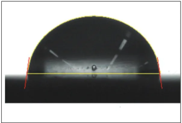
Wettability analyses were performed by contact angle measurement and surface free energy cal- culation. Contact angle measurements were per- formed with a contact angle measuring system from Phoenix 300 (Surface Electro Optics Co., Korea), equipped with a video charge-coupled device (CCD) camera and Image XP ver 5.6 soft- ware. Contact angle were measured with dis- tilled water, formamide, bromonaphtalene employ- ing the sessile drop method and subsequently sur- face free energy were calculated (Table I). Contact angles were obtained on the photographs as fol- lows: 10μl droplets of each liquid were posi- tioned on test specimens by means of a 1mL syringe with a blunt point. A tangent to the droplet was drawn from the point of air-fluid-sol- id phase intersection. Contact angles between this tangent line and the test specimen surface were calculated from enlarged photonegatives of the droplets (Fig. 1). Measurements of contact angle
Fig. 1.Contact angle measurement.
were performed five times each test solution at room temperature to provide adequate replications for statistical analysis. Surface free energy was cal- culated using the Lewis Acid/Base method for 3 liquids.
4. Statistical analysis
Experiments were repeated three to five times.
Statistical evaluation of data was performed using the software package SPSS/PC statisticsTM 13.0(SPSS Inc., Chicago, IL, USA). Data were expressed as mean values and standard deviation of each group of samples. Statistics were per- formed by analysis of variance (ANOVA), followed by and Scheffe’s method as a post-hoc compar- ison. A differences of a p<0.05 was considered to be statistically significant.
RESULTS
1. Surface morphology
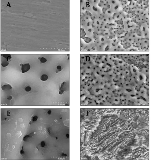
Figure 2 displays the finding of Scanning elec- tron microscopy (SEM) of disks. The machined disk revealed uniformly smooth surface with typi- cal machining grooves and ridge as produced by manufacturing instruments. The examination of anodized samples showed smooth surface exhibit- ing multiple micropores. The topography of the specimens in anodized and Ca-P deposited was
similar to that in group AN in terms of the num- ber and size of pores. However, tiny homoge- neously distributed crystalline particles were shown attached on specimens in group CaP/AN.
The SLA surface had a duplex surface morphol- ogy of a coarser microstructure and a finer microstructure. Much finer micropits were super- imposed onto the rougher indentations or cavities.
Micropits of the SLA surfaces had a sharp edge and pyramidal aspect of the protrusions.
2. Surface roughness
Table II showed results of the surface roughness measurements. The roughness parameters of disks showed significant differences in the Sa
values of each specimen (p < 0.05). The order of surface roughness values were, from the low- est to the highest, machined group, anodized group, anodized and Ca-P deposited group, and sandblasted and acid etched group.
3. X-ray diffraction (XRD)
Figure 3 showed the result of XRD analysis. The TF-XRD patterns of the machined Ti substrate and SLA surface expressed only titanium peaks. The TF-XRD pattern of anodized Ti substrate exhib- ited anatase and amorphous oxide. The TF-XRD patterns of group CaP/AN sample exhibited calcium phosphorous crystalline structure.
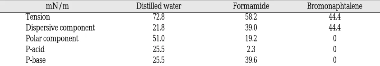
Table I. Surface energy components and parameters of test liquids used in contact angle and surface energy determination
mN/m Distilled water Formamide Bromonaphtalene
Tension 72.8 58.2 44.4
Dispersive component 21.8 39.0 44.4
Polar component 51.0 19.2 0
P-acid 25.5 2.3 0
P-base 25.5 39.6 0
4. Surface contact angle
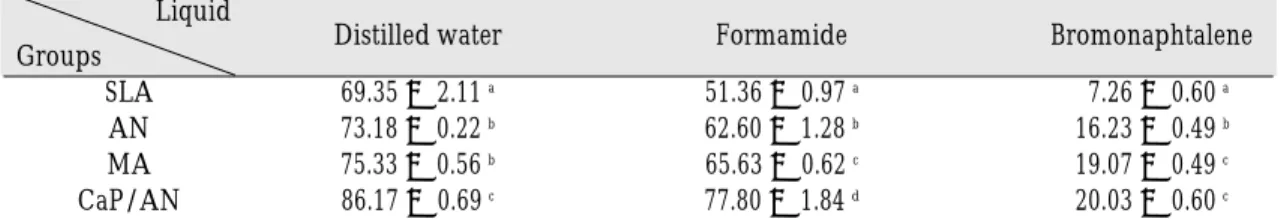
Table III gives the contact angles obtained by the sessile drop method on the different surfaces.
The mean contact angles increased in the order of group SLA, group AN, group MA, and group CaP/AN. There was no significant difference between group AN and group MA for water
contact angles, and significant difference between group MA and group CaP/AN for bromonaph- talene values (p < 0.05). There were significant dif- ferences among four groups for formamide val- ues (p < 0.05). In the group SLA, the shape of the drops was highly asymmetric and not stable during the measuring period.
Fig. 2.SEM photographs of each specimen: (A) machined (× 5,000), (B) anodized (× 5,000), (C) anodized (× 30,000), (D) anodized-CaP coated (× 5,000), (E) anodized-CaP coated (× 30,000), and (F) sand- blasted and acid etched surface (× 5,000).
A B
C D
E F
Table II. Surface roughness of the titanium disks
Group Sa(㎛) Smax(㎛)
MA 0.297 ± 0.006 a 3.071 ± 0.818 a
AN 0.549 ± 0.020 b 8.031 ± 0.737 b
CaP/AN 0.641 ± 0.087 c 8.740 ± 0.737 b
SLA 1.185 ± 0.033 d 14.646 ± 0.541 c
Data were expressed as mean values and standard deviations. Statistical significance was tested by ANOVA (p <
0.05). The same letters indicate nonsignificant differences between groups on Scheffe’s multiple comparison tests.
Table III. Contact angles (degree) of liquid drops on the samples Liquid
Groups Distilled water Formamide Bromonaphtalene
SLA 69.35 ± 2.11 a 51.36 ± 0.97 a 7.26 ± 0.60 a
AN 73.18 ± 0.22 b 62.60 ± 1.28 b 16.23 ± 0.49 b
MA 75.33 ± 0.56 b 65.63 ± 0.62 c 19.07 ± 0.49 c
CaP/AN 86.17 ± 0.69 c 77.80 ± 1.84 d 20.03 ± 0.60 c
Data were expressed as mean values and standard deviations. Statistical significance was tested by ANOVA (p <
0.05). The same letters indicate nonsignificant differences between groups on Scheffe’s multiple comparison tests.
Fig. 3. TF-XRD patterns of each specimen: (A) machined, (B) sandblasted and acid etched surface, (C) anodized, and (D) anodized-CaP coated surfaces.
Fig. 4. Surface Free Energy (mN/m) of the Samples:
Data were expressed as mean values and standard devi- ations. Statistical significance was tested by ANO- VA (p < 0.05). The same letters indicate nonsignificant differences between groups on Scheffe’s multiple comparison tests.
apatite anatase Ti
20 30 40 50 60
2ΘΘ/ Degree
Intensity
5. Surface free energy
Figure 4 showed the results of surface free energy (SFE). Values for surface free energy var- ied between 30.98 mN/m and 39.59 mN/m decreasing in the order SLA, AN, MA, and CaP/AN. There was no significant difference between group AN and group MA (p < 0.05).
DISCUSSION
This study was perform to characterize the surface properties which affect the biologic responses on surface of a new CaP/AN implant materials with a thin calcium phosphorus layer on titanium by means of anodization and ion beam- assisted deposition(IBAD). And their character- istics were compared with those of surfaces that prepared with different roughnesses and sur- face physicochemical properties. After anodiza- tion and IBAD treatment, the porous structure remained on the surface and many small crystals appeared. The crystals were calcium phosphorous, and they were about several hundred nanometers in size, which induced roughness on the micro- porous surface. The main composition of this oxide film is TiO2in anatase and amorphous forms. The contact angles for test liquids on anodized-CaP coated surface were higher than on other surfaces, and its surface free energy was low- er than that of cp-Ti.
Cell behavior on biomaterial surfaces depends upon implant cell interactions, correlated with sur- face properties. Surface chemical composition, roughness, texture, morphology, and hydrophilic- ity strongly affect cellular responses in contact with the implants.16-19 Several recent studies have shown that the surface energy of biomaterials strongly influences the initial cell attachment and spreading of osteoblastic cells on the bio- material surface.20-27Hallab et al.22thought that sur- face energy might be a more important determinant
of cell adhesion and proliferation, and might be more useful than surface roughness for generat- ing cell adhesion and cell colonization on the engineered tissue scaffolds. Den Braber et al.23eval- uated the effect of parallel surface microgrooves and surface energy on cell growth. The most significant conclusion was that physicochemi- cal parameters such as wettability and surface free energy influence cell growth but play no mea- surable role in the shape and orientation of cells on microtextured surfaces. Meyer et al.24quanti- fied the rate of cell attachment and revealed a con- stant dependence on a material within the first 7 h of culture. This may reflect the wettability.
After 24 h of incubation, however, measurement of cell attachment revealed less significant dif- ferences between the materials. Other research teams observed similar results. Webb et al.25have stated that, when material surfaces are exposed to dilute serum, fibroblast attachment and fibrob- last spreading are greater on hydrophilic sur- faces compared to hydrophobic surfaces. However, differences between the hydrophilic surfaces in water wettability influenced cell attachment but not spreading or cytoskeleton organization.
Redey et al.26 studied osteoclast adhesion and activity on synthetic hydroxyapatite, carbonated hydroxyapatite, and natural calcium carbonate and the relationship to surface free energies. Surface energy was found to play an essential role in osteoclast adhesion, whereas osteoclast spread- ing was found to depend on surface chemistry, especially on protein adsorption and newly formed apatite layers. Ponsonnet et al.27also sug- gested that fibroblast proliferation cannot be directly correlated to the water contact angles.
Roughness (in the micron range) seems to be the major parameter for cell proliferation. Indeed, only smooth surfaces (roughness less than 1㎛) showed significant proliferation, whatever the wet- tability.
In general, it has been hypothesized that an
increase in surface roughness results in an increase in contact angle. It was surprising that the high roughness did not always indicate the high con- tact angle. Lim et al.28confirmed that for cp Ti, the contact angle increased linearly with Ra when contact angle was greater than 45 degrees, while it decreased linearly with Ra when contact angle was less than 45 degrees. In present study, results for only CaP/AN group is agreement with the work of Lim et al. The SLA surfaces revealed highest roughness value among the samples but had a lowest contact angle. This result indicates that the SLA surface was more hydrophilic and exhib- ited markedly improved wettability. It seems that this parameter is related with the increase spe- cific surface area. And the open microtopography of SLA surface is more likely at the origin of more hydrophilicity, probably due to capillary forces. Because of the anisotropy of the differ- ent surface topographies in sandblasted surface, the shape of the drops was highly asymmetric and not stable during the measuring period. It was found that the water contact angle on SLA surface was strongly dependent on surface microtopog- raphy and micromorphology.29,30
The surface crystallography should also be considered as an important factor at the same time.
The contact angle appears to be related to the crys- talline structure of the oxide films formed on titanium and it alloys. Lim et al.28also suggested that the group in which the contact angle increased linearly with Rahad only a rutile type oxide lay- er, while the group that decreased linearly with Rahad a mixture of dominant rutile and anatase oxides. In our experiment, the crystalline structure of oxide film didn’t seem to affect the surface wet- tability. AN specimens had only anatase type oxides and CaP/AN specimens had anatase type oxides and calcium phosphorous crystalline structures. The wettability of AN group was not different with machined surface and that of CaP/AN was decreased. The chemical composition
and surface chemistry of titanium alloys con- tribute to these results. Other surface properties should also be considered as of importance for bio- logical response and may be more critical para- meters for biocompatibility than surface rough- ness itself.
Schakenraad et al.31concluded poor cell spread- ing of various cell types on low surface free energy substrata and good cell spreading on high surface free energy substrata. The inflection point of the curves, indicating the change from poor cell spreading to good cell spreading, is around 57 mN/m. This limit is higher than the SFE val- ues obtained in the present study and the curve corresponding to cell proliferation indicates that, before the inflection point in the 31.0-39.6 mN/m range, the relationship between SFE and prolif- eration can be reversed: proliferation can be higher for lower SFE in CaP/AN group. Webb et al.25and Ponsonnet et al.27considered that surfaces with a moderate wettability favor a higher cell adhesion behavior.
When dental root implant material is implant- ed into bone tissue, many biological reactions occur between its interface and the bone. The surfaces of dental root implants are usually in con- tact with a variety of ions, blood, serum pro- teins, bone morphogenetic protein, and noncol- lagenous bone proteins (e.g., bone sialoprotein, osteopontin, osteocalcin) which are present in the tissue fluid in vivo, and adsorption of these pro- teins is a function of the surface energy of the TiO2 matrix formed on the titanium surface and calcium phosphorous layer. Furthermore, the chemical or physical state of the surface may affect the adsorp- tion of ions and proteins that support cell attach- ment.32Adsorption of proteins always precedes to cell adhesion, which required for cellular adherence to the implant material surface, and wet- tability on the surface of the implant material is also thought to be important.24Van Oss33 stud- ied that proteins adsorb at significantly lower
concentrations to hydrophilic than to hydropho- bic substrates whereas cells prefer hydrophilic sur- faces. Groth and Altankov34studied the role of tyro- sine phosphorylation during fibroblast spreading on surfaces with different values of wettability. The authors concluded that the effect of hydrophobic substrata on cells with respect to adhesion and pro- liferation is due to a transfer of signals via integrins from the substratum to the cell interior. The pre- adsorption of fibronectin on hydrophobic substrata may provide better initial conditions, thus improv- ing the tissue compatibility of the material. In our study, anodization and IBAD treatments result- ed in a higher surface hydrophobicity of the original Ti surface. Calcium phosphate ceramics have been shown to have a very high adsorption capacity for serum proteins compared with oth- er materials.35-39It is well admitted that the cellular response to a material is only a secondary event and the presence of adsorbed protein layer is essential in mediating cell response to the mate- rial.
CONCLUSION
1. Anodized and calcium phosphate coated specimen showed multiple micropores and tiny homogeneously distributed crystalline par- ticles.
2. Calcium phosphate deposition induced rough- ness on the microporous anodized surface.
3. Anodized and calcium phosphate deposited group was found to have anatase type oxides and exhibited calcium phosphorous crys- talline structures.
4. Hydrophobicity was increased in the anodized and calcium phosphate deposited specimen.
Further study of cellular and tissue responses, particularly long-term studies of cell culture or ani- mal studies, would also help elucidate the improved biocompatibility and bioactivity in titanium modified by combining method of the
anodization and nano-scale calcium phosphate coating using ion beam-assisted deposition.
REFERENCES
1. Brunette DM, Tengvall P, Textor M, Thomsen P.
Titanium in medicine. Springer; Berlin; 2001.
2. Lausmaa J, Mattson L, Rolander U, Kasemo B.
Chemical composition and morphology of titani- um surface oxides. Mater Res Soc Symp Proc 1986;55:351-359.
3. Eisenbarth E, Velten D, Schenk-Meuser K, Linez P, Biehl V, Duschner H, Breme J, Hildebrand HF.
Interactions between cells and titanium surfaces.
Biomol Eng 2002;19:243-250.
4. Schreckenbach JP,Marx G, Schlottig F, Textor M,Spencer ND. Characterization of anodic spark- converted titanium surfaces for biomedical ap- plications. J Mater Sci Mater Med 1999;10:453-7.
5. Cook SD, Kay JF, Thomas KA, Jarcho M. Interface mechanics and histology of titanium and hy- droxyapatite-coated titanium for dental implant ap- plications. Int J Oral Maxillofac Implants 1987;2:15- 22.
6. Moroni A, Caja VL, Egger EL, Trinchese L, Chao EYS. Histomorphometry of hydroxyapatite coat- ed and uncoated porous titanium bone implants.
Biomaterials 1994;15:926-930.
7. Hulshoff JEG, van Dijk K, de.Ruijter JE, Rietveld FJR, Ginsel LA, Jansen JA. Interfacial phenomena:
An in vitro study of the effect of calcium phosphate (Ca-P) ceramic on bone formation. J Biomed Mater Res 1998;40:464-474.
8. de Brujin JD, Bovell YP, Davies JE, van Blitterswijik CA. Osteoclastic resorption of calcium phosphates in potentiated in postosteogenic culture. J Biomed Mater Res 1994;28:105-112.
9. Jung YC, Han CH, Lee IS, Kim HE. Effects of ion beam-assisted deposition of hydroxyapatite on the osseointegration of endosseous implants in rabbit tibiae. Int J Oral Maxillofac Implants 2001;16:809-818.
10. Park YS, Yi KY, Lee IS, Han CH, Jung YC. The Effects of Ion Beam Assisted Deposition of Hydroxyapatite on the Grit-blasted Surface of Endosseous Implants in Rabbit Tibiae. Int J Oral Maxillofac Implants 2005;20:31-3.
11. Keller JC, Dougherty WJ, Grotendorst GR, Wrightman JP. In vitro cell attachment to charac- terized cpTi surfaces. J Adhesion 1989;28:115-133.
12. Keller JC, Draughn RA, Wrightman JP, Dougherty WJ. Characterization of sterilized CP titanium implant surfaces. Int J Oral Maxillofac Implants 1990;
5:360-369.
13. Rich A, Harris AK. Anomalous preferences of cultured macrophages for hydrophobic and rough- ened substrata. J Cell Sci 1981;50:1-7.
14. Buser D, Schenk RK, Steinemann S, Fiorellini JP, Fox
CH, Stich H. Influence of surface characteristics on bone integration of titanium implants. A histo- morphometric study in miniature pigs. J Biomed Mater Res 1991;25:889-902.
15. van Kooten TG, Schakenraad JM, van der Mei HC, Busscher HJ. Influence of substratum wetta- bility on the strength of adhesion of human fi- broblasts. Biomaterials 1992;13(13):897-904.
16. Kipadi KL, Chang PL, Bellis SL. Hydroxyapatite binds more serum protein, purified integrins, and osteoblast precursor cells than titanium or steel. J Biomed Mater Res 2001;57:258-267.
17. Schwartz Z, Lohmann CH, Oefinger J, Bonewald LF, Dean DD, Boyan BD. Implant surface charac- teristics modulate differentiation behavior of cells in the osteoblastic lineage. Adv Dent Res 1999;13:38- 48.
18. Deligaianni DD, Katsala ND, Koutsoukos PG, Missirlis YF. Effect of surface roughness of hy- droxyapatite on human bone marrow cell adhesion, proliferation, differentiation, and detachment strength. Biomaterials 2001;22:87-96.
19. Hatton S, Andrade JD, Hibbs JB Jr, Gregonis DE, King RN. Fibroblast cell proliferation on charged hydroxyethyl methacrylate copolymers. J Colloid Interface Sci 1985;104:73-81.
20. Lampin M, Warocquier-Clerout R, Legris C, Degrange M, Sigot-Luizard MF. Correlation between substratum roughness and wettability, cell ad- hesion, and cell migration. J Biomed Mater Res 1997;36:99-108.
21. Redey SA, Nardin M, Bernache-Assolant D, Rey C, Delannoy P, Sedel L, Marie PJ. Behavior of human osteoblastic cells on stoichiometric hydroxyap- atite and type A carbonate apatite: role of sur- face energy. J Biomed Mater Res 2000;50:353-364.
22. Hallab NJ, Bundy KJ, O’Connor K, Moses RL, Jacobs JA. Evaluation of metallic and polymeric bio- material surface energy and surface roughness characteristics for directed cell adhesion. Tissue Eng 2001;7:55-71.
23. Den Braber ET, de Ruijter JE, Smits HTS, Ginsel LA, Von Recum AF, J.A. Jansen JA. Effect of parallel sur- face microgrooves and surface energy on cell growth, J. Biomed Mater Res 1995;29:511-518.
24. Meyer U, Szulczewski DH, Moller K, Heide H, Jones DB. Attachment kinetics and differentiation of osteoblasts on different biomaterials. Cell Mater 1993;3:129-140.
25. Webb K, Hlady V, Tresco P. Relative importance of surface wettability and charged functional groups on NIH 3T3 fibroblast attachment, spread- ing, and cytosquelette organization J Biomed Mater Res 1998;41:422-430.
26. Redey SA, Razzouk S, Rey C, Bernache-Assollant D, Leroy G, Nardin M, Cournot G. Osteoclast ad- hesion and activity on synthetic hydroxyapatite, car- bonated hydroxyapatite, and natural calcium car- bonate: relationship to surface energies. J Biomed Mater Res 1999;45:140-147.
27. Ponsonnet L, Reybier K, Jaffrezic N, Comte V, Lagneau C, Lissac M, Martelet C. Relationship between surface properties (roughness, wettabil- ity) of titanium and titanium alloys and cell behavior.
Materials Science and Engineering C 2003:23;551- 560.
28. Lim YJ, Oshida Y, Andres CJ, Barco MT. Surface Characterizations of Variously Treated Titanium Materials. Int J Oral Maxillofac Implants 2001;16:333- 342.
29. Rupp F, Scheideler L, Rehbein D, Axmann D, Geis-Gerstorfer J. Roughness induced dynamic changes of wettability of acid etched titanium implant modifications. Biomaterials 2004;25(7- 8):1429-38.
30. Cao H, Yang X, Wu D, Zhang X. Observation of topography and analysis of surface contamination of titanium implant after roughness treatment.
Sheng Wu Yi Xue Gong Cheng Xue Za Zhi 2007;
24(2):372-5.
31. Schakenraad JM, Busscher HJ, Wildevuur ChRH, Arends J. Thermodynamic aspects of cell spread- ing on solid substrata. J Cell Biophys 1988;13:75-91.
32. Ellingsen JE. A study on the mechanism of protein adsorption to TiO2. Biomaterials 1991;12:593-596.
33. Van Oss CJ. Hydrophobicity of biosurfaces-Origin, quantitative determination and interaction ener- gies. Colloids Surf B 1995;5:91-110.
34. Groth T, Altankov G, Studies on cell biomaterial in- teraction: role of tyrosine phosphorylation during fibroblasts spreading on surfaces varying in wet- tability, Biomaterials 1995;17:1227-1234.
35. Villareal DR, Sogal A, Ong JL. Protein adsorp- tion and ostebolast responses to different calcium phosphate surfaces. J Oral Implantol 1998;24:67-73.
36. Rosengren A, Pavlovic E, Oscarsson S, Krajewski A, Ravaglioli A, Piancastelli A. Plasma protein adsorption pattern on characterized ceramic bio- materials. Biomaterials 2002;23:1237-1247.
37. Hughes Wassell DT, Hall RC, Embery G.
Adsorption of bovine serum albumin onto hy- droxyapatite. Biomaterials 1995;16:697-702.
38. Takemoto S, Kusudo Y, Tsuru K, Hayakawa S, Osaka A, Takashima S. Selective protein adsorp- tion and blood compatibility of hydroxy-carbon- ate apatites. J Biomed Mater Res 2004;69A:544- 551.
39. Zheng H, Chittur KK, Lacefield WR. Analysis of bovine serum albumin adsorption on calcium phosphate and titanium surfaces. Biomaterials 1999; 20:377-384.
Reprint request to:
CHANG-WHEKIM, D.D.S., M.S.D., Ph.D.
DEPARTMENT OFPROSTHODONTICS,COLLEGE OFDENTISTRY, SEOULNATIONALUNIVERSITY
28-1, YEONGUN-DONG,CHONGNO-GU,SEOUL, 110-749, KOREA