ABSTRACT
Purpose: This study aimed to evaluate the biocompatibility and the mechanical properties of ultraviolet (UV) cross-linked and biphasic calcium phosphate (BCP)-added collagen membranes and to compare the clinical results of ridge preservation to those obtained using chemically cross-linked collagen membranes.
Methods: The study comprised an in vitro test and a clinical trial for membrane evaluation. BCP- added collagen membranes with UV cross-linking were prepared. In the in vitro test, scanning electron microscopy, a collagenase assay, and a tensile strength test were performed. The clinical trial involved 14 patients undergoing a ridge preservation procedure. All participants were randomly divided into the test group, which received UV cross-linked membranes (n=7), and the control group, which received chemically cross-linked membranes (n=7). BCP bone substitutes were used for both the test group and the control group. Cone-beam computed tomography (CBCT) scans were performed and alginate impressions were taken 1 week and 3 months after surgery. The casts were scanned via an optical scanner to measure the volumetric changes. The results were analyzed using the nonparametric Mann-Whitney U test.
Results: The fastest degradation rate was found in the collagen membranes without the addition of BCP. The highest enzyme resistance and the highest tensile strength were found when the collagen-to-BCP ratio was 1:1. There was no significant difference in dimensional changes in the 3-dimensional modeling or CBCT scans between the test and control groups in the clinical trial (P>0.05).
Conclusions: The addition of BCP and UV cross-linking improved the biocompatibility and the mechanical strength of the membranes. Within the limits of the clinical trial, the sites grafted using BCP in combination with UV cross-linked and BCP-added collagen membranes (test group) did not show any statistically significant difference in terms of dimensional change compared with the control group.
Keywords: Alveolar bone loss; Artificial membranes; Biocompatible materials;
Periodontal atrophy; Ultraviolet rays
Research Article
Received: Jan 10, 2020 Revised: Apr 4, 2020 Accepted: Apr 27, 2020
*Correspondence:
Sungtae Kim
Department of Periodontology, Dental Research Institute, Seoul National University School of Dentistry, 101 Daehak-ro, Jongno-gu, Seoul 03080, Korea.
E-mail: [email protected] Tel: +82-2-2072-4712 Fax: +82-2-744-0051
†Jung-Tae Lee and Dajung Lee contributed equally to this study.
Copyright © 2020. Korean Academy of Periodontology
This is an Open Access article distributed under the terms of the Creative Commons Attribution Non-Commercial License (https://
creativecommons.org/licenses/by-nc/4.0/).
ORCID iDs Jung-Tae Lee
https://orcid.org/0000-0001-5383-3004 Yoonsub Lee
https://orcid.org/0000-0001-5105-711X Dajung Lee
https://orcid.org/0000-0002-6977-6515 Yusang Choi
https://orcid.org/0000-0003-2369-3199 Jinyoung Park
https://orcid.org/0000-0002-3357-1964 Sungtae Kim
https://orcid.org/0000-0001-6361-4104
Jung-Tae Lee 1,†, Yoonsub Lee 2, Dajung Lee 2,†, Yusang Choi 3, Jinyoung Park 3, Sungtae Kim 2,*
1 Department of Periodontics, One-Stop Specialty Center, Seoul National University Dental Hospital, Seoul, Korea
2 Department of Periodontology, Dental Research Institute, Seoul National University School of Dentistry, Seoul, Korea
3Department of Bio Team, Implant Research Institute, Dentis Co., Ltd., Daegu, Korea
Evaluation of the mechanical properties and clinical efficacy of biphasic calcium phosphate-added collagen membrane in ridge preservation
Periodontal Science
Funding
This study was supported by a grant from the Korea Health Technology R&D Project through the Korea Health Industry Development Institute (KHIDI) funded by the Ministry of Health and Welfare, Republic of Korea (grant number: HI15C1535).
Author Contributions
Conceptualization: Sungtae Kim; Formal analysis: Jung-Tae Lee, Dajung Lee;
Investigation: Jung-Tae Lee, Yoonsub Lee, Dajung Lee, Yusang Choi, Jinyoung Park; Methodology: Sungtae Kim; Project administration: Sungtae Kim; Writing - original draft: Jung-Tae Lee, Yoonsub Lee, Dajung Lee;
Writing - review & editing: Sungtae Kim.
Conflict of Interest
No potential conflict of interest relevant to this article was reported.
INTRODUCTION
Following tooth extraction, atrophic changes in the alveolar bone are found at the extracted sites [1,2]. The horizontal dimensional change (29%–63%) is more distinct during healing of the alveolar bone than the vertical dimensional change (11%–22%) [3]. Furthermore, alveolar ridge resorption is more pronounced in the buccal plate than in the lingual plate at post- extraction sites [2,4]. An especially noticeable dimensional change is expected when some parts of the bony walls are missing [5,6]. In periodontally compromised teeth with missing parts of the bony walls, a more significant dimensional change can be expected following extraction. Restorative treatment becomes more challenging due to this characteristic change in the alveolar ridge following tooth loss [1,7].
Ridge preservation has been performed to reduce the dimensional changes in the alveolar ridge following tooth extraction [8,9]. Post-extraction dimensional changes decreased when ridge preservation was performed [3,10]. With the help of ridge preservation, more favorable clinical outcomes can be expected, and an ideal implant position for an esthetically/
functionally acceptable restoration can be achieved [11,12]. Various bone graft materials and surgical methods have been introduced to increase the predictability of clinical results.
Bone grafting is performed to compensate for the dimensional decrease of the alveolar ridge following tooth extraction by maintaining space that is replaced with newly formed bone [13].
A previous study demonstrated that, irrespective of the type of bone grafting material used, ridge preservation was more effective with bone grafting than without [14]. In guided tissue regeneration, barrier membranes have been used to prevent the unwanted rapid growth of epithelial cells into periodontal defects [15,16]. These membranes could also be effective for securing grafted particles in the extraction sockets during ridge preservation procedures [17,18].
Resorbable collagen membranes have one major disadvantage: a faster degradation rate through enzymatic activity, which could result in the early collapse of the ridge contour and epithelial ingrowth [19]. Therefore, a cross-linking procedure is required to prolong the degradation of the resorbable membrane. Various cross-linking methods have been developed to this end. Enzymatically and chemically cross-linked membranes have exhibited a slower degradation rate than non-cross-linked membranes [19]. However, various cytotoxic effects of chemically cross-linked membranes (glutaraldehyde and formaldehyde) have been reported, including interference with attachment, the proliferation of periodontal ligament fibroblasts, and reduced biocompatibility [20,21]. Other cross-linking methods have thus been developed to overcome the weak points of chemical cross-linking.
Physical cross-linking methods with ultraviolet (UV) irradiation or dehydrothermal (DHT) treatment have been developed as alternatives. Unlike chemically cross-linked membranes, physically cross-linked membranes do not have any residue. UV irradiation (254 nm) on collagen membranes initiates free radical formation, which triggers cross-linking, whereas DHT treatment on collagen membranes removes bound water molecules from the collagen molecules. The resulting condensation reactions strengthen the collagen membranes.
The mechanical properties (tensile strength and resistance to degradation) of collagen membranes improve with UV and DHT treatments [22,23].
Biphasic calcium phosphate (BCP) has been used successfully for ridge preservation procedures [24]. BCP is composed of hydroxyapatite (HA) and beta-tricalcium phosphate
(β-TCP). BCP is widely used in the reconstruction of bone defects as a healing biomaterial, due to its similar composition to the inorganic components of natural bone [25]. β-TCP is resorbed early during the healing phase and is replaced by newly formed bone, whereas HA is resorbed slowly and can be used as an effective scaffold for new bone formation. To achieve the optimal degradation rate of BCP for each clinical situation, the HA/β-TCP ratio can be changed [26]. In fact, various HA/β-TCP ratios can be applied for manufacturing BCP-added collagen membranes. Collagen is one of the most widely used scaffold materials based on its excellent biocompatibility and biodegradability [27]. A previous study showed that the surface area of the collagen scaffold could be increased by combining it with HA [28]. This binding procedure resulted in gradual cell adhesion. Another study demonstrated that a nano-TCP-coated collagen scaffold had high compressive strength [29].
This study aimed to evaluate the biocompatibility and mechanical properties of UV cross- linked and BCP-added collagen membranes and to compare the clinical results of ridge preservation to those achieved using chemically cross-linked collagen membranes.
MATERIALS AND METHODS
Materials
A 1% concentration of type I atelocollagen solution (SK Bioland Company, Cheonan, Korea) was stirred at room temperature for 2 hours and then kept overnight at 4°C. BCP (Ovis BCP, Dentis Co., Ltd., Daegu, Korea) (HA:β-TCP=20:80) was mixed with the type I atelocollagen solution. The BCP/atelocollagen composite was then stirred at room temperature for 2 hours before being placed into a 90-mm-diameter polystyrene Petri dish and lyophilized at
−45°C for 64 hours. The BCP/atelocollagen sheets were pressed with 10 tons of pressure for 10 s using a presser (SAIT, Incheon, Korea) and were exposed to UV light (254 nm, 28 mW/
cm2) for 3 minutes with a distance of 85 mm in a UV cross-linker (AhTech LTS, Anyang, Korea). The cross-linked BCP/atelocollagen sheets were then treated with 0.1 M NaOH for 30 minutes and washed with distilled water 5 times for 15 minutes each. The cross-linked BCP- atelocollagen sheets were also lyophilized at −45°C for 24 hours and then pressed with 10 tons of pressure for 10 seconds. Membrane samples with different collagen-to-BCP ratios (1:1, 2:1, only collagen) were prepared (total n=9).
In vitro tests
Scanning electron microscopy (SEM) observations
The membrane surface and cross-sectional images were characterized through SEM (ALS2300C, Ceron Technology Corporation, Uiwang, Korea) with an accelerating voltage of 15–20 kV.
Collagenase assay
Phosphate-buffered saline (PBS) solution (0.025 M; pH 7.4) was prepared (Welgene, Daegu, Korea). The acidity was adjusted to a pH of 7.0 with the addition of 0.36 mM CaCl2 solution.
The weight of the Petri dish and membrane used in the collagenase test was measured (125 collagen digestion units [CDU]/mg, C0130-1G, Sigma-Aldrich, St. Louis, MO, USA). Five CDU of collagenase per milliliter was prepared with 0.025 M PBS. Samples of the membrane (n=9) were also prepared by adding 0.025 M PBS at a rate of 2 cm2/mL. The membrane samples in the Petri dishes were treated with enzymes at 100 rpm in a 37°C dryer for 3 hours. After confirming the shape of the membranes, the supernatant was removed. These samples were dried in a dryer at 60°C for 14 hours. Enzyme resistance was calculated by measuring the
weight difference of the membrane before and after enzyme treatment. These samples were measured at 1-hour intervals.
Tensile strength test
UV cross-linked membranes with different collagen-to-BCP ratios were subjected to standard, uniaxial tensile tests. The specimens were fixed to the bottom of a tensile strength tester jig (model LRXPlus-E, Lloyd Instruments, Bognor Regis, England). The tensile strength of each specimen (n=9) was measured under strain at a rate of 1 mm/min up to the breaking point.
Clinical trial Study population
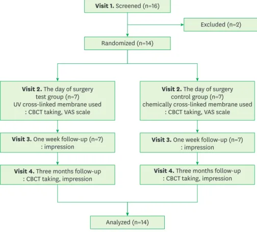
Fourteen patients visited the Department of Periodontology of Seoul National University Dental Hospital in Seoul, Korea from June 2017 to April 2018. All participants were at least 20 years of age and had bony defects in posterior molars in the maxilla or the mandible as a result of chronic periodontitis. Patients with uncontrolled hypertension or diabetes, pregnancy, systemic diseases contraindicating the use of surgical treatment, hyperthyroidism, or hypothyroidism were excluded. Two of the original 16 patients were excluded after withdrawing their consent and refusing an appointment after they had been screened. None of the subsequently enrolled patients dropped out (Figure 1). The patients' general information (e.g., age, sex, and jaw position) is shown in Table 1.
Visit 1. Screened (n=16)
Randomized (n=14)
Excluded (n=2)
Analyzed (n=14) Visit 2. The day of surgery
test group (n=7) UV cross-linked membrane used
: CBCT taking, VAS scale
Visit 3. One week follow-up (n=7) : impression
Visit 4. Three months follow-up : CBCT taking, impression
Visit 3. One week follow-up (n=7) : impression
Visit 4. Three months follow-up : CBCT taking, impression Visit 2. The day of surgery
control group (n=7)
chemically cross-linked membrane used : CBCT taking, VAS scale
Figure 1. Procedure flowchart.
CBCT: cone-beam computed tomography, VAS: visual analog scale.
Surgical procedure and postoperative care
This research was designed as a pilot clinical trial. The design was approved by the Research Ethics Committee of Seoul National University Dental Hospital (Approval No. CDE17001).
All protocols were performed according to the Declaration of Helsinki and the Consolidated Standards of Reporting Trials 2010 statements. Written informed consent was obtained from all the patients, who voluntarily participated in the study. The surgical procedure in this study, except the membranes and the follow-up periods, was the same as in a previous study [30].
In the test group, UV cross-linked membranes (Ovis BCP/Collagen MEMBRANE, Dentis Co., Ltd.) were applied, whereas chemically cross-linked membranes (collagen membrane, Genoss, Suwon, Korea) were applied in the control group. A total of 14 participants were randomly divided into test (n=7) and control groups (n=7). The test and control groups were assigned at a ratio of 1:1, with a pre-generated randomization table and a number of identification codes. The patients, clinicians, and investigators had no information about the assigned groups.
One periodontist (S.T.K) performed all the surgical procedures. The patients received oral hygiene instructions before surgery. The surgical sites were treated under local anesthesia (2% lidocaine with 1:100,000 epinephrine). With no flap elevation, the teeth were extracted, and the extraction sockets were then curetted with the removal of any tissue remnants. The buccal bone of the extraction sockets was found to be either intact or partially resorbed.
The sockets were irrigated with normal saline solution, filled with bone graft material (Ovis BONE BCP, Dentis Co., Ltd.) and covered with a membrane. A releasing incision was not performed at the site, and then secondary healing was induced.
Amoxicillin, aceclofenac, and chlorhexidine were administered to all participants for 3 to 5 days. Stitch-out was performed 7 to 10 days after surgery. The patients were recalled at 1 week (visit 3, V3) and at 3 months (visit 4, V4) after surgery for the measurements.
Evaluation of dimensional changes through 3-dimensional model scanning
A study model was created using alginate impressions at 1 week (V3) and at 3 months (V4) after surgery. An optical scanner (Freedom, DOF, Seoul, Korea) was used to scan the casts, whereas computer-aided design software (DentalCAD, EGS, Lazzaro, Italy) was used for the visualization and analysis. Software (Polyworks, Innovmetric, Quebec, Canada) was Table 1. General characteristics
Characteristics Test group Control group P value
Number 7 7
Age, (yr) 59.4±6.4 52.9±17.0 0.620
Sex 0.383
Male 2 (33.3) 4 (66.7)
Female 5 (62.5) 3 (37.5)
Jaw position 0.383
Upper 5 (62.5) 3 (37.5)
Lower 2 (33.3) 4 (66.7)
Right/left 0.710
Right 3 (57.1) 4 (42.9)
Left 4 (42.9) 3 (57.1)
Values are presented as mean±standard deviation or number (%). Test group: ultraviolet cross-linked membranes; control group: chemically cross-linked membranes.
also used to superimpose the V3 and V4 models of each patient. A region of interest (ROI) was determined at the upper-middle region of the gingiva (the mucogingival junction). The ROI was applied on the image of the V3 model. The mean value of the surface vector was then calculated, and the ROI was scanned perpendicularly to this vector. The volume was then measured. The V4 model was measured in the same manner as with the V3 model.
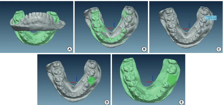
Dimensional changes were calculated by dividing the difference between the volumes of the V3 and V4 models by the surface vector value (Figure 2).
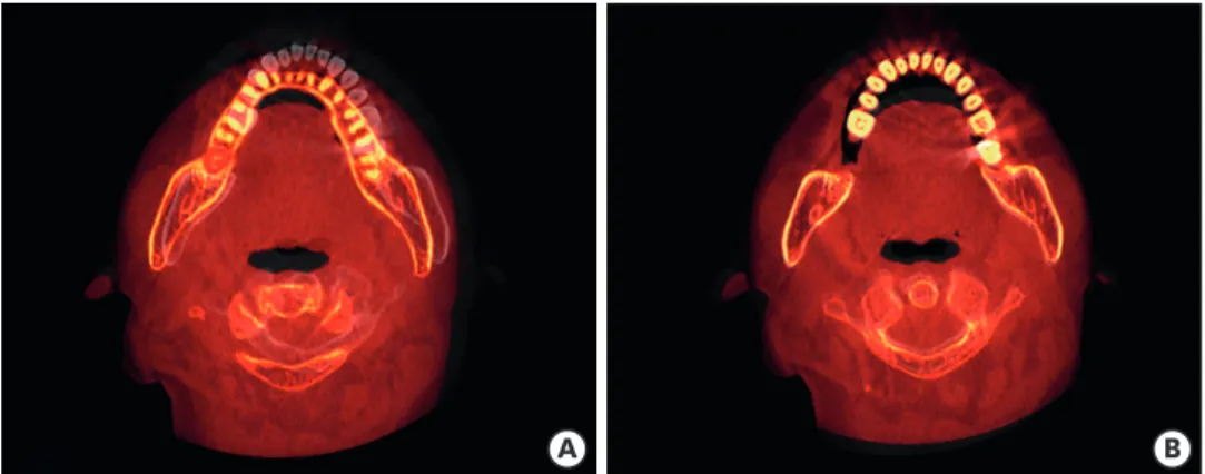
Evaluation of bone tissue quantity by cone-beam computed tomography (CBCT) CBCT images with a scan time of 7 seconds at 120 kV and 10 mA were taken on the day of operation (visit 2, V2) and 3 months after V2 (V4). OnDemand3D software (Cybermed Inc., Daejeon, Korea) was used to correct any positional discrepancies between the V2 and V4 CBCT images. The 2 images were merged and the merged image was resliced with a resolution of 0.1 mm. The V4 images were then adjusted to the V2 position. The V2 and V4 images in the same range were set through 3-dimensionally reconstructed processes. The amount of change between the 2 different time points was measured using the volume ratio of the divided regions (Figure 3).
Data analysis
This study compared UV cross-linked and chemically cross-linked membranes. Due to the small sample size, all analyses were conducted using the nonparametric Mann-Whitney U test (SPSS version 25.0, IBM Corp., Armonk, USA). The cutoff for significance was a P value of 0.05.
A B C
D E
Figure 2. ROI and calibration of the dimensional changes between V3 and V4 in the scanned models. (A) Scanned models before superimposition (gray: V3 scan data, green: V4 scan data). (B) Scanned models after superimposition. (C) ROI in the V3 model. (D) Volume of the ROI in the V3 model. (E) Volume of the ROI in the V4 model.
ROI: region of interest, V3: visit 3, V4: visit 4.
RESULTS
In vitro tests SEM observations
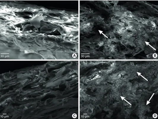
The collagen membranes with and without BCP showed a dense porous structure. BCP particles filled the porous space in the collagen membranes with BCP (Figures 4 and 5).
A B
Figure 3. Compensation of the CBCT data obtained on different dates. (A) Two axial views of the CBCT scan at V3 and V4 before superimposition; the primary data (V2) of the gray tone did not match the secondary data (V4) of the red tone. (B) Two axial views of the CBCT scan after superimposition.
CBCT: cone-beam computed tomography, V2: visit 2, V3: visit 3, V4: visit 4.
A B
C D
10 µm 10 µm
5 µm 5 µm
Figure 4. Scanning electron microscopy observations of the membrane surface. (A) Collagen membrane with no BCP added. (B) Collagen membrane with BCP added at the same ratio as collagen. (C) Collagen membrane with no BCP added. (D) Collagen membrane with BCP added at the same ratio as collagen. The arrow indicates BCP particles.
BCP: biphasic calcium phosphate.
Collagenase assay
The collagen membranes with BCP were divided according to the collagen-to-BCP ratio. The fastest degradation rate was found in the collagen membranes with no BCP. The collagen membrane with a collagen-to-BCP ratio of 1:1 exhibited the highest enzyme resistance (Figure 6).
A B
C D
30 µm 30 µm
10 µm 10 µm
Figure 5. Scanning electron microscopy observations of the membrane cross-section. (A) Collagen membrane with no BCP added. (B) Collagen membrane with BCP added at the same ratio as collagen. (C) Collagen membrane with no BCP added. (D) Collagen membrane with BCP added at the same ratio as collagen. The arrow indicates BCP particles.
BCP: biphasic calcium phosphate.
0 30 40 60 50 80 70
20 10 90
Only collagen Collagen-to-BCP ratio
2:1 Collagen-to-BCP ratio 1:1 Collagenase (%)
Figure 6. Collagenase assay of ultraviolet cross-linked collagen membranes with 3 different collagen-to-BCP ratios. The fastest degradation rate was found in the collagen membrane with no BCP, whereas the collagen-to- BCP ratio of 1:1 exhibited the highest enzyme resistance.
BCP: biphasic calcium phosphate.
Tensile strength test
It was observed that preparing collagen and BCP at a ratio of 1:1 resulted in the maximum load value and tensile strength (Figure 7).
Clinical trial
Evaluation of dimensional changes through 3-dimensional model scanning
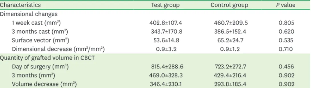
The dimensional changes in the study models are shown in Table 2. The volume tended to decrease in both the test and control groups. The mean of the dimensional decrease between the V3 and V4 models was not significantly different between the test (0.9±3.2 mm3/mm2) and control groups (0.9±1.2 mm3/mm2) (P>0.05).
Evaluation of bone tissue quantity by CBCT
The results for the normalized quantity of bone tissue in the CBCT data are presented in Table 2. The mean of the dimensional decrease did not significantly differ between the test (346.4±230.1 mm3) and control groups (293.8±185.4 mm3) (P>0.05). There was less bone tissue at V4 than at V2 in both the test and control groups.
DISCUSSION
The present study evaluated the biocompatibility and the mechanical properties of UV cross-linked and BCP-added collagen membranes and compared the clinical results of ridge
0 15 20 25
10
5
Only collagen Collagen-to-BCP ratio
2:1 Collagen-to-BCP ratio 1:1 Maximum load (n)
Tensile strength (Mpa)
Figure 7. Tensile strength test of the ultraviolet cross-linked membrane with 3 different collagen-to-BCP ratios.
The collagen-to-BCP ratio of 1:1 exhibited the highest tensile strength.
BCP: biphasic calcium phosphate.
Table 2. Clinical changes after ridge preservation
Characteristics Test group Control group P value
Dimensional changes
1 week cast (mm3) 402.8±107.4 460.7±209.5 0.805
3 months cast (mm3) 343.7±170.8 386.5±152.4 0.620
Surface vector (mm2) 53.6±14.8 65.2±24.7 0.535
Dimensional decrease (mm3/mm2) 0.9±3.2 0.9±1.2 0.710
Quantity of grafted volume in CBCT
Day of surgery (mm3) 815.4±288.6 723.2±272.7 0.456
3 months (mm3) 469.0±328.3 429.4±216.4 0.902
Volume decrease (mm3) 346.4±230.1 293.8±185.4 0.902
Values are presented as mean±standard deviation. Test group: ultraviolet cross-linked membranes; control group: chemically cross-linked membranes.
CBCT: cone-beam computed tomography.
preservation with those achieved using chemically cross-linked collagen membranes. In the in vitro test, dense porous structures were shown in the collagen membranes both with and without BCP. The collagen membrane with a collagen-to-BCP ratio of 1:1 exhibited the highest enzyme resistance and tensile strength. Model scanning and CBCT scans showed volumetric reductions in both the test and control groups, but with no significant difference between the groups.
In this clinical study, ridge preservation with bone grafting and collagen membranes led to reductions in the vertical and horizontal dimensions of the ridge. This finding could be explained in light of the results of previous studies [31,32]. Neither of the membranes could completely prevent resorption of the alveolar bone after extraction of the tooth. Volumetric changes of the edentulous ridge were evaluated in the present study. No significant differences were found between the 2 types of membranes with respect to changes in the volume, width, or height of the edentulous alveolar ridge after tooth extraction. These results indicate that both types of membrane for ridge preservation could successfully prevent the ingrowth of soft tissue and the collapse of the contour of the buccal/coronal area of the alveolar ridge, even if the bone graft contributed more than the membrane. The dimensional changes of the alveolar bone were compensated for through this ridge preservation procedure using barrier membranes. In the extraction sockets, both membranes remained in the sockets long enough to secure the bone graft material in position and to prevent soft tissue ingrowth.
The duration of the UV irradiation affects cross-linking. The BCP/atelocollagen sheets were exposed to 254 nm UV radiation for 3 minutes in the present study. Previous studies reported that residues capable of cross-linking were affected by UV irradiation within 30 minutes and that UV irradiation beyond 30 minutes did not affect further cross-linking [23]. After more than 4 hours of UV irradiation, collagen molecules were degraded [22]. BCP was applied to control the degradation time of the collagen membranes by modifying the HA/β-TCP ratio.
Given the in vitro test results, a 1-to-1 ratio of collagen and BCP was used, which ensured maximum tensile strength and maximum resistance to resorption. The membrane used in this study consisted of HA and β-TCP at a 20:80 ratio. A recent study applying the same membrane as this study demonstrated that the membrane showed high biocompatibility and resistance to degradation [33]. A study of a mouse model using the same proportion of BCP materials combined with human mesenchymal stem cells demonstrated that these materials resulted in stem cell-induced bone formation [34].
Most previous studies have evaluated ridge alteration after ridge preservation with direct measurements (e.g., changes in buccolingual width and vertical height) [35,36]. In the present study, we evaluated ridge alteration 3-dimensionally by superimposing the data obtained through CBCT and 3-dimensional scanner imaging, which allowed for detailed comparisons. The bone quantity in the CBCT images decreased from baseline to 3 months in both groups. This decrease is in line with the results of previous studies [35,37].
Considering the small sample size and the risk of the normality test being underpowered, a similar study (bone grafting) with the same participants was used to set the sample size [38].
Another study of this issue was also referenced [39]. Nonetheless, the present pilot study showed that a newly devised membrane might be a good candidate for ridge preservation.
It could be concluded that the biocompatibility and the mechanical strength of collagen membranes improved after the addition of BCP and UV cross-linking. The findings of this study will provide a basis for identifying a proper target population in future research.
Within the limits of the clinical trial, the sites grafted using BCP in combination with UV cross-linked and BCP-added collagen membranes did not show any statistically significant difference in terms of dimensional change compared with the control group.
REFERENCES
1. Schropp L, Wenzel A, Kostopoulos L, Karring T. Bone healing and soft tissue contour changes following single-tooth extraction: a clinical and radiographic 12-month prospective study. Int J Periodontics Restorative Dent 2003;23:313-23.
PUBMED
2. Araújo MG, Lindhe J. Dimensional ridge alterations following tooth extraction. An experimental study in the dog. J Clin Periodontol 2005;32:212-8.
PUBMED | CROSSREF
3. Tan WL, Wong TL, Wong MC, Lang NP. A systematic review of post-extractional alveolar hard and soft tissue dimensional changes in humans. Clin Oral Implants Res 2012;23 Suppl 5:1-21.
PUBMED | CROSSREF
4. Sanz M, Cecchinato D, Ferrus J, Pjetursson EB, Lang NP, Lindhe J. A prospective, randomized-controlled clinical trial to evaluate bone preservation using implants with different geometry placed into extraction sockets in the maxilla. Clin Oral Implants Res 2010;21:13-21.
PUBMED | CROSSREF
5. Araújo MG, Lindhe J. Ridge preservation with the use of Bio-Oss collagen: a 6-month study in the dog.
Clin Oral Implants Res 2009;20:433-40.
PUBMED | CROSSREF
6. Lee JS, Jung JS, Im GI, Kim BS, Cho KS, Kim CS. Ridge regeneration of damaged extraction sockets using rhBMP-2: an experimental study in canine. J Clin Periodontol 2015;42:678-87.
PUBMED | CROSSREF
7. Zhao L, Wei Y, Xu T, Zhang B, Hu W, Chung KH. Changes in alveolar process dimensions following extraction of molars with advanced periodontal disease: a clinical pilot study. Clin Oral Implants Res 2019;30:324-35.
PUBMED | CROSSREF
8. Araújo MG, Lindhe J. Socket grafting with the use of autologous bone: an experimental study in the dog.
Clin Oral Implants Res 2011;22:9-13.
PUBMED | CROSSREF
9. Barone A, Ricci M, Tonelli P, Santini S, Covani U. Tissue changes of extraction sockets in humans: a comparison of spontaneous healing vs. ridge preservation with secondary soft tissue healing. Clin Oral Implants Res 2013;24:1231-7.
PUBMED
10. Vignoletti F, Matesanz P, Rodrigo D, Figuero E, Martin C, Sanz M. Surgical protocols for ridge preservation after tooth extraction. A systematic review. Clin Oral Implants Res 2012;23 Suppl 5:22-38.
PUBMED | CROSSREF
11. Park SH, Song YW, Sanz-Martín I, Cha JK, Lee JS, Jung UW. Clinical benefits of ridge preservation for implant placement compared to natural healing in maxillary teeth: a retrospective study. J Clin Periodontol 2020;47:382-91.
PUBMED | CROSSREF
12. Ramanauskaite A, Borges T, Almeida BL, Correia A. Dental implant outcomes in grafted sockets: a systematic review and meta-analysis. J Oral Maxillofac Res 2019;10:e8.
PUBMED | CROSSREF
13. Dahlin C, Linde A, Gottlow J, Nyman S. Healing of bone defects by guided tissue regeneration. Plast Reconstr Surg 1988;81:672-6.
PUBMED | CROSSREF
14. Avila-Ortiz G, Chambrone L, Vignoletti F. Effect of alveolar ridge preservation interventions following tooth extraction: a systematic review and meta-analysis. J Clin Periodontol 2019;46 Suppl 21:195-223.
PUBMED | CROSSREF
15. Nyman S, Gottlow J, Karring T, Lindhe J. The regenerative potential of the periodontal ligament. An experimental study in the monkey. J Clin Periodontol 1982;9:257-65.
PUBMED | CROSSREF
16. Nyman S, Lindhe J, Karring T, Rylander H. New attachment following surgical treatment of human periodontal disease. J Clin Periodontol 1982;9:290-6.
PUBMED | CROSSREF
17. Barone A, Aldini NN, Fini M, Giardino R, Calvo Guirado JL, Covani U. Xenograft versus extraction alone for ridge preservation after tooth removal: a clinical and histomorphometric study. J Periodontol 2008;79:1370-7.
PUBMED | CROSSREF
18. Jung RE, Ioannidis A, Hämmerle CH, Thoma DS. Alveolar ridge preservation in the esthetic zone.
Periodontol 2000 2018;77:165-75.
PUBMED | CROSSREF
19. Rothamel D, Schwarz F, Sager M, Herten M, Sculean A, Becker J. Biodegradation of differently cross- linked collagen membranes: an experimental study in the rat. Clin Oral Implants Res 2005;16:369-78.
PUBMED | CROSSREF
20. Bunyaratavej P, Wang HL. Collagen membranes: a review. J Periodontol 2001;72:215-29.
PUBMED | CROSSREF
21. Rothamel D, Schwarz F, Sculean A, Herten M, Scherbaum W, Becker J. Biocompatibility of various collagen membranes in cultures of human PDL fibroblasts and human osteoblast-like cells. Clin Oral Implants Res 2004;15:443-9.
PUBMED | CROSSREF
22. Weadock KS, Miller EJ, Bellincampi LD, Zawadsky JP, Dunn MG. Physical crosslinking of collagen fibers:
comparison of ultraviolet irradiation and dehydrothermal treatment. J Biomed Mater Res 1995;29:1373-9.
PUBMED | CROSSREF
23. Lee JE, Park JC, Hwang YS, Kim JK, Kim JG, Suh H. Characterization of UV-irradiated dense/porous collagen membranes: morphology, enzymatic degradation, and mechanical properties. Yonsei Med J 2001;42:172-9.
PUBMED | CROSSREF
24. Sohn B, Hwang M, Kim S, Kim HI, Ku Y. Ridge preservation using basic fibroblast growth factor-2 and collagenated biphasic calcium phosphate in beagle dogs. J Periodontal Implant Sci 2017;47:381-7.
PUBMED | CROSSREF
25. Cai H, Yao Y, Xu Y, Wang Q, Zou W, Liang J, et al. A Col I and BCP ceramic bi-layer scaffold implant promotes regeneration in osteochondral defects. RSC Adv 2019;9:3740-8.
CROSSREF
26. Bouler JM, Pilet P, Gauthier O, Verron E. Biphasic calcium phosphate ceramics for bone reconstruction: a review of biological response. Acta Biomater 2017;53:1-12.
PUBMED | CROSSREF
27. Zhang D, Wu X, Chen J, Lin K. The development of collagen based composite scaffolds for bone regeneration. Bioact Mater 2017;3:129-38.
PUBMED | CROSSREF
28. Sionkowska A, Kozłowska J. Properties and modification of porous 3-D collagen/hydroxyapatite composites. Int J Biol Macromol 2013;52:250-9.
PUBMED | CROSSREF
29. Ibara A, Miyaji H, Fugetsu B, Nishida E, Takita H, Tanaka S, et al. Osteoconductivity and biodegradability of collagen scaffold coated with nano-β-TCP and fibroblast growth factor 2. J Nanomater 2013;2013:1-11.
CROSSREF
30. Chang H, Kim S, Hwang JW, Kim S, Koo KT, Kim TI, et al. Comparative, randomized, double-blind clinical study of alveolar ridge preservation using an extracellular matrix-based dental resorbable membrane in the extraction socket. J Periodontal Implant Sci 2017;47:165-73.
PUBMED | CROSSREF
31. Marconcini S, Giammarinaro E, Derchi G, Alfonsi F, Covani U, Barone A. Clinical outcomes of implants placed in ridge-preserved versus nonpreserved sites: a 4-year randomized clinical trial. Clin Implant Dent Relat Res 2018;20:906-14.
PUBMED | CROSSREF
32. Barone A, Orlando B, Cingano L, Marconcini S, Derchi G, Covani U. A randomized clinical trial to evaluate and compare implants placed in augmented versus non-augmented extraction sockets: 3-year results. J Periodontol 2012;83:836-46.
PUBMED | CROSSREF
33. Hong I, Khalid AW, Pae HC, Cha JK, Lee JS, Paik JW, et al. Distinctive bone regeneration of calvarial defects using biphasic calcium phosphate supplemented ultraviolet-crosslinked collagen membrane. J Periodontal Implant Sci 2020;50:14-27.
PUBMED | CROSSREF
34. Arinzeh TL, Tran T, Mcalary J, Daculsi G. A comparative study of biphasic calcium phosphate ceramics for human mesenchymal stem-cell-induced bone formation. Biomaterials 2005;26:3631-8.
PUBMED | CROSSREF
35. Cardaropoli D, Tamagnone L, Roffredo A, Gaveglio L, Cardaropoli G. Socket preservation using bovine bone mineral and collagen membrane: a randomized controlled clinical trial with histologic analysis. Int J Periodontics Restorative Dent 2012;32:421-30.
PUBMED
36. Jung RE, Philipp A, Annen BM, Signorelli L, Thoma DS, Hämmerle CH, et al. Radiographic evaluation of different techniques for ridge preservation after tooth extraction: a randomized controlled clinical trial. J Clin Periodontol 2013;40:90-8.
PUBMED | CROSSREF
37. Thalmair T, Fickl S, Schneider D, Hinze M, Wachtel H. Dimensional alterations of extraction sites after different alveolar ridge preservation techniques - a volumetric study. J Clin Periodontol 2013;40:721-7.
PUBMED | CROSSREF
38. Le B, Borzabadi-Farahani A, Nielsen B. Treatment of labial soft tissue recession around dental implants in the esthetic zone using guided bone regeneration with mineralized allograft: a retrospective clinical case series. J Oral Maxillofac Surg 2016;74:1552-61.
PUBMED | CROSSREF
39. Dall' Acqua AM, Sachetti A, Santos LJ, Lemos FA, Bianchi T, Naue WS, et al. Use of neuromuscular electrical stimulation to preserve the thickness of abdominal and chest muscles of critically ill patients: a randomized clinical trial. J Rehabil Med 2017;49:40-8.
PUBMED | CROSSREF