S
ince the long-term high success rate of the screw type implants was documented in the lit- eratures, osseointegrated implants has become one of the reliable treatment options in dentistry1,2. After osseointegration occurred, maintenance of osseoin- tegration is the key factor for long-term clinical suc- cess of the implants. Bone is a living tissue that remodels, i.e. changes its geometry, density, and structure. Bone remodelling is a sequence of bio- logical processes which are mediated by a num- ber of factors, mechanical load, hormonal effects,nutrition and neuronal influences3. The effect of mechanical loading to bone was well described by Wolff and Frost4. He stated that the change in the function of a bone causes a change in the internal and external conformation of the bone. This process called bone adaptation. Not only the strain level, but also the rate of strain change, the number of strain cycles and the strain distribution change influence the bone response5,6.
It is very difficult to analyze actual clinical conditions in in vitro experiments. Bone adaptation J Korean Acad Prosthodont : Volume 38, Number 3, 2000
The influence of screw type and osseointegration ratio on stress distribution in two different
endosseous implants
Jung-Suk Han, DDS, MS, PhD
Department of Prosthodontics, College of Medicine, Ewha Womans University, Seoul, Korea
The purpose of this study is to examine the effect of partial osseointegration situation on bone loading patterns around two different free-standing screw shaped implants (Nobel Biocare, Gothenburg, Sweden and Degussa-Huls, Hanau, German). Two dimensional axisymmetric Finite element models of two implants(10mm length and 4mm diameter) were created according to different bone quantity, quality and osseointegration ratio in maxilla and mandible bone. At the same time uni-cortical and bi-cortical fixation were analyzed.
Generally, full bond case showed less stress than partial bond case in overall area and mandibular model showed less amount of stress than that of maxilla model. Maximum stress of the Branemark implant is higher than that of ANKYLOS regardless of bonding ratio at crestal and apex region. However, more stress concentration was noted in ANKYLOS implant at screw body area especially in mandible. The effect of bicortical fixation on crestal bone stress reduction is dramatical in mandible however, there was no significant effect in max- illary case. The effect of partial bond on stress distribution was more significant at screw body and apex region than in crestal region. Partial bond cases demonstrated greater stress accumulation in trabecular bone than cortical bone.
It is concluded that the more accurate model of implant and bone which affects stress and strain distribution is needed to mimic in vivo behavior of implants.
to altered loading condition around the implants is not only influenced by the mechanical stimuli but also by cascade biological responses7-9. However, general information of load distribution can be obtained through nondestructive method, such as finite element method which analyze stress by mathematical calculations. A number of clinical situations were investigated by using finite element analysis so far10,11. However, the real clinical situation of implant bone interface in the jaw bone was not considered. Osseointegration to implant material is not occurred perfectly.
The contact ratio of bone to implant varies accord- ing to types of bone, implant surface conditions and functional stress etc. The purpose of this study is to examine the effect of partial osseoin- tegration situation on bone loading patterns around two different free-standing screw shaped implants (Nobel Biocare, Gothenburg, Sweden and Degussa-Huls, Hanau, German).
MATERIALS AND METHODS
Two dimensional axisymmetric finite element models of a Branemark(Nobel Biocare, Gothenburg, Sweden) and a ANKYLOS (Degussa-Huls, Hanau,
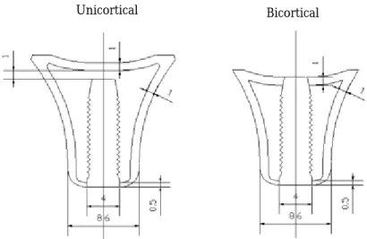
German) implant surrounded by different bone quantity and quality according to arch shape were created respectively as shown in Fig. 1 and Fig. 2. The ANSYS 5.5 software was used for modelling and analyses. To compare the same dimension, 10mm length and 4mm diameter of each implant was modelled. Axisymmetric mod- els were used to study the bone loading pat- terns of bicortical and unicortical anchorage cas- es of both maxilla and mandible bone (Fig. 1 and 2). The average dimensions of each jaw bone were measured in dissected skull. For bicorti- cal fiaxation case, the implant apex is placed 1mm deep in the apical cortex.
To mimic the clinical situations, the different bonding ratios of bone-implant interface were giv-
Table 1. The properties of each bone and implant.
Implant 117 GPa 0.3
Cortical
bone 13.7 GPa 0.3
Cancellous
bone 1.37 GPa 0.3
Young’s Poisson’s
modulus ratio
Fig. 1.Dimensions of maxilla model and implant.
Unicortical Bicortical
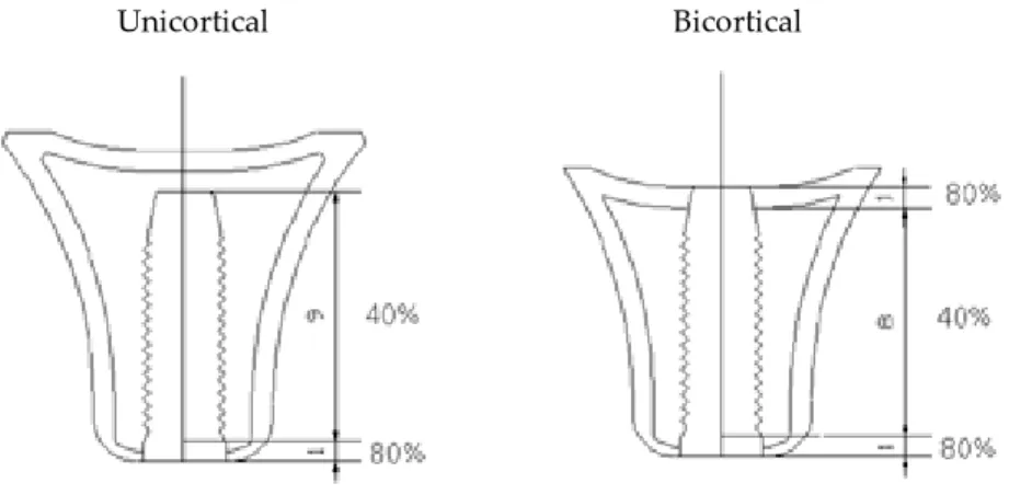
en to cortical and cancellous bone each. The bonding ratio was assumed as shown in Fig. 3 and Fig. 4 based on several autopsy studies. A 0.05mm thin layer was modelled around entire implant body and the very small Youngs modulus was assigned to given numbers of elements according to bonding ratio.
The material properties are given in Table 1. The rep-
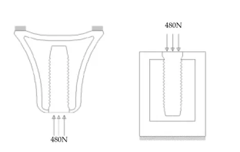
resentative figure of the finite element model is shown for reference in Fig. 5. The 480N axial force was applied to each model. The bottom of the mandible model and top of the maxilla model were fixed to pre- vent any displacement of bone during loading situa- tion (Fig. 5). Finally, Von Mises stress of each case at 0.05mm apart from interface was obtained under the various conditions.
Fig. 2. Dimensions of mandibular model and implant.
Fig. 3. Given bonding ratio of cortical and trabecular bone in maxilla.
Unicortical
Unicortical
Unicortical
Bicortical
RESULTS
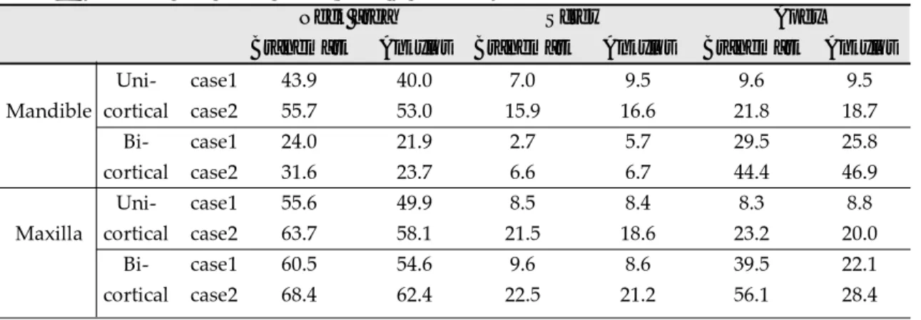
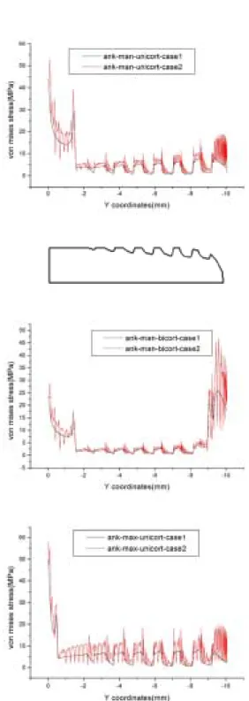
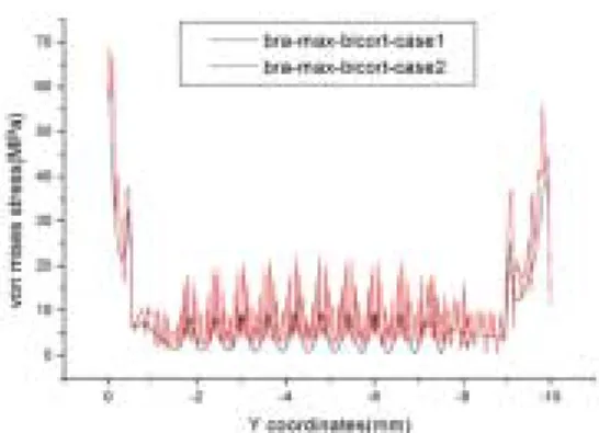
Table 2�3 shows the Von mises stress distrib- ution in cortical and trabecular bone under axial load of 480 N for a full bond case and a partial bond case respectively. It represents the results of bicortical fixation and unicortical fixation also.
Generally, full bond case showed less stress
than partial bond case in overall area and mandibu- lar model showed less amount of stress than that of maxilla model. In comparison of full and partial bonding situation, Branemark and ANKY- LOS implant showed 12% to 27% and 8% to 33%
more stress concentration respectively at crestal region in partial bond situation. The amount of maximum stress at screw body area increased 127%
Fig. 5.Fixation area and loading condition.
Fig. 4.Bonding ratio of cortical and trabecular bone in mandible.
Unicortical Bicortical
480N
480N
Table 2. Maximum Von Mises stress(MPa) in implant.
Uni- case1 43.9 40.0 7.0 9.5 9.6 9.5
Mandible cortical case2 55.7 53.0 15.9 16.6 21.8 18.7
Bi- case1 24.0 21.9 2.7 5.7 29.5 25.8
cortical case2 31.6 23.7 6.6 6.7 44.4 46.9
Uni- case1 55.6 49.9 8.5 8.4 8.3 8.8
Maxilla cortical case2 63.7 58.1 21.5 18.6 23.2 20.0
Bi- case1 60.5 54.6 9.6 8.6 39.5 22.1
cortical case2 68.4 62.4 22.5 21.2 56.1 28.4
Neck area Screw Apex
Branemark Ankylos Branemark Ankylos Branemark Ankylos
Table 3. Average Von Mises stress(MPa) in implant.
Uni- case1 20.2 19.0 2.9 3.1 5.5 5.1
Mandible cortical case2 25.1 23.5 5.0 4.8 9.4 11.4
Bi- case1 11.6 10.3 1.5 2.8 15.2 21.5
cortical case2 13.7 12.4 2.0 3.3 18.3 28.9
Uni- case1 28.4 24.8 3.9 4.1 5.1 4.0
Maxilla cortical case2 36.2 32.2 7.4 6.8 9.7 10.1
Bi- case1 31.0 27.4 4.2 4.8 12.6 18.2
cortical case2 38.8 35.1 7.7 7.5 16.9 21.7
Neck area Screw Apex
Branemark Ankylos Branemark Ankylos Branemark Ankylos
to 153% in Branemark implant and 18% to 147%
in ANKYLOS implant respectively. At the apex, the difference of maximum stress between Branemark and ANKYLOS was 42% to 180%
and 29% to 127%.
Maximum stress of the Branemark implant is higher than that of ANKYLOS regardless of bonding ratio at crestal and apex region. However, more stress concentration was noted in ANKYLOS implant at screw body area especially in mandible (Table 2).
The effect of bicortical fixation on crestal bone stress reduction is dramatical in mandible how- ever, there was no significant effect in maxil- lary case (Table 2).
There is almost 50% stress reduction effect at cer- vical cortex in mandibular bicortical fixation case. However, there is less than 10% stress reduction effect of bicortical fixation in maxilla sit- uation. The effect of bond ratio is more significant in maxilla than mandible regardless of implant shape.
A huge difference in stress values between the cer- vical cortex and trabecular bone was noticed regard- less of bone configuration especially in mandible. In trabecular bone stress and strain peaks occurred at the tips of the screw thread. The pattern of stress is different to screw designs however, the amount of stress is sim- ilar to regardless of screw shape in maxillary trabec- ular bone.
Fig. 6. Comparison of full bond and partial bond case.
Case 1 - full bond Case 2 - partial bond
Fig. 7. Comparison of Branemark and Ankylos implant.
Case 1 - full bond Case 2 - partial bond
DISCUSSION
Some assumptions and simplifications have been made with regard to the material properties and model geometry. Bone tissue is assumed to have uniform isotropic elastic properties, although bone will exhibit a more complex situation in vivo. Implant bone interface conditions, as indi- cated, have a very strong influence on the bone loading patterns around the implant. The level and mode of stress and strain distribution are highly affected by the interface conditions10. Even though the amount of stress is not a actual number, the general tendency of stress distribution can be analyzed in this model.
To mimic real clinical situation of osseointegration ratio, the average osseointegration ratio based on several autopsy study was applied to cortical
and cancellous bone model in this study12-16. It is also meaningful to analyse stress at various loca- tions of a implant body such as crestal, screw body and apex region.
The full bond case showed less stress accumu- lation on overall implant surfaces.
The effects of partial bonding is greater in screw body and apex area than in crestal region.
The amount of stress is greater in crest and apex than screw body area. It seems that bone quality and bone amount exaggerated the stress accu- mulation in each jaw model. Especially in mandible model where the lower cortical layer is thicker than upper cortical layer more stress was concentrat- ed at apex area than crest. Consequently the effect of bicortical fixation was great in mandible.
There are two mechanical explanations of crestal bone resorption phenomenon. One is overload-
ing of the implant and the other is a stress shield- ing effect17-19. In unicortical fixation case, stress accu- mulation seems to be a plausible explanation however, stress shielding effect resulting from too rigid anchorage seems to be a possible explana- tion in bicortical fixation case17. At the moment, there are many suggested reasons for crestal bone resoption such as surgical trauma, biolog- ical width and stress. So far, no conclusive expla- nation of marginal bone loss was reported. As reported in previous studies, this study sup- ported that the amount of stress and strain can be significantly affected by bone quality, configuration and bonding ratio also.
Generally, stress accumulated at crest and apex area. There was no significant advantage of bicor- tical fixation at crestal and screw body region in maxilla as reported earlier11. It may be postulat- ed that thin layer of cortical bone, soft bone qual- ity and surrounding bone configuration of max- illa contributed the results. Recent long-term clinical observation study also indicated that no evidence of advantage of bicortical fixation of 10 mm implants on implant success17. The margin- al bone loss was alomost identical regardless of fixation method for 15 years17.
However, bicortical fixation reduced stress at cre- stal and screw body region dramatically in mandible(43% to 60%). It also showed even stress distribution. Shape and lower cortical bone thick- ness of mandible may contribute to stress reduc- tion at crestal and screw body. On the contrary to maxilla, the shape and thickness of cortical bone of mandible is quite different, especially the thickness of cortical bone may affect the stress dis- tribution mainly.
The average stress at screw body area demon- strated no significant difference between two implants (less than 10%). Although the average amount of stress is different according to screw design, it has not as much effect as bone quality and thickness on stress distribution.
CONCLUSIONS
Following conclusions were drawn within the limits of this study.
1. Full bond case showed less stress than partial bond case in overall area.
2. The effect of partial bond on stress distribution was more significant at screw body and apex region than in crestal region.
3. Partial bond cases demonstrated greater stress accumulation in trabecular bone than cortical bone.
4. Branemark implant demonstrated more stress accumulation than ANKYLOS implant at cre- stal and apex area. However, ANKYLOS implant showed more stress at screw body area in full bond mandible case only.
5. The stress distribution effect of bicortical fix- ation was great in mandible however, there was no siginificant effect in maxilla.
Acknowledgment
The author would like to express appreciation for the assistance of professor Joo-Ho Choi, Department of Aeronaufical & Mechanical Engineering, Hankook Aviation University.
REFERENCES
1. Adell R, Eriksson B, Lekholm M, Branemark P-I and Jemt T. A long term follow-up study of osseoin- tegrated implants in the treatment of totally eden- tulous jaws. Int J Oral Maxillofac Implants 1990;5:347-359.
2. Lekhom U, Gunne J, Henry P, Higuchi K, Linden U, Bergstrom C, van Steenberghe. Survival of the Branemark implant in partially edentulous jaws: A 10-year prospective multicenter study. Int J Oral Maxillofac Implant 1999;14:639-645.
3. Betram J, Swartz S. “Law of Bone Transformation”:
a case of crying Wolff? Biological Review 1991;66:245-273.
4. Frost, HM. A determinant of bone architecture: the minimum effective strain. Clinical Orthopaedics 1983;175:286-292.
5. Lanyon LE, Goodship AE, Pye CJ, MacFie JH.
Mechanically adaptive bone remodelling. J of Biomechanics 1982;15:141-154.
6. Rubin CT, Lanyon LE. Regulation of bone for- mation by applied dynamic loads. J of Bone and Joint Surgery 1984;66-A:397-402.
7. Shenk R, Hunziker EB. Histologic and ultra- structural features of fracture healing. In Brighton CT, Friedlander G, Lane JM(eds). Bone Formation and Repair. Rosemount, IL:American Academy of Orthopaedic Surgeons, 1994:117-146.
8. Roberts WE. Bone tissue interface. J Dent Educ 1988;52:804-809.
9. Isidor F. Histological evaluation of peri-implant bone at implant subjected to occlusal overload or plaque accumulation. Clin Oral Impl Res 1997;8:1-9.
10. Van Oosterwyck et al. The influence of bone me- chanical properties and implant fixation upon bone loading around oral implants. Cli Oral Impl Res 1998;9:407-418.
11. Choi JH, Seo KY, Choi JH and Han JS. Effects of bone engagement type and implant length on stress distribution: A three dimensional finite el- ement analysis. J Korean Acad Prosthodont 1999;37(5);687-697.
12. Rohrer MD, Sobczak RR, Prasad HS and Morris HF.
Postmortem histological evaluation of mandibu- lar titanium and maxillary hydroxyapatite coated implant from 1 patient. Int J Oral Maxillofac Implants 1999;14:579-586.
13. Dominici JT, Olsen JW, Rohrer MD, Morris HF.
Postmortem histological evaluation of hydrox- yapatite coated cylinder and titanium alloy basket implants in situ for 37 months in the posterior mandible. Implant Dent 1997;6:215-222.
14. Rohrer MD, Bulard RA, Patterson MK. Maxillary and mandibular titanium implants 1 year after surgery: Histologic examination in a cadever. Int
J Oral Maxillofac Implants 1995;10:466-473.
15. Nystrom E, Kahnberg K, Albrektsson T. Treatment of the severely resorbed maxilla with bone graft and titanium implants. Histologic review of autopsy specimens. Int J Oral Maxillofac Implants 1993;8:162- 172.
16. Wilson TG Jr, Schenk R, Buser D, Cochran D.
Implants placed in immediate extraction sites: A report of histologic and histometric analyses of hu- man biopsies. Int J Oral Maxillofac Implants 1998;13:333-341.
17. Ivanoff CJ, Grondahl K, Bergstrom C, Lekholm U and Branemark PI. Influence of bicortical or monocortical anchorage on maxillary implant sta- bility: A 15-year retrospective study of Branema가 system implants. Int J Oral Maxillofac Implants 2000;15:103-110.
18. Hoshaw S, Bunski J, Cochran G. Mechanical over- loading of Branemark implants affects interfacial bone modeling and remodeling. Int J Oral Maxillofac Implants 1994;9:345-360.
19. Quirynen M, Naert I, van Steenberghe D. Fixture design and overload influence marginal bone loss and fixture success in the Branemark system. Clin Oral Implants Res 1992;3:104-111.
Reprint request to:
DR.JUNG-SUKHAN
DEPARTMENT OFPROSTHODONTICS,EWHAWOMANS UNIVERSITY 911-1 MOK-DONG,YANGCHEON-GU SEOUL,158-710 KOREA E-mail:[email protected].