단순 낭종을 포함하여 유방의 낭성 종양은 40세 이상의 여 성에서 흔한 질환으로 초음파 검사를 통하여 쉽게 발견할 수 있다. 단순낭종은 악성 잠재력은 없으나 고형성분이 혼합되어 있는 낭성 병변의 경우 진단에 딜레마를 갖게 된다(1, 2). 초 음파상 낭성 병변을 가지는 유방암은 전체 유방암의 0.3-2.0%
로 드물다(3). 저자들은 유방 낭성 질환의 초음파양상을 분류 하고 병리소견과 비교하여 초음파 양상에 따른 양성과 악성의 특징적 소견을 알아보고 각 분류에 따른 처치법을 제시하고자 하였다.
대상과 방법
2002년 6월부터 2004년 6월까지 유방초음파를 시행 받은 3364명의 환자 중 낭성병변을 가진 330명의 330개의 병변을 대상으로 하였으며 후향적으로 분석하였다. 추적검사를 하지
않은 환자들과 추적 초음파만 시행한 환자들을 제외하였고 병 리적으로 확진된 예는 113 예였다 병리적 확진은 세침 흡인 (n =78), 핵 생검(n=24), 절개 생검(n=11) 등을 시행하였 으며 초음파 소견과 비교하였다. 113명의 환자 중 만져지는 병변은 64예(56%)였으며 만져지지는 않으나 유즙 분비나 통 증이 있는 경우 또는 환자가 확인을 원하는 경우는 초음파 유 도하에 세침흡인 이나 핵생검을 시행하였다. 환자의 평균연령 은 44.6세(15-7세)였으며 모두 여자 환자고 병변의 평균 크 기는 23 mm(5-140 mm)였다. 초음파 검사는 7-10 MHz 선 형 탐촉자를 사용하여 전체 유방에서 시행되었고 기종은 GE LogiQ 700 Expert Series(GE Medical systems, Milwaukee, Wisconsin, U.S.A.)과 ATL(Advanced Technology Laboratories, Bothell, WA) HDI 5000이었다.
각각의 낭성병변들은 Berg 등(1)이 사용한 분류를 약간 변 형하여 단순 낭종(simple cysts)은 경계가 잘 그려지는 무에 코의 종괴로 음향증강을 가지는 경우로 정의하였고, 군집 낭 종(clustered cysts)은 고형성분이 없는 군집된 무에코의 별 개의 작은 낭종들로 정의하였으며, 얇은 격벽을 가지는 낭종 (cysts with thin septa)은 0.5 mm 미만의 얇은 격벽이 낭종
낭성 유방 질환의 초음파 소견에 따른 양성과 악성의 감별
1장윤우・김동훈1, 4・권귀향・구동억・이민혁2・이동화3
목적: 유방 낭성 질환의 초음파소견을 분류하고 병리소견과 비교하여 초음파 분류에 따른 양 성과 악성의 특징적인 소견을 알아보고 각 분류에 따른 처치법을 제시하고자 하였다.
대상과 방법: 2002년 6월부터 2004년 6월까지 유방 초음파상 낭성병변을 가지며 병리적으로
확진된 113예를 후향적으로 분석하였다. 낭성 병변은 단순 낭종, 군집 낭종, 얇은 격벽을 가지 는 낭종, 합병 낭종, 두꺼운 벽/ 격벽 혹은 결절을 가지는 낭종, 복합 고형 낭종 등으로 구분하 였다. 각 병변의 병리결과를 조사하여 각 분류에 따른 양성과 악성의 빈도를 비교하였다.
결과: 113개의 병변중 17예는 단순낭종, 10예는 군집 낭종, 19예는 얇은 격벽을 가지는 낭종, 24예는 합병 낭종이었으며 모두 조직 검사에서 양성이었다. 두꺼운 벽/격벽 혹은 결절을 가지 는 낭종 16예 중 5예(31.3%)와 복합 고형낭종 27예 중 17예(63%)가 악성으로 총 43예의 고 형성분을 가지는 낭성종괴 중 22예(51.2%)가 악성이었다. 고형성분이 있는 낭성 종괴 43예의 모양과 경계를 분석하였을 때 둥글거나 난원형을 보인 36예 중 17예(47%)가 악성이었으며 경계가 잘 그려지는 27예 중 10예(37%)가 악성이었다.
결론: 고형 성분이 없는 단순낭종, 군집낭종, 얇은 격벽을 가지는 낭종과 증상이 없는 합병낭종 은 모두 양성이었다. 증상을 가지는 합병 낭종은 세침흡인 후 적절한 치료를 시행하여야 한다.
고형 성분을 가지는 낭성 종괴는 반드시 조직학적 검사를 통한 확진이 필요하다.
1순천향대학교 서울병원 영상의학과
2순천향대학교 서울병원 외과
3순천향대학교 서울병원 병리과
4조선대학교병원 영상의학과
이 논문은 2005년 1월 25일 접수하여 2005년 12월 29일에 채택되었음.
내부에 포함되는 경우로 정의하였다. 합병 낭종(complicated cysts)은 균질한 저에코를 보이는 단순낭종으로 Breast Imaging Reporting and Data System(BI-RADS) (4)의 정의 에 의하여 분류하였으며 물-파편층 혹은 움직이는 내부파편 을 가지는 경계가 그려지는 낭종도 포함하였다. 두꺼운 벽/ 격 벽 혹은 결절을 가지는 낭종(cystic masses with thick wall/
septa or nodules)은 0.5 mm 이상의 두꺼운 벽 혹은 격벽을 가지거나 50% 이상의 낭성성분을 가지는 종괴로 정의하였으 며 복합 고형 낭종(complex solid and cystic masses)은 50%
이상의 고형성분을 가지며 내부 혹은 주변부에 낭성 성분을 가지는 경우로 구분하였다(Table 1). 초음파상 보이는 낭성 종괴들은 위의 분류에 따라 2명의 숙련된 영상의학과 전문의 가 합의하에 구분하였고 병리 소견과 비교하였다. 낭성종괴중
고형성분을 가지는 종괴인 두꺼운 벽/ 격벽 혹은 결절을 가지 는 낭종과 복합 고형낭종 총 43예에 대하여 모양과 경계를 분 석하였으며 병리소견과 비교하였다.
결 과
각각의 분류와 검사방법은 Table 1에 요약하였다. 대부분의 단순낭종은 그대로 두거나 추적검사를 하였으나, 증상을 가지 거나 환자가 확인을 원했던 17예의 단순낭종은 병리소견상 낭 종(n=16), 섬유성 낭성질환(n=1)으로 모두 양성으로 확진되 었다. 10예의 군집 낭종은 낭종(n=1)과 섬유성 낭성질환(n=9) 이었으며 19예의 얇은 격벽을 가지는 낭종도 낭종(n=12) 혹 은 섬유 낭성 질환(n=7)으로 모두 양성이었다 (Figs. 1, 2), 합 병 낭종은 24 예로 병리소견상 낭종(n=2) 섬유성 낭성질환 (n=6), 농양(n=15), 점액낭성종양(mucocele- like tumor) (n=1)으로 모두 양성이었다 (Fig. 3). 열감이나 통증 등의 증 상이 있던 경우가 16예로, 15예는 세침흡인(n=13)과 절개생 검(n=2)에서 농양으로 확진되었으며 외과적 배농술과 항생제 로 치료를 시행하였다. 만져지는 예가 17예였으며 7예의 만져 지지 않는 종괴는 세침흡인(n=2)상 제거되었으며 핵생검 (n=5)상 모양이 변하며 크기가 작아졌다. 16 예의 두꺼운 벽 / 격벽 혹은 결절을 가지는 낭종은 11 예에서 낭종(n=1), 섬 유성 낭성질환(n=1), 농양(n=5), 점액낭성종양(mucocele- like tumor) (n=1), 유두종(n=3)의 양성이었고 5예는 침윤성 유방암(infiltrative ductal carcinoma, IDC) (n=4)과 유두상암 종(papillary carcinoma) (n=1)으로 악성이었다 (Fig. 4). 27 예의 복합 고형낭종은 10예에서 양성이었으며 섬유성 낭성질 환(n=5), 농양 (n=1), 섬유선종(n=2), 유두종(n=2)이었고 17예는 침윤성 유방암 (n=9) (Fig. 5), 화생성 유방암 (metaplastic carcinoma) (n=4), 악성 엽상종양(malignant
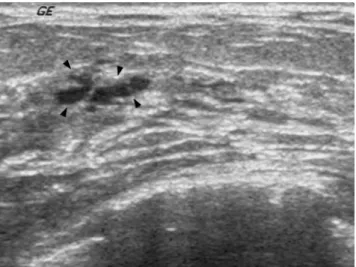
Fig. 1. A 44-year-old woman with clustered cysts. Transverse US scan shows an aggregate of small cysts (arrowheads) with- out discrete solid component. The lesion was incidentally de- tected during screening examination. Aspiration cytology re- vealed fibrocystic change.
Fig. 2. A 27-year-old woman with cyst with thin septa. Radial US scan shows a cyst with thin (<0.5 mm) septa (arrowheads) that otherwise met the criteria for a simple cyst. The cyst dis- appeared after aspiration and the pathologic result was benign cyst contents.
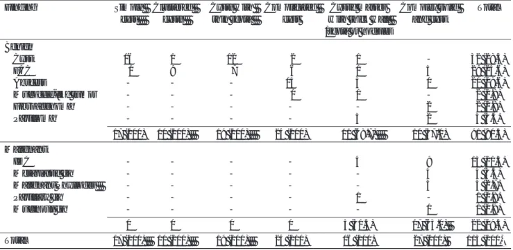
Table 1. Subclassification, Method of Sampling, & Rates of Malignancy of 113 Cystic Lesions
Sonographic feature Aspirtaion CNB Excision Rate of Malignant Simple cysts (n=17) 14 02 01 - Clustered cysts (n=10) 04 04 02 - Cysts with thin septa 14 03 02 -
(n=19)
Complicated cysts (n=24) 17 05 02 - Cystic masses with thick 11 01 04 5 (23)0
wall/ septa or nodules (n=16)
Complex solid and cystic
masses (n=27) 18 09 - 17 (77)0 Total (n=113) 78 24 11 22 (100) Note.- Data are the number of lesions. Data in parentheses are percentages.
CNB: core needle biopsy
phyllodes tumor) (n=3), 점액암(mucinous carcinoma) (n=1)의 악성이었다(Table 2).
고형성분을 가지는 두 개의 분류인 두꺼운 벽/ 격벽 혹은 결 절을 가지는 낭종과 복합 고형 낭종 43예 중 악성은 22예 (51.2%)로 모두 만져지는 종괴였다. 초음파상에서 43예의 모
양과 경계를 분석하였을 때 둥글거나 난원형을 보인 36예 중 17예(47%)가 악성이었으며 경계가 잘 그려지는 27예 중 10 예(37%)가 악성이었다(Table 3). 모양과 경계를 종합하여 분 석하였을 때 둥글고 난원형이면서 경계가 잘 그려지는 예는 10예(22%)가 악성이었다. 2예를 제외한 20예에서 수술을 시 행하였고 유방절제술 7예, 유방 보존술 4예, 사분절제술 6예, 종괴 절제술이 3예에서 시행되었다.
고 찰
유방 초음파상 단순 낭종은 40세 이상의 여성에서 흔하게 발견되며 BI-RADS에서 category 2, 양성으로 분류된다. 단 순 낭종은 악성 잠재력은 없으므로 증상이 없다면 침습적인 검사가 요구되지 않으나 환자가 통증이 있거나 낭종이 매우 커서 만져질 경우 선택적으로 세침 흡인술을 시행할 수 있다 (4-8). 세침흡인시 흡인액이 노랗게 투명하거나 암녹색이라 면 일반적으로 단순 낭종으로 여겨 반드시 세포검사를 보내지 는 않으나 흡인액이 붉은 경우, 환자의 요구 혹은 암이나 비 전형병변의 과거력이 있을 경우 세포검사를 보낸다(1). 단순 낭종과 내부에코를 가지거나 고형성분을 가지는 낭종을 감별 하는 것은 크기가 작거나 깊이 위치할 경우 어려울 수 있으나 최근 고해상도 탐촉자의 사용과 조직 조화영상 (tissue harmonic image) 의 사용으로 내부 에코를 줄일 수 있어 도 움이 된다(9). 유방초음파상 낭종을 진단하는 정확도는 거의 100%로 보고되고 있으며 고해상도 탐촉자를 사용하면 2-3 mm의 작은 낭종도 발견할 수 있다(1). 그러나 공간 혼합영상 (spatial compounding image)을 사용하는 경우는 반점이나 다 른 잡음 원인을 줄여서 종괴 내부를 더 잘 볼 수 있는 장점이 있으나 신호대 잡음비의 증가로 후향 음향강조를 평가하기는 어렵다(10).
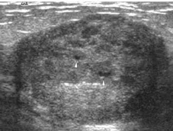
Fig. 5. A 37-year-old woman with complex solid and cystic mass. Transverse US scan shows well-circumscribed, oval sol- id mass with internal small cystic components (arrowheads).
The pathologic result was infiltrative ductal carcinoma of anaplastic type.
Fig. 3. A 27-year-old woman with complicated cysts.
Transverse US scan shows well-defined, oval masses with ho- mogeneous internal echoes (arrowheads) with tubular struc- ture of radial US scan (not seen). Aspiration yielded mucinous material with atypical cell and following excisional biopsy re- vealed mucocele-like tumor with foci of intraductal papilloma.
Fig. 4. A 64-year-old woman with cystic mass with thick septa and nodule. Transverse US scan shows a well-circumscribed, oval cystic mass with thick septa (arrowheads) and mural nodular component. Fluid-debris level is also seen. Aspiration yielded bloody fluid and following core needle biopsy revealed low-grade intraductal papillary carcinoma.
고형성분이 없는 군집낭종은 양성으로 간주하며 추적검사를 시행한다. 이들 병변은 이출성 이형성증(apocrine meta- plasia) 이거나 섬유낭성변화인 경우가 많으며 이출성 이형성 증에서 소엽이 융합되어 낭종이 되는 일련의 과정으로 얇은 격벽을 가지는 낭종으로 발전할 수 있다(11). 저자들의 연구 에서도 군집낭종을 보인 9예 중 8예가 섬유낭성변화였으며 모 두 양성이었다.
합병 낭종은 초음파 영상만으로 고형종괴와 감별이 어려운 경우가 많으며 흉벽에 가까이 위치하는 병변일수록 고형종괴 와 감별이 어렵고 세침흡인 검사 등을 통하여 확인하여야 한 다 (1, 3, 5, 8). 본 연구에서는 내부에 균질한 내부 저에코를 가지는 낭종이나 고형성분없이 내부에 물-파편층이나 떠다니 는 파편들을 포함하는 종괴도 합병 낭종으로 분류하였고 총 24 예 중 15예(62.5%)가 농양이었다. 임상적으로 농양이 의
심되는 경우 세침흡인을 시행하여 확인한 후 항생제를 사용하 거나 증상에 따라 외과적 배농을 시행할 수 있다. Venta 등 (8)의 연구에서 308개의 합병 낭종중 0.3%만이 악성이었으 며 합병 낭종은 악성가능성이 낮으므로 다른 양성가능성 병변 들과 같이 규칙적인 추적검사를 시행하여도 된다고 하였다.
Buchberger 등(12)은 133개의 병변중 악성은 없었으며 Kolb 등(13)도 126개의 병변중 악성은 없었다고 보고하였다. 저자 들의 연구에서는 24 예의 합병 낭종은 모두 양성이었으며 1 예는 유두양 종양 부위를 가지는 점액성 종양 이었다(Fig. 3).
저에코나 물-파편층을 가지는 우연히 발견된 만져지지 않는 합병 낭종은 양성 가능성 병변으로 분류하며 추적검사를 하며, 증상이 있는 합병 낭종은 임상적인 양상에 따라 치료하여야 하고 일반적으로 세침 흡인 검사를 시행하며 농양, 혈종, 지방 괴사, 유선낭종등과 감별해야 한다.
0.5 mm 이상의 두꺼운 벽/ 격벽 혹은 결절을 가지는 낭종 의 경우 악성의 가능성이 있으며 조직검사로 확인해야 한다.
Berg 등(1)은 35%에서 악성으로 진단되었다고 하였으며 이 중 86%는 고분화 침윤성 유선암이었다고 하였고 초음파상 경 계가 잘 그려지는 종괴로 보인다고 하였다. 고분화암에서 낭 성 성분들은 세침 흡인시 괴사나 세포를 가지지 않아 진단이 어려우며 핵 생검등을 통하여 벽이나 고형성분에서 조직을 얻 어야 악성으로 진단할 수 있고 이때 낭성 성분들은 괴사를 시 사한다고 하였다. 저자들의 연구에서는 두꺼운 벽/ 격벽을 가 지거나 고형 결절을 가지는 병변 16예 중 악성으로 진단된 경 우가 5 예(31.3%) 였으며 침윤성 유선암이 4예(80%), 악성 유두암이 1예였다. 농양, 이출성 이형성증, 염증 혹은 낭종이 나 유선 파열, 혈종 등이 두꺼운 벽을 가지는 낭종으로 나타 날 수 있다. 지방괴사도 두꺼운 벽을 가지는 낭성병변이나 복 Table 3. Sonographic Findings of 43 Cystic Masses with Solid
Component and Rates of Malignancy
No. of lesion No. of Malignant lesion Shape Round 09 (100) 07 (78)
Oval 27 (100) 10 (37) Irregular 07 (100) 05 (71) Margin Circumscribed 27 (100) 10 (37) Indistinct 02 (100) 002 (100) Angular 03 (100) 02 (67) Microlobulated 11 (100) 08 (73) Spiculated 00 (100) 0 (0) Total No. 43 (100) 0.22 (51.2) Note.- Data are the number of lesions. Data in parentheses are percentage.
Table 2. Correlation of Pathologic Outcome & Sonographic Features for 113 Cystic Lesions
Finding Simple Clustered Cysts with Complicated Cystic masses Complex solid Total cyst cysts thin septa cyst with thick wall and cyst
/septa or nodules Benign
Cyst 16 1 12 02 1 - 32 (28.3)
FCC 01 9 07 06 1 5 29 (25.6)
Abscess - - - 15 5 1 21 (18.6)
Mucocele-like tumor - - - 01 1 - 2 (1.8)
Fibroadenoma - - - 2 2 (1.8)
Papilloma - - - - 3 2 5 (4.4)
17 (100) 10 (100) 19 (100) 24 (100) 11 (68.7) 10 (37.0) 91 (80.5) Malignant
IDC - - - - 4 9 13 (11.5)
Metaplastic ca - - - 4 4 (3.5)
Malignant Phyllodes - - - 3 3 (2.7)
Papillary ca - - - - 1 - 1 (0.9)
Mucinous ca - - - 1 1 (0.9)
0 0 0 0 5 (31.3) 17 (63.0) 22 (19.5)
Total 17 (100) 10 (100) 19 (100) 24 (100) 16 (100) 27 (100) 113 (100)0.
Note.- Data are the number of lesions. Data in parentheses are percentages.
FCC: fibrocystic disease, IDC: infiltrative ductal carcinoma, ca: carcinoma
합성 고형 낭성종괴로 보일 수 있다(1, 14). 악성의 경우 외 상이나 유선의 폐색 혹은 감염 등으로 출혈이 유발되면 짧은 시간에 크기가 커질 수 있다. 임상적으로 외상력이 있거나 염 증을 가지는 두꺼운 벽을 가지는 낭종의 경우 짧은 기간(2- 3개월)의 추적검사를 시행해야하며 조금이라도 병변이 커지는 양상을 보일 경우 악성가능성을 확인하기 위해 즉시 조직검사 를 시행하여야 한다(1).
복합성 고형 낭성종괴에서 편심성 낭성 부위는 늘어난 유선, 소엽 혹은 괴사에 의한 것으로 알려졌다. Jackson 등(15)은 섬유선종에서 드물게 편심성 낭성 부위를 보일 수 있다고 보 고하였다. 섬유선종에서 낭성부위가 존재하는 것은 엽상종의 가능성을 제시한다. Liberman 등(16)은 악성 엽상육종에서 낭 성성분을 가지는 경우가 더 많다고 보고하였다. 엽상육종은 내 부에 물이 차있는 홈을 가지는 것이 특징으로 알려져 있다.
Berg 등(1)은 악성종앙이 편심성 낭성부위를 가질 때 저분화 혹은 고분화 침윤성 유방암, 침윤성 소엽암, 미세침윤암등의 병 리소견을 감별하는데 도움이 되지 않는다고 하였다. 저자들의 연구에서는 복합고형낭종을 보이는 종괴는 침윤성 유방암의 분화 정도에 따른 차이는 없었으나 화생성 유방암, 악성 엽상 암, 점액암등 낭성 성분을 보일 수 있는 악성종양들이었다. 낭 성 병변을 가지는 유방암은 전체 유방암의 0.3-2.0%를 차지 하며 90%의 경우 진단 당시 고형성분의 크기가 3 cm 이상으 로 알려졌다. 예후는 일반적인 유방암에 비해 좋은 것으로 되 어 있으며 내부에 포함된 낭성 성분은 출혈 등의 낭성 변성이 다(3, 17-19). 낭성유방암이 발생할 수 있는 기전으로는 낭 성 질환이 있는 부위에 유방암의 침윤이 생기는 경우거나 고 분화 유방암이 낭성변화를 하는 것 등으로 알려져 있다(3, 19, 20). 유두암 혹은 유두암종도 낭성종양으로 보일 수 있으며 낭 성 유두암은 전체 유방암의 0.3%를 차지한다(21).
Berg 등(1)은 낭성 종양의 경계를 조사하였을 때 만져지지 않으나 초음파상 경계가 그려지는 둥글고 난원형의 병변중 악 성은 없었으나 만져지면서 경계가 그려지며 둥글거나 난형 혹 은 소엽을 가지는 병변은 54개 중 12개(22%)가 악성이라고 하였다. 저자들의 연구에서도 만져지지 않는 종괴로 경계가 그 려지는 둥글고 난원형의 병변 33개 중 악성은 없었고 만져지 며 경계가 잘 그려지고 둥글거나 난원형 혹은 소엽의 모양을 가지는 병변은 45개 중 10개(22%)가 악성이었다.
이 연구의 제한점은 후향적 연구로 병변들의 선택에 병리적 으로 확인된 증례들만을 선택하여 낭성병변의 일반적인 추이 를 관찰하는데 제한점이 있다는 것, 병리적으로 확진된 낭성 병변들의 추적검사를 포함하지 못하였다는 점 그리고 분류된 각각의 병변의 수가 적어 통계적인 유의성을 확인하는데 제한 이 있다는 점이다. 하지만, 유방 낭성 질환들의 모양에 따른 분 류를 병리소견과 비교하여 양성과 악성 소견을 예측할 수 있 으며 각각의 양상에 따른 치료방법을 제시할 수 있다는 점에 서 의미가 있을 것으로 생각된다.
결론적으로 초음파상 단순낭종은 양성으로 간주해도 된다.
군집낭종, 얇은 격벽을 가지는 낭종과 증상이 없는 합병 낭종 의 경우 모두 양성이었다. 증상이 있는 합병 낭종은 세침 흡
인 등을 통한 확인이 필요하며 임상적인 상태에 따라 치료를 시행하여야 한다. 고형 성분을 가지는 낭성유방병변인 두꺼운 벽/ 격벽 혹은 결절을 가지는 낭종과 복합성 고형낭성종괴는 경계가 잘 그려지는 둥글고 난원형의 종괴라도 반드시 조직 검사가 필요하다.
참 고 문 헌
1. Berg WA, Campassi CI, Ioffe OB. Cystic lesions of the breast: sono- graphic-pathologic correlation. Radiology 2003;227:183-191 2. Bassett LW. Imaging of breast masses. Radiol Clin North Am 2000;
38:669-691
3. Omori LM, Hisa N, Ohkuma K, Fujikura Y, Hiramatsu K, Enomoto I, et al. Breast masses with mixed cystic-solid sonograph- ic appearance. J Clin Ultrasound 1993;21:489-495
4. American College of Radiology. Breast imaging reporting and data system (BI-RADSTM) ultrasound. Reston, VA: American College of Radiology, 2003
5. Mendelson EB, Berg WA, Merritt CR. Toward a standardized breast ultrasound lexicon, BI-RADS: ultrasound. Semin Roentgenol 2001;36:217-225
6. Hilton SV, Leopold GR, Olson LK, Willson SA. Real-time breast sonography: application in 300 consecutive patients. AJR Am J Roentgenol 1986;147:479-486
7. American College of Radiology. Breast Imaging Reporting and Data System. 3rd ed. Reston, VA: American College of Radiology, 1998 8. Venta LA, Kim JP, Pelloski CE, Morrow M. Management of com-
plex breast cysts. AJR Am J Roentgenol 1999;173:1331-1336 9. Sklair-Levy M, Muradali D, Kulkarini S. Linear transducer har-
monic imaging: improved characterization of breast cysts com- pared to conventional sonography. AJR Am J Roentgenol 2001;176:
6-7
10. Merritt C, Piccoli C, Forsberg F, Wilkes A, Cavanaugh B, Lee S.
Real-time spatial compound imaging of the breast: clinical evalua- tion of mass. Radiology 2000;217:491-492
11. Warner JK, Kumar D, Berg WA. Apocrine metaplasia: mammo- graphic and sonographic appearances. AJR Am J Roentgenol 1998;
170:1375-1379
12. Buchberger W, DeKoekkoek-Doll P, Springer P, Obrist P, Dunser M. Incidental findings on sonography of the breast: clinical signifi- cance and diagnostic workup. AJR Am J Roentgenol 1999;173:921- 927
13. Kolb TM, Lichy J, Newhouse JH. Occult cancer in women with dense breasts: detection with screening US-diagnostic yield and tu- mor characteristics. Radiology 1998;207:191-199
14. Soo MS, Kornguth PJ, Hertzberg BS. Fat necrosis in the breast:
sonographic features. Radiology 1998;206;261-269
15. Jackson VP, Rothschild PA, Kreipke DL, Mail JT, Holden RW. The spectrum of sonographic findings of fibroadenoma of the breast.
Invest Radiol 1986;21:34-40
16. Liberman L, Bonaccio E, Hamele-Bena D, Abramson AF, Cohen MA, Dershaw DD. Benign and malignant phyllodes tumors: mam- mographic and sonographic findings. Radiology 1996;198:121-124 17. Kersschot EA, Hoste MV, Dochez CL, van Marck EA, De Schepper
AM, Van Goethem ML. Intracystic carcinoma of the breast. Rofo 1986;144:728-279
18. Reuter K, D’Orsi CJ, Reale F. Intracystic carcinoma of the breast:
the role of ultrasonography. Radiology 1984;153:233-234
19. Czernobilsky B. Intracystic carcinoma of the female breast. Surg Gynecol Obstet 1967; 124:93-98
20. Ravichandran D, Carty NJ, al-Talib RK, Rubin C, Royle GT, Taylor I. Cystic carcinoma of the breast: a trap for the unwary.
Ann R Coll Engl 1995;77:123-126
21. Knelson MH, el Yousef SJ, Goldberg RE, Ballance W. Intracystic
papillary carcinoma of the breast:mammographic, sonographic, and MR appearance with pathologic correlation. J Comput Assist Tomogr 1987;11:1074-1076
J Korean Radiol Soc 2006;54:441-446
Address reprint requests to : Yun-Woo Chang, M.D., Department of Radiology, Soonchunhyang University Hospital 657 Hannam-dong, Yongsan-gu, Seoul 140-743, Korea.
Tel. 82-2-709-9396 Fax. 82-2-709-3928 E-mail: [email protected]
Differentiation of Benign and Malignant Cystic Lesions of the Breast according to Sonographic Findings
1Yun-Woo Chang, M.D., Dong Hun Kim, M.D.1, 4, Kui Hyang Kwon, M.D., Dong Erk Goo, M.D., Min Hyuk Lee, M.D.2, Dong Wha Lee, M.D.3
1Department of Radiology, 2General Surgery, 3Pathology Soonchunhyang University Hospital
Purpose: To classify the ultrasonographic findings of cystic lesions of the breast and correlated them with the pathology, to evaluate the characteristic features of cystic masses in benign and malignant tumors, and to de- termine the appropriate level of patient management according to the ultrasonographic findings.
Materials and Methods: From June 2002 through to June 2004, the ultrasonographic findings of 113 pathologi- cal proven cystic breast lesions were reviewed retrospectively. The cystic lesions were classified as simple cysts, clustered cysts, cysts with thin septa, complicated cysts, cystic masses with a thick wall/ septa or nod- ules, and complex solid and cystic masses. The ultrasonographic findings of each type of cystic lesion of the breast were compared with the pathology and evaluated according to whether they were benign or malignant.
Results: Of the 113 lesions, there were 17 simple cysts, 10 clustered cysts and 19 cysts with thin septa. Twenty four cases of complicated cysts were found to be benign. Five (31.3%) of the 16 cases of cystic masses with a thick wall / septa or nodules and 17 (63%) of the 27 cases of complex solid and cystic masses were found to be malignant. The shape and margin of the 43 cases of cystic masses with a solid component were analyzed.
Seventeen out of 36 sonographical round or oval shaped masses and 10 out of 27 sonographical circumscribed margins were found to be malignant.
Conclusion: The simple cysts, clustered cysts, cyst with thin septa and non-symptomatic complicated cysts de- tected by sonography were all benign. Symptomatic complicated cysts should be aspirated and treated appro- priately. Cystic masses with a solid component should be examined by a biopsy with a pathological confirma- tion.
Index words :Breast, cysts Breast, US Breast, diseases