© 2011 The Korean Academy of Medical Sciences.
This is an Open Access article distributed under the terms of the Creative Commons Attribution Non-Commercial License (http://creativecommons.org/licenses/by-nc/3.0) which permits unrestricted non-commercial use, distribution, and reproduction in any medium, provided the original work is properly cited.
pISSN 1011-8934 eISSN 1598-6357
A Case of Hepatic Portal Venous Gas as a Complication of Endoscopic Balloon Dilatation
The development of hepatic portal venous gas (HPVG) is rare but it might be associated with serious disease and poor clinical outcome. Recently, several iatrogenic causes of HPVG have been reported. HPVG as a complication of endoscopic balloon dilatation is a
previously unreported event. We experienced a case of HPVG after endoscopic balloon dilatation in a 31 yr-old man with pyloric stricture due to corrosive acids ingestion. The patient was treated conservatively with fluid resuscitation, antibiotics and Levin tube with natural drainage. Five days later, the follow-up CT scan showed spontaneous resolution of HPVG. This case reminded us the clinical importance and management strategy of HPVG.
We report here a case of iatrogenic HPVG with a review of relevant literature.
Key Words: Hepatic Portal Venous Gas; Endoscopic Balloon Dilatation; Pyloric Stricture Chang Geun Lee, Hyoun Woo Kang,
Min Keun Song, Jae Hak Kim, Jun Kyu Lee, Yun Jeong Lim, Moon-Soo Koh and Jin Ho Lee Department of Internal Medicine, Dongguk University Ilsan Hospital, Seoul, Korea Received: 20 February 2011 Accepted: 21 June 2011 Address for Correspondence:
Hyoun Woo Kang, MD
Department of Internal Medicine, Dongguk University Ilsan Hospital, 29 Dongguk-ro, Ilsandong-gu, Goyang 411-773, Korea Tel: +82.31-961-7128, Fax: +82.31-961-9309
E-mail: gangmali@naver.com
DOI: 10.3346/jkms.2011.26.8.1108 • J Korean Med Sci 2011; 26: 1108-1110
CASE REPORT
Gastroenterology & Hepatology
INTRODUCTION
The development of hepatic portal venous gas (HPVG) is rare but clinically important radiographic finding (1, 2). It has been reported to have an association with numerous abdominal con- ditions, including inflammatory bowel diseases, bowel obstruc- tion, and bowel necrosis (2). Recently HPVG was also reported as a rare complication of endoscopic and radiologic procedures such as endoscopic retrograde cholangiopancreatography (ERCP), liver transplantation, esophageal variceal band ligation and per- cutaneous endoscopic gastrostomy (PEG) (2-5). HPVG as a com- plication of endoscopic balloon dilatation has not been previ- ously reported. Here we present a case of HPVG following en- doscopic balloon dilatation in a patient with pyloric stricture.
CASE DESCRIPTION
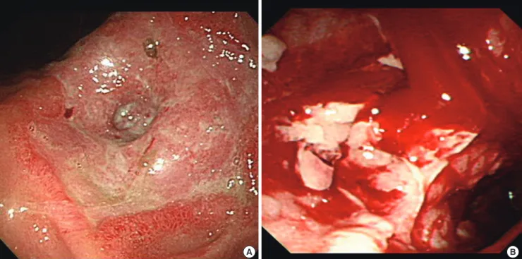
A 31-yr-old man was admitted with corrosive acids ingestion on July 13, 2010. Endoscopic findings showed long and deep linear ulceration and black pigmented mucosal change in the esoph- agus and stomach. Four weeks later the patient complained of abdominal distension and vomiting. Significant pyloric stricture was found in follow up endoscopy (Fig. 1A). The next day, we conducted endoscopic balloon dilatation, through an 8 mm bal- loon for 40 seconds. After endoscopic balloon dilatation, pyloric stricture was improved and endoscopy passage was possible.
There was no evidence of perforation and only mild mucosal
injury was observed (Fig. 1B). However, 1 hr after the balloon dilatation procedure, he complained of diffuse abdominal pain.
His blood pressure was 95/65 mmHg, pulse rate was 110 times per min. The abdomen was soft and slightly distended with whole abdomen tenderness. However, no involuntary guarding or re- bound tenderness was detected. Laboratory data showed white blood cell count of 8,930/µL, hemoglobin of 10.0 g/dL, C-reac- tive protein of 0.16 mg/dL, lactate dehydrogen level of 333 U/L.
Abdominal CT showed large amounts of HPVG in the peripher- ies of the liver (Fig. 2A). The patient was moved to intensive care unit and an emergency operation was considered. However, we decided to go on conservative treatment because there was no clinical evidence of bowel necrosis or perforation and vital signs returned to normal range. The patient was monitored closely and received fluid replacement, Levin tube with natural drain- age, broad-spectrum antibiotics. Five days later, we performed follow-up abdominal CT; hepatic portal gas showed complete spontaneous resolution (Fig. 2B). The patient was then trans- ferred to the gastro-intestinal surgical team for elective surgery for gastrojejunostomy, and was discharged in good condition without any complication of hepatic portal venous gas.
DISCUSSION
In this report, we first described a case of HPVG following by en- doscopic balloon dilatation. HPVG was first described by Wolfe and Evens in 1955 in infants with necrotizing enterocolitis (NEC)
Lee CG, et al. • Hepatic Portal Venous Gas after Endoscopic Balloon Dilatation
http://jkms.org 1109
DOI: 10.3346/jkms.2011.26.8.1108
(6). It has been reported in many abdominal conditions such as inflammatory bowel disease, diverticulitis, intraabdominal sep- sis, pneumatosis intestinalis, pancreatitis, bowel obstruction and suppurative cholangitis (2, 7). It also has been reported as iatro- genic complications of as endoscopic retrograde cholangiopan- creatography (ERCP), liver transplantation, esophageal variceal band ligation and percutaneous endoscopic gastrostomy (PEG)
(2-5). HPVG following endoscopic balloon dilatation has not been previously reported.
HPVG most often represents an ominous clinical finding. The clinical presentation of HPVG is usually consistent with acute abdomen but occasionally shows vague abdominal symptoms and nonspecific laboratory findings (1, 8). The major predispos- ing factor of the development of HPVG is the damage of intesti-
A B
Fig. 1. Endoscopic findings. (A) Severe pyloric stricture was observed, followed by a caustic lye injury 4 weeks later. (B) After endoscopic balloon dilatation, pyloric stricture was improved and endoscopy passage was possible. Only mild mucosal injury was found.
A B
Fig. 2. Enhanced abdomen CT findings. (A) Enhanced abdominal CT finding after balloon dilatation. Massive hepatic portal venous gas is observed. (B) Enhanced abdominal CT five days later is shown. Spontaneous resolution of amounts of hepatic portal venous gas is observed.
Lee CG, et al. • Hepatic Portal Venous Gas after Endoscopic Balloon Dilatation
1110 http://jkms.org DOI: 10.3346/jkms.2011.26.8.1108
nal mucosa combined with bowel dilatation or bacterial gas pro- duction (1, 8). Most authors agree that the formation of HPVG is a multiplex event in which two or three of these conditions are combined (1, 8). We speculated that the main cause of HPVG in our case was gastric dilatation. Increased intraluminal pressure forces intraluminal gas to pass through a damaged bowel wall, allowing the intraluminal gas to enter the portal circulation. Like this, the development of HPVG secondary to gastric dilatation has been reported in cases of ileus, barium enema and blunt ab- dominal trauma (9).
Originally the diagnosis of HPVG was based on plain abdom- inal radiography, primarily left lateral decubitus views (10). How- ever, along with the development of CT scan, we found CT is more sensitive than plain film radiography. The pattern for HPVG in CT scan is described as a tubular lucency branching from the porta hepatis to the liver capsule (10, 11). The gas travels periph- erally in the portal vein consequent to the centrifugal flow of blood, and the branching pattern with a peripheral distribution extending to within 2 cm of the liver capsule (10, 11). Addition- ally, in abdomen CT finding, HPVG must be differentiated from gas in the biliary tracts such as pneumobilia, which tends to move with the centripetal flow of bile toward the hilum, and whose central air lucencies do not extend to within 2 cm of the liver cap- sule (10).
Gas in the portal venous system was once regarded as a fatal event (12). However, in most clinical settings, the finding of HPVG alone cannot be a predictor of mortality or an indication for emer- gency exploration. The key point when making a decision of its surgical management is the presence of peritonitis or bowel per- foration (13, 14). In our case, there was no clinical evidence of peritonitis or bowel perforation. Therefore, we decided to go on conservative treatment. In addition, if there is a gastric dilatation due to endoscopy, inserting of a Levin tube for gastric decom- pression plays an essential role in rapid removal of portal vein gas (9).
In conclusion, we report the first case of HPVG followed by endoscopic balloon dilatation. Gas in the portal vein in our case may occur as a transient incidental finding with gastric dilata- tion and mucosal damage. HPVG resulting from iatrogenic causes, the amount of portal vein gas may be not an indication of surgical management and medically stable patients without sign of bowel peritonitis or perforation could be effectively man- aged with supportive care.
REFERENCES
1. Abboud B, El Hachem J, Yazbeck T, Doumit C. Hepatic portal venous gas: physiopathology, etiology, prognosis and treatment. World J Gas- troenterol 2009; 15: 3585-90.
2. Fred HL, Mayhall CG, Harle TS. Hepatic portal venous gas. A review and report on six new cases. Am J Med 1968; 44: 557-65.
3. Bobba RK, Arsura EL. Hepatic portal and mesenteric vein gas as a late complication of percutaneous endoscopic gastrostomy tube placement in an elderly patient. Dig Dis Sci 2005; 50: 411-4.
4. Alqahtani S, Coffin CS, Burak K, Chen F, MacGregor J, Beck P. Hepatic portal venous gas: a report of two cases and a review of the epidemiology, pathogenesis, diagnosis and approach to management. Can J Gastroen- terol 2007; 21: 309-13.
5. Ahmed K, Atiq M, Richer E, Neff G, Kemmer N, Safdar K. Careful obser- vation of hepatic portal venous gas following esophageal variceal band ligation. Endoscopy 2008; 40(Suppl 2): E103.
6. Wolfe JN, Evans WA. Gas in the portal veins of the liver in infants: a roent- genographic demonstration with postmortem anatomical correlation.
Am J Roentgenol Radium Ther Nucl Med 1955; 74: 486-8.
7. Park HC, Lee WS, Joo SY, Park SY, Joo YE, Kim HS, Choi SK, Rew JS. He- patic portal venous gas associated with acute pancreatitis: reports of two cases and review of literature. Korean J Gastroenterol 2007; 50: 131-5.
8. Peloponissios N, Halkic N, Pugnale M, Jornod P, Nordback P, Meyer A, Gillet M. Hepatic portal gas in adults: review of the literature and presen- tation f a consecutive series of 11 cases. Arch Surg 2003; 138: 1367-70.
9. Lamberto S, Vinci S, Salamone I, Racchiusa S. Intrahepatic portal vein gas. Good prognosis in patients with gastric dilatation. Case report. Ra- diol Med 2001; 101: 508-10.
10. Schindera ST, Triller J, Vock P, Hoppe H. Detection of hepatic portal ve- nous gas: its clinical impact and outcome. Emerg Radiol 2006; 12: 164-70.
11. Chevallier P, Peten E, Souci J, Chau Y, Padovani B, Bruneton JN. Detec- tion of portal venous gas on sonography, but not on CT. Eur Radiol 2002;
12: 1175-8.
12. Bani-Hani KE, Heis HA. Iatrogenic gastri dilatation: a rare and transient cause of hepatic-portal venous gas. Yonsei Med J 2008; 49: 669-71.
13. Nelson AL, Millington TM, Sahani D, Chung RT, Bauer C, Herti M, War- shaw AL, Conrad C. Hepatic portal venous gas. Arch Surg 2009; 144: 575- 81.
14. Kinoshita H, Shinozaki M, Tanimura H, Umemoto Y, Sakaguchi S, Taki- fuji K, Kawasaki S, Hayashi H, Yamaue H. Clinical features and manage- ment of hepatic portal venous gas: four case reports and cumulative re- view of the literature. Arch Surg 2001; 136: 1410-4.