INTRODUCTION
Transient mid- and basal ventricular ballooning is a new variant of transient left ventricular (LV) ballooning similar to transient LV apical ballooning syndrome, mainly due to emotional and physical stress (1-4). Its exact mechanism of pathogenesis remains unknown. We report transient mid- and basal ventricular ballooning associated with catechola- mines in two patients. These cases showed similar clinical courses to transient LV apical ballooning syndrome and cat- echolamine-mediated myocardial dysfunction, which might be a potential mechanism of this syndrome.
CASE REPORT Case 1
A 32-yr-old man presented to our emergency department with palpitation and squeezing chest pain. His initial blood pressure was 190/112 mmHg and electrocardiogram showed sinus tachycardia of 120/min. The initial electrocardiogram showed slight ST segment elevation in the inferior leads (lead II, III, and aVF). Echocardiography demonstrated a dilated mid portion of the LV and LV ejection fraction was 35%
(Fig. 1A, B). However, the motion of the LV apex was nor- mal. During the echocardiographic examination, his heart rate decreased suddenly to 70/min from 122/min. Serum
level of creatinine kinase (CK) was 177 IU/L (normal range:
55-170 IU/L), CK-MB was 7.8 ng/mL (normal range: 0-4 ng/mL), and troponin I was 1.74 ng/mL (normal range: 0-0.05 ng/mL). The serum level of N-terminal-pro-B type natri- uretic peptide (NT-pro-BNP) was increased up to 733 pg/
mL (normal range: <200 pg/mL).
We performed emergent cardiac catheterization to rule out acute myocardial infarction. The coronary arteries were free of any organic stenosis and LV showed severe hypokine- sia in the mid to basal portion. However, the LV apical wall motion was preserved (Fig. 2).
To rule out pheochromocytoma, we performed abdominal computerized tomography (CT). An abdominal CT showed an approximately 7.8×8.3×10.0 cm large septated cystic mass with irregular wall enhancement of the right adrenal gland (Fig. 3A). His serum catecholamine levels were reported as follows: epinephrine, 436.1 pg/mL (normal range: 0-140 pg/mL) and norepinephrine, 509.7 pg/mL (normal range:
0-450 pg/mL). The urinary catecholamine levels were as fol- lows: epinephrine, 1,530.6 μg/day (normal range: 10-20 μg/
day), norepinephrine, 1,048.8 μg/day (normal range: 15-80 μg/day) and metanephrine, 22,107.4 μg/day (normal range:
52-341 μg/day). From these findings, catecholamine induced cardiomyopathy showing midventricular ballooning was our final diagnosis.
The patient was treated with oral furosemide, an oral alpha blocker (prazosin) and intravenous nitrate infusion. The low dose of beta blocker was initiated after alpha blockade. The
898
Address for correspondence Jae-Hyeong Park, M.D.
Division of Cardiology, Department of Internal Medicine, Chungnam National University Hospital, 640 Daesa-dong, Jung-gu, Daejeon 301-721, Korea Tel : +82.42-259-8237, Fax : +82.42-259-8238 E-mail : jaehpark@cnuh.co.kr
Key Words : Ventricular Dysfunction, Left; Catecholamines; Pheochromocytoma
Received : 23 July 2007 Accepted : 28 November 2007
follow up echocardiography after three days of treatment showed marked improvement of LV systolic function and decreased LV dimension. The LV ejection fraction was 59%, with no regional wall motion abnormalities (Fig. 1C, D).
He underwent successful surgical resection and the patho- logic findings of the excised adrenal gland were compatible with pheochromocytoma (Fig. 3B).
Case 2
A 47-yr-old man with no cardiac history presented with
hypotension after resection of an inverted papilloma of the left nasal cavity. He was treated with subcutaneous epinephrine (epinephrine HCl 1 mg/ample) injection, about 0.2-0.3 mL, to the nasal mucosa to prevent excessive bleeding. After the injection, his systolic blood pressure and heart rate transient- ly increased to 170 mmHg and 140/min, respectively, then decreased to 50 mmHg and 70/min, respectively. Electro- cardiogram showed normal sinus rhythm and no significant change of his initial electrocardiogram.
Echocardiography demonstrated a dilated LV, akinesia of the basal to mid portion of the LV with sparing of apex and
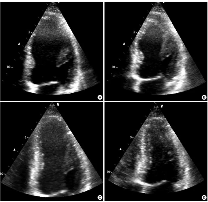
Fig. 1. An apical four-chamber view of the left ventricle in the emergency department is shown at end-diastole (A) and end-systole (B). At end-systole, mid- and basal ventricular ballooning was noted. The follow-up echocardiographic study was performed after three days of treatment. The left ventricular dimension was decreased and ballooning had disappeared (C at end-diastole and D at end-systole).
A B
C D
LV ejection fraction was 28% (Fig. 4A, B). Serum level of CK was 248 IU/L (normal range: 55-170 IU/L), CK-MB was 9.1 ng/mL (normal range: 0-4 ng/mL), and troponin I was 5.4 ng/mL (normal range: 0-0.05 ng/mL). The serum level of NT-pro-BNP was increased up to 4,277 pg/mL (nor- mal range: <200 pg/mL). No coronary angiography was performed because of the low probability of coronary arteri- al disease and a normal preoperative treadmill exercise test.
A follow-up echocardiogram obtained after two days of con- servative treatment showed normalized LV size and func- tion (Fig. 4C, D). The LV ejection fraction was 56%, with
no regional wall motion abnormalities. Treatment with β- blocker and angiotensin converting enzyme inhibitor was initiated and continued for about one month after normal- ization of LV function.
DISCUSSION
We report two cases of transient mid- and basal ventricu- lar ballooning syndrome associated with catecholamines.
Transient mid- and basal ventricular ballooning is a new vari-
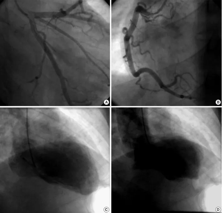
Fig. 2. The coronary angiogram showed normal left (A) and right coronary arteries (B). The left ventriculogram in the right anterior oblique view showed at end-diastole (C) and end-systole (D). At end-systole, mid- and basal ventricular ballooning was noted. However, LV api- cal motion was normal.
A B
C D
ant of the transient LV apical ballooning syndrome, which is also known as ‘‘stress cardiomyopathy’’, and includes tran- sient cardiac contractile abnormalities and heart failure pre- cipitated by acute emotional or physical stress. However,
the involvement of the LV’s mid- and basal ventricle with sparing of the apical segment is the unique finding of this variant (1, 2). Proposed potential mechanisms of transient LV apical ballooning are multivessel epicardial spasm, micro-
Fig. 3. (A) Abdominal computerized tomography showed an approximately 7.8×8.3×10.0 cm sized large septated cystic mass with irreg- ular wall enhancement of the right adrenal gland (arrow). (B) Large and pink cells are arranged in nests with capillaries between them in the high power field image, which is compatible with pheochromocytoma.
A 50 μm B
Fig. 4. An apical four-chamber view of the left ventricle after operation is shown at end-diastole (A) and end-systole (B). At end-systole, the mid-and basal ventricular ballooning was noted with sparing of the LV apex. A follow-up echocardiographic study was performed after two days. The left ventricular dimension had decreased and ballooning was no longer present (C at end-diastole and D at end-systole).
A B
C D
segment is unknown. One possible explanation is different distribution of sympathetic nerves (9) and dissimilar densi- ty of sympathetic nerves in the heart (10), which make the apex more vulnerable to a sudden increase in circulating cat- echolamine levels. In our cases, the patients showed severe mid- and basal ventricular dysfunction from transient eleva- tion of blood catecholamine level probably due to hemor- rhagic necrosis of the adrenal pheochromocytoma and injec- tion of epinephrine, respectively. The variations in segmen- tal involvement regardless of coronary anatomy in patients with excessive catecholamine levels may suggest different susceptibility to sympathetic stimulation from individual to individual.
REFERENCES
1. Hurst RT, Askew JW, Reuss CS, Lee RW, Sweeney JP, Fortuin FD, Oh JK, Tajik AJ. Transient midventricular ballooning syndrome: a new variant. J Am Coll Cardiol 2006; 48: 579-83.
2. Yasu T, Tone K, Kubo N, Saito M. Transient mid-ventricular bal-
Neurohumoral features of myocardial stunning due to sudden emo- tional stress. N Engl J Med 2005; 352: 539-48.
6. Lacy CR, Contrada RJ, Robbins ML, Tannenbaum AK, Moreyra AE, Chelton S, Kostis JB. Coronary vasoconstriction induced by mental stress (simulated public speaking). Am J Cardiol 1995; 75:
503-5.
7. Bolli R, Marban E. Molecular and cellular mechanisms of myocar- dial stunning. Physiol Rev 1999; 79: 609-34.
8. Takizawa M, Kobayakawa N, Uozumi H, Yonemura S, Kodama T, Fukusima K, Takeuchi H, Kaneko Y, Kaneko T, Fujita K, Honma Y, Aoyagi T. A case of transient left ventricular ballooning with pheo- chromocytoma, supporting pathogenetic role of catecholamines in stress-induced cardiomyopathy or takotsubo cardiomyopathy. Int J Cardiol 2007; 114: e15-7.
9. Pierpont GL, DeMaster EG, Cohn JN. Regional differences in ad- renergic function within the left ventricle. Am J Physiol 1984; 246:
H824-9.
10. Kawano H, Okada R, Yano K. Histological study on the distribu- tion of autonomic nerves in the human heart. Heart Vessels 2003;
18: 32-9.