JCS
Journal of Chest SurgeryRecent Evidence and Initial Experiences of Transcatheter Edge- to-Edge Repair of the Mitral Valve in South Korea
Sung-Jin Hong, M.D., Jung-Sun Kim, M.D., Ph.D., Geu-Ru Hong, M.D.
Division of Cardiology, Severance Cardiovascular Hospital, Yonsei University Health System, Seoul, Korea
ARTICLE INFO
Received
January 28, 2021
AcceptedFebruary 9, 2021
Corresponding authorJung-Sun Kim
Tel
82-2-2228-8457
Fax82-2-393-2041
E-mail[email protected]
ORCIDhttps://orcid.org/0000-0003-2263-3274
As a percutaneous technique for the reduction of mitral regurgitation, the MitraClip sys- tem (Abbott Vascular, Abbott Park, IL, USA) for transcatheter edge-to-edge repair of the mitral valve was developed in 1998 and first used in 2003. Its main advantage is being less invasive than surgery, because it can be performed through a transcatheter approach without any hemodynamic compromise. Recent studies have shown that this procedure reduces symptoms and improves functional capacity with low complication rates. Two randomized clinical trials have investigated the use of this technique for functional mitral regurgitation. The Korean Ministry of Food and Drug Safety approved its use for degener- ative mitral regurgitation in 2019, and this procedure started to be performed in Korea in January 2020. Its use for functional mitral regurgitation was also approved in Korea in 2020.
In this article, recent evidence on transcatheter edge-to-edge repair of the mitral valve and our initial experiences in Korea will be reviewed.
Keywords: Mitral regurgitation, Transcatheter edge-to-edge repair, Heart valve disease, Mitral valve repair
Copyright©The Korean Society for Thoracic and Cardiovascular Surgery. 2021. All right reserved.
This is an Open Access article distributed under the terms of the Creative Commons Attribution Non-Commercial License (http://creativecommons.org/licenses/
Introduction
As a percutaneous technique to reduce mitral regurgita- tion (MR), the MitraClip system (Abbott Vascular, Abbott Park, IL, USA) for transcatheter edge-to-edge repair (TEER) of the mitral valve was developed in 1998 and first used in 2003. The main advantage of the TEER procedure that it is less invasive than surgery because it can be per- formed via the femoral venous approach without any he- modynamic compromise. Furthermore, unlike other per- cutaneous cardiovascular interventions such as coronary or aortic valve interventions, contrast medium is not re- quired.
Based on recent results and evidence from randomized clinical trials, the use of MitraClip system was permitted in Europe (Conformité Européene, CE Mark) in 2008. In the United States, the Food and Drug Administration ap- proved it in 2013 for degenerative MR, and in 2018 for functional MR. The Korean Ministry of Food and Drug Safety approved its use for degenerative MR in 2019, and this procedure started to be performed in Korea in January
2020. Its use for functional MR was also approved in Korea in 2020. In this article, recent evidence regarding TEER and our initial experiences in Korea will be reviewed.
Recent clinical trials and evidence for TEER
After the phase I feasibility study of the Endovascular Valve Edge-to-edge Repair Study (EVEREST) trial with 55 patients [1,2], the EVEREST II trial randomized 279 pa- tients with 3+ or 4+ MR to receive TEER with the Mitra- Clip or open mitral valve surgery in a 2:1 ratio [3]. Patients with both degenerative and functional MR were enrolled, and functional MR was present in 27% of patients [3]. In the surgery group, 86% of patients were treated with mitral valve repair and 14% had mitral valve replacement. The primary endpoint, a composite of freedom from death, surgery for mitral valve dysfunction, and 3+ or 4+ MR at 12 months, was achieved in 55% of patients in the percuta- neous repair group and 73% of those in the surgical group (p=0.007) [3]. This difference was mainly driven by the
https://doi.org/10.5090/jcs.21.010 pISSN: 2765-1606 eISSN: 2765-1614 J Chest Surg. 2021;54(3):165-171
Collective
of Current
Reviews,
Lectures
https://doi.org/10.5090/jcs.21.010
JCS
need for surgery for mitral valve dysfunction (20% in the percutaneous repair group versus 2% in the surgical repair group, p<0.001). However, major adverse events within the first 30 days occurred more frequently in the surgery group, mainly driven by the need for blood transfusions (13% versus 45%, p<0.001). Thus, though percutaneous re- pair was less effective at reducing MR than surgery, TEER was superior in terms of safety and showed similar im- provements in clinical outcomes [3]. According to the sec- ondary analyses, percutaneous repair resulted in greater preservation of left ventricular (LV) function and a smaller decrease in LV dimensions compared with surgery. When the EVEREST II population was followed for 5 years, the primary endpoint still favored the surgical group (64.3% in the surgical group and 44.2% in the percutaneous repair group) [4]. However, there was no significant difference in the rate of death (21% versus 27%), and freedom from mi- tral valve surgery or reoperation remained the same after percutaneous repair or surgery beyond 6 months after the procedure. Improvements in symptoms and LV dimensions were maintained at 5 years in both groups [4]. The EVER- EST II trial became the landmark trial for commercial ap- proval in the United States, particularly for degenerative MR. Functional MR is frequently present in patients with ad- vanced heart failure. Optimal medical and cardiac resyn- chronization therapy have been proposed as first-line strat- egies for functional MR. Surgical intervention may be considered in patients who are refractory to currently available medical or cardiac resynchronization therapy as a class IIb indication because the beneficial effect of mitral valve surgery has not been proven by prospective random- ized clinical trials [5,6]. Meanwhile, transcatheter mitral intervention can be better option with a less invasive strat- egy in high-risk population with surgery and comparable strategy to mitral valve surgery. In 2018, two important randomized trials that included the patients with function- al MR and heart failure, the MITRA-FR (Percutaneous Re- pair with the MitraClip Device for Severe Functional/
Secondary Mitral Regurgitation) trial and COAPT (Car- diovascular Outcomes Assessment of the MitraClip Percu- taneous Therapy for Heart Failure Patients with Function- al Mitral Regurgitation) trial, were published [7,8]. It is interesting to note that these 2 trials gave different results, as the MITRA-FR trial failed to prove superiority for the primary endpoint, but the COAPT trial proved it. The MI- TRA-FR study randomized 307 patients with symptomatic LV dysfunction and significant secondary MR to either medical therapy or medical therapy combined with the
MitraClip procedure, and 92% of patients achieved an MR grade of 2+ immediately after the procedure [7]. There was no difference in the primary outcome of all-cause death and unplanned re-hospitalization for heart failure at 1 year, which occurred in 55% of the intervention group and 51%
of the control group (odds ratio, 1.16; p=0.53) [7]. In the other COAPT trial, a total of 614 patients with symptomat- ic heart failure and moderate-to severe or severe secondary MR to medical therapy or medical therapy and MitraClip repair [8]. The primary outcome was the rate of hospital- ization for heart failure within 24 months, which was 35.8% per patient-year in the device group as compared with 67.9% in the control group (hazard ratio, 0.53;
p<0.001) [8]. Moreover, the powered secondary endpoint of death from any cause within 24 months was significantly lower, occurring in 29.1% of the patients in the device group versus 46.1% in the control group (hazard ratio, 0.62;
p<0.001). All prespecified secondary endpoints including quality of life and functional assessments significantly im- proved in the MitraClip arm [9,10].
The following reasons have been proposed for these dis- cordant results [11-13]. First, the definitions of severe MR were different between European and American guide- lines. MR was more severe in the COAPT trial than in the MITRA-FR trial. Second, the LV end-diastolic volumes were smaller in the COAPT trial than in the MITRA-FR trial (101 versus 135 mL/m
2). Third, more aggressive guide- line-directed medical therapy was overseen by the screen- ing committee in the COAPT trial. Fourth, the number of patients receiving more than 1 clip was higher in the COAPT trial, possibly explaining the higher proportion of patients with moderate-to-severe or severe residual MR at 1 year in the MITRA-FR trial (17% versus 5% in COAPT).
In light of the apparently discordant results from 2 recent
randomized controlled trials of mitral valve repair, the
concept of the characterization of MR as proportionate or
disproportionate to LV size was proposed to differentiate
patients who can benefit from the TEER procedure as part
of optimal treatment for chronic heart failure and systolic
dysfunction [11,12]. Patients with heart failure, an LV ejec-
tion fraction (LVEF) of 30%, an LV end-diastolic volume of
220 to 250 mL, and a regurgitation fraction of 50% would
be expected to have an effective regurgitant orifice area of
approximately equal to 0.3 cm
2independent of specific
tethering abnormalities of the mitral valve leaflets [11]. The
MR in these patients is proportionate to the degree of LV
dilatation and can respond to drugs and devices that re-
duce LV end-diastolic volume. In contrast, patients with an
effective regurgitant orifice area of 0.3 to 0.4 cm
2, but with
Sung-Jin Hong, et al. Transcatheter Edge-to-Edge Repair of MV JCS
an LV end-diastolic volume of only 160 to 200 mL, exhibit degrees of MR that are disproportionately higher than would be predicted by LV end-diastolic volume. Thus, these patients were proposed to preferentially benefit from interventions directed at the mitral valve [11-13].
Recommendations and indications for TEER according to current guidelines
For the assessment of the feasibility of TEER, a rigorous echocardiographic assessment and multidisciplinary team consensus is required [14]. The anatomically favorable fea- tures of TEER are non-commissural pathology (medial, middle, lateral segments), no or minimal calcification, mean trans-mitral gradient <4 mm, mitral valve area ≥4 cm
2, and a grasping zone length >10 mm [14]. For primary MR, flail width <15 mm, flail gap <10 mm, single segment pathology, and normal leaflet thickness are also favorable features. For secondary MR, coaptation depth <11 mm and coaptation length (overlap length) ≥2 mm are favorable.
The 2020 American College of Cardiology/American
Heart Association guideline, for the patients with primary MR, recommended that TEER is reasonable in severely symptomatic patients (New York Heart Association [NYHA] class III or IV) with primary severe MR and high or prohibitive surgical risk if the mitral valve anatomy is favorable for the repair procedure and the patient’s life ex- pectancy is at least 1 year, as a class IIa recommendation [5]. It is notable that the previous 2017 European Society of Cardiology guideline recommended that TEER may be considered in patients with symptomatic severe primary MR who fulfil the echocardiographic criteria of eligibility and are judged inoperable or at high surgical risk by the heart team, avoiding futility, as a class IIb recommenda- tion [6].
As for chronic secondary MR, TEER is reasonable (class IIa) in patients with appropriate anatomy as defined on transesophageal echocardiography and with an LVEF be- tween 20% and 50%, LV end-systolic diameter ≤70 mm, and pulmonary artery systolic pressure ≤70 mm Hg, in pa- tients with chronic severe secondary MR related to LV sys- tolic dysfunction (LVEF <50%) who have persistent severe
A B C
D E F
G H I
R R
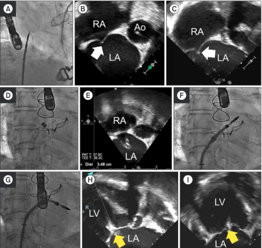
Fig. 1. Step-by-step procedure. (A–
C) Trans-septal pucture. Tenting of
the atrial septum (white arrow) can
be seen in transesophageal echocar-
diography (TEE) images of short-axis
and 4-chamber views. (D, E) Intro-
duction of a 24F orientable guiding
catheter to the left atrium (LA). (F)
Introduction of a steerable clip de-
livery catheter. (G–I) Advance of the
device into the left ventricle (LV) and
grasping of the mitral valve (yellow
arrow) under the left ventricular out-
flow tract view and intercommisural
view on TEE images. RA, right atri-
um; Ao, aorta.
https://doi.org/10.5090/jcs.21.010
JCS
symptoms (NYHA class II, III, or IV) while on optimal guideline-directed medical therapy for heart failure (stage D) [5]. Furthermore, we expect that the RESHAPE-HF2 (NCT02444338) and MATTERHORN (NCT02371512) tri- als might provide more concrete evidence and appropriate indications for the application of the MitraClip device for functional MR and the concept of disproportionate MR.
Step-by-step procedure
TEER is performed under general endotracheal anesthe- sia. To access the left atrium, a trans-septal puncture is performed via the femoral vein. One or two Perclose Pro- Glide devices (Abbott Vascular, Abbott Park, IL, USA) might be deployed in a pre-close fashion in the right com- mon femoral vein followed by introduction of an 18F sheath to help hemostasis after the procedure. Trans-septal puncture is performed in a posterior and mid-to-superior location on the interatrial septum using transesophageal echocardiographic guidance after confirmation of an ade- quate distance from the puncture location to the mitral annular plane (Fig. 1A–C). After trans-septal puncture, a 24F orientable guiding catheter is percutaneously intro- duced (Fig. 1D, E), and a steerable clip delivery catheter enables orientation of the clip (Fig. 1F). Throughout the entire procedure, transesophageal echocardiographic guid- ance including real-time 3-dimensional images is import- ant for the septal puncture, introduction of clip delivery system, positioning, and precision-targeting of the free edges of the opposing leaflets at the site of regurgitation.
The device is then advanced into the left ventricle and while pulling back the catheter, the mitral valve leaflets are grasped (Fig. 1G–I).
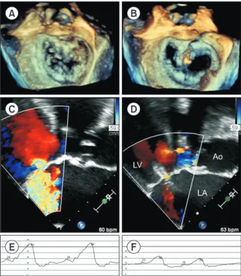
Once optimal grasping has been undertaken, the clip is closed to create a double orifice (Fig. 2A, B). An echocar- diographic assessment is performed for adequate leaflet in- sertion, residual MR with color Doppler (Fig. 2C, D), and new trans-valvular gradients avoiding mitral stenosis prior to the final deployment. Direct measurements of the left atrial mean pressure and V wave provide complementary hemodynamic data to guide treatment decision-making (Fig. 2E, F). Multiple clips can be implanted to optimize imperfect results on a case-by-case basis if the gradients and anatomy allow. Hemodynamics usually remain very stable during the procedure and the recovery time is short.
Our initial experience and the results from major registries
At Severance Cardiovascular Hospital, between February 2020 and January 2021, a total of 17 patients with moderate to severe or severe MR underwent TEER with the Mitra- Clip NT system (Abbott Vascular), which is currently available in Korea. The patients’ median age was 81 years (interquartile range, 76–86 years) and 10 patients (59%) were women. The proportion of patients with NYHA III and IV functional status was 53% and 47%, respectively.
The comorbidities were as follows: atrial fibrillation, 11 pa- tients (65%); hypertension, 15 patients (88%); diabetes, 5 patients (29%); and chronic kidney disease, 10 patients (59%). At the time of the procedure, 1 patient (6%) was in shock. The median EuroSCORE II was 5.0% (interquartile range, 2.3%–6.9%). The median Society for Thoracic Sur- geons (STS) scores for mitral valve repair and mitral valve
Fig. 2. Case example comparing before and after the procedure.
(A) Before the procedure, A2 prolapse can be seen on 3-dimen- sional transesophageal echocardiography (TEE) images. (B) Cre- ation of a double orifice after clip closure. (C) Severe mitral regur- gitation (MR) on the left ventricular outflow tract view of the TEE image before the procedure. (D) After the clip closure, MR was reduced to a trivial degree. (E) Before the procedure, the mean left atrial (LA) pressure was 35 mm Hg with a prominent V wave. (F) After the procedure, the mean LA pressure was 21 mm Hg with a decreased V wave. Ao, aorta.
A B
C D
E F
Sung-Jin Hong, et al. Transcatheter Edge-to-Edge Repair of MV JCS
replacement were 6.8% (interquartile range, 4.0%–9.1%) and 9.0% (interquartile range, 8.0%–12.0%), respectively.
The overwhelming majority of patients (88%) had STS scores ≥8% for mitral valve replacement. One patient had severe right ventricular dysfunction with severe tricuspid regurgitation. One patient had severe heart failure with chronic constrictive pericarditis.
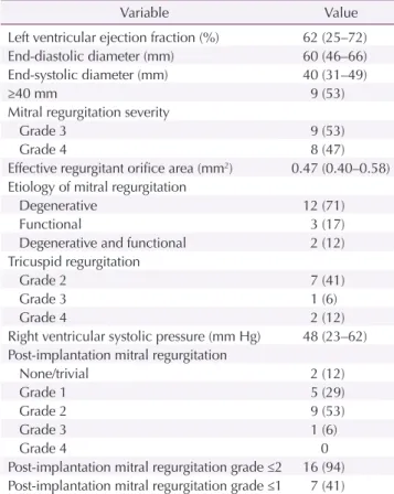
Echocardiographic data are presented in Table 1. The median LVEF and end-diastolic diameter were 62% and 60
mm, respectively. Before the procedure, 9 patients had grade 3+ MR and 8 patients had grade 4+ MR. Fig. 3A presents the procedural time (from the initiation of the trans-septal puncture to device deployment). It took a me- dian of 129 minutes (interquartile range, 79–211 minutes;
minimum, 70 minutes; maximum, 253 minutes). Twelve patients (71%) had 1 clip implanted and 5 patients (29%) required 2 clips; the average number of devices used was 1.29. Post-implantation MR severity is presented in Table 1, and post-implantation MR grade ≤2 could be achieved in 16 patients (94%) (Fig. 3B). The procedural complications and device-related adverse events were as follows: cardiac Table 1. Echocardiographic data (n=17)
Variable Value
Left ventricular ejection fraction (%) 62 (25–72)
End-diastolic diameter (mm) 60 (46–66)
End-systolic diameter (mm) 40 (31–49)
≥40 mm 9 (53)
Mitral regurgitation severity
Grade 3 9 (53)
Grade 4 8 (47)
Effective regurgitant orifice area (mm
2) 0.47 (0.40–0.58) Etiology of mitral regurgitation
Degenerative 12 (71)
Functional 3 (17)
Degenerative and functional 2 (12)
Tricuspid regurgitation
Grade 2 7 (41)
Grade 3 1 (6)
Grade 4 2 (12)
Right ventricular systolic pressure (mm Hg) 48 (23–62) Post-implantation mitral regurgitation
None/trivial 2 (12)
Grade 1 5 (29)
Grade 2 9 (53)
Grade 3 1 (6)
Grade 4 0
Post-implantation mitral regurgitation grade ≤2 16 (94) Post-implantation mitral regurgitation grade ≤1 7 (41) Values are presented as median (interquartile range) or number (%).
Table 2. Summary of major registries of the transcatheter edge-to-edge repair procedure
Variable No. of
patients Age (yr) EuroSCORE (%)
STS score
a)(%)
Etiology of MR (%) Acute procedural success (%)
30-day mortality
(%)
1-year mortality Degenerative Functional (%)
ACCESS-EU [15] 567 74±10 22±18 - 23 77 91 3.4 11.8
TCVT [16] 628 74±10 20±17 - 28 72 95 2.9 15.3
TRAMI [17] 749 76 (71–81) 20 (12–31) 6 (4–11) 28 71 97 4.7 20.3
STS/ACC TVT [18] 2,952 82 (74–86) - 9 (6–14) 86 9 92 5.8 25.8
Asia-Pacific Registry [19] 142 71±12 17±15
b)7±8 53 47 94 5.6 -
Values are presented as number, mean±standard deviation, or median (interquartile range).
MR, mitral regurgitation; TCVT, Transcatheter Valve Treatment Sentinel Pilot Registry; TRAMI, transcatheter mitral valve interventions; STS, Society of Thoracic Surgeons; ACC TVT, American College of Cardiology Transcatheter Valve Therapy.
a)
For mitral valve replacement.
b)Logistic EuroSCORE; the others are EuroSCORE II.
Fig. 3. Our initial experiences with the MitraClip (Abbott Vascular, Abbott Park, IL, USA). (A) Total time (minutes) between the initi- ation of trans-septal puncture to device deployment. (B) Severity of mitral regurgitation before and after the TEER procedure. TEER, transcatheter edge-to-edge repair.
300 240 180 120 60
17 Initiationoftrans-septalpuncture todevicedeploymenttime(min)
Case no.
0
Grade 1 Grade 2 Grade 3 Grade 4 Baseline
Post-TEER
20 40 60 80 100
Proportion of the patients (%) 0
A
B
1 2 3 4 5 6 7 8 9 10 11 12 13 14 15 16