http://dx.doi.org/10.5090/kjtcs.2014.47.2.106 ISSN: 2233-601X (Print) ISSN: 2093-6516 (Online)
Department of Thoracic and Cardiovascular Surgery, Seoul St. Mary’s Hospital, The Catholic University of Korea College of Medicine Received: August 23, 2013, Revised: October 15, 2013, Accepted: October 15, 2013
Corresponding author: Hyun Song, Department of Thoracic and Cardiovascular Surgery, Catholic Cancer Center, Seoul St. Mary’s Hospital, The Catholic University of Korea College of Medicine, 222 Banpo-daero, Seocho-gu, Seoul 137-701, Korea
(Tel) 82-2-2258-2858 (Fax) 82-2-594-8644 (E-mail) [email protected]
C
The Korean Society for Thoracic and Cardiovascular Surgery. 2014. All right reserved.
CC
This is an open access article distributed under the terms of the Creative Commons Attribution Non-Commercial License (http://creative- commons.org/licenses/by-nc/3.0) which permits unrestricted non-commercial use, distribution, and reproduction in any medium, provided the original work is properly cited.
Hybrid Coronary Revascularization Using Limited Incisional Full Sternotomy Coronary Artery Bypass Surgery in Multivessel
Disease: Early Results
Joonkyu Kang, M.D., Hyun Song, M.D., Ph.D., Seok In Lee, M.D., Mi Hyung Moon, M.D., Ph.D., Hwan Wook Kim, M.D., Ph.D., Gyun Hyun Jo, M.D., Ph.D.
Background: There are several modalities of coronary artery revascularization for multivessel coronary artery disease. Hybrid coronary revascularization (HCR) with minimally invasive direct coronary artery bypass grafting was introduced for high-risk patients, and recently, many centers have been using it. Limited incisional full sternotomy coronary artery bypass (LIFCAB) involves left internal thoracic artery (LITA)-to-left anterior descending coronary ar- tery (LAD) anastomosis through a sternotomy with a minimal skin incision; it could be considered another techni- que for minimally invasive LITA-to-LAD anastomosis. Our center has performed HCR using LIFCAB, and in this pa- per, we report our short-term results, obtained in the past 3 years. Methods: The medical records of 38 patients from May 2010 to June 2013 were analyzed retrospectively. The observation period after HCR was 1 to 37 months (average, 18.3±10.3 months). The patency of revascularization was confirmed with postoperative coronary angio-computerized tomography or coronary angiography. Results: There were 3 superficial wound complications, but no mortalities. All the LITA-to-LAD anastomoses were patent in the immediate postoperative and follow-up stud- ies, but stenosis was detected in 3 cases of percutaneous coronary intervention. Conclusion: HCR using LIFCAB is safe and yields satisfactory results from the viewpoint of revascularization for multivessel disease.
Key words: 1. Myocardial revascularization 2. Coronary artery bypass 3. Minimally invasive surgery
INTRODUCTION
Coronary artery revascularization for multivessel disease can be managed through various modalities, and the patency of left internal thoracic artery (LITA)-to-left anterior descend- ing artery (LAD) anastomosis is the best among the options.
Recent reports have noted a 10-year patency rate of 95% to 98% [1,2]. The patency of percutaneous coronary intervention (PCI) is enhanced by using drug-eluting stents (DESs). In the
Randomized Comparison of a Sirolimus-Eluting Stent with a Standard Stent for Coronary Revascularization trial, PCI using DESs yielded free rates from target lesion revascularization at 1, 3, and 5 years of 99%, 93.8%, and 89.7%, respectively [3].
Hybrid coronary revascularization (HCR) for multivessel
disease was introduced in the mid-1990s [4]. HCR is a com-
bination method of grafting surgery using LITA of the LAD
lesion with PCI of the remaining lesions. Because the proce-
dure-related morbidity and mortality are lower than those
Table 1. Demographic data of patients
Demographics Value
Male Female Mean age (yr) No. of diseased vessels Two-vessel disease Three-vessel disease Mean EuroSCORE
Mean left ventricular ejection fraction (%) Good (>50)
Moderate (35–50) Poor (<35) Hypertension Diabetes mellitus Hyperlipidemia Stable angina Unstable angina Myocardial infarction
29.0 (76.3) 9.0 (23.7) 64.9±9.52
14.0 (36.8) 24.0 (63.2) 3.71±2.73 58.5±8.88 30.0 (78.9) 7.0 (18.4) 1.0 (2.63) 28.0 (73.7) 21.0 (55.3) 8.0 (21.1) 18.0 (47.4) 16.0 (42.1) 4.0 (10.5) Values are presented as mean±standard deviation or number (%).
Fig. 1. Skin incision for limited incisional full sternotomy coronary artery bypass surgery.
with conventional coronary artery bypass grafting (CABG), interest in HCR has recently increased. Previous studies re- ported that the mortality rate associated with HCR was low.
In particular, for high-risk patients, HCR was found to be safer and more effective [5,6].
Minimally invasive direct coronary artery bypass grafting (MIDCAB) is usually performed through a 5- to 10-cm left anterolateral thoracotomy. There are advantages of this proce- dure compared with conventional CABG, which are better cosmetic outcomes, no morbidity of cannulation or car- diopulmonary bypass, and faster recovery [7,8]. However, in the cases wherein patients were hemodynamically unstable during the operation, switching to conventional CABG was difficult. The conversion rate to conventional sternotomy dur- ing MIDCAB was 1.8%, and the risk of morbidity and mor- tality increased with the conversion [9]. Therefore, we began with the safer minimally invasive coronary artery bypass sur- gery of limited incisional full sternotomy coronary artery by- pass surgery (LIFCAB). LIFCAB involved LITA-to-LAD anastomosis through a sternotomy with a minimal skin incision. The surgeon could create a classic view through the sternotomy, and it was easy to convert to conventional CABG when the patient was hemodynamically unstable. We have used LIFCAB since 2010 and performed HCR using
LIFCAB. Therefore, in this paper, we report the short-term results of HCR using LIFCAB.
METHODS
From May 2010 to June 2013, 78 patients were treated with LIFCAB, whereas 38 multivessel disease patients under- went treatment with HCR. The medical records of these 38 patients were analyzed retrospectively. They included 29 males and 9 females. The mean values of the EuroSCORE and left ventricular ejection fraction were 3.71%±2.73% and 58.50%±8.88%, respectively. The demographic data are sum- marized in Table 1.
HCR was defined as LITA-to-LAD grafting and PCI to a non-LAD lesion during the same hospitalization period. We included patients with high risk for PCI of LAD lesions be- cause of diffuse stenosis or severe calcification. PCI was pos- sible in lesions other than LAD.
LIFCAB involved off-pump CABG surgery through a full sternotomy and small skin incision (range, 5 to 7 cm) (Fig.
1). A long sternal blade was used for performing full sternot- omy under the small incision (Fig. 2).
A Swan-Ganz catheter was inserted via the right internal
jugular vein, and the cardiac output was monitored conti-
nuously. In the supine position, a midline incision was made
Fig. 2. Long sternal blade.
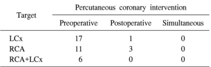
Table 2. Target of percutaneous coronary intervention Target Percutaneous coronary intervention
Preoperative Postoperative Simultaneous LCx
RCA RCA+LCx
17 11 6
1 3 0
0 0 0 LCx, left circumflex artery; RCA, right coronary artery.
from the point of 1 to 1.5 cm above the internipple line to about 6 cm lower, around the xyphoid process. The subcuta- neous tissue was softly separated from the muscle layer with electrocautery until the sternal notch, thus making it easier to perform a full sternotomy. The sternal notch was dissected easily with a right-angled clamp without the fear of bleeding.
After the entire sternum was exposed, sternotomy was per- formed as the second step. First, after marking the median line of the sternum with electrocautery, usual sternotomy was performed from the xyphoid process to the upper end of the skin incision. A small separation of the sternum was retracted with an Army-Navy retractor, and upper sternotomy was per- formed with an oscillating saw (Fig. 2). The fibrous band in the sternal notch was incised for achieving full separation of the sternum. A full pericardiotomy was performed to evaluate the gross state of the heart. With the help of an ordinary re- tractor, the LITA was harvested as pedicled graft by using a harmonic scalpel, and in some cases, the proximal LITA was harvested using a 5-mm thoracoscope. After harvesting the LITA, off-pump coronary artery bypass grafting to the LAD was performed in the usual manner. A small (24Fr) chest tube and a hemovac drain were sufficient for the post- operative drainage. Five or six sternal wires (one or two on the manubrium) were placed for stabilizing the sternum. The subcutaneous tissue was fixed to the pectoralis muscle with one or two interrupted sutures, while not allowing for the stagnation of tissue fluid or blood, which could lead to tissue swelling. The wound was closed considering cosmesis (Fig.
1).
PCI was performed before the operation for 34 patients and after the operation for 4 patients. The patency of the re- vascularization procedure was confirmed with postoperative coronary angio-computerized tomography (CT) or coronary angiography before discharge. The short-term outcomes with respect to patency were determined in the outpatient depart- ment at 6 or 12 months after HCR or when new onset symp- toms developed. In addition, occurrences of mortality and perioperative morbidities were surveyed. All data were col- lected retrospectively, and this study was approved by the in- stitutional review bard of Seoul St. Mary’s Hospital.
RESULTS
All cases except one were performed electively. In one emergency case, PCI was performed on the right coronary ar- tery before the operation due to acute myocardial infarction.
In all cases, HCR was completed without major complica- tions or mortality, but there were two superficial wound in- fections and one case of wound dehiscence. The target ves- sels for PCI are listed in Table 2. The mean number of DESs used for PCI was 1.34±0.71, and the diameter and the length of the lesion were 3.08±0.65 mm and 21.66±8.13 mm, respectively.
The conversion to CABG using CPB was unnecessary. The
mean operation time was 135.66±33.44 minutes. Antiplatelet
agents aspirin (100 mg) and clopidogrel (75 mg) were con-
tinued before the operation for patients who underwent PCI
first. The medication was stopped only on the operative day
and was restarted immediately after extubation. Most patients
could be weaned from mechanical ventilation within 6 hours
postoperatively. There were no mortalities or complications
such as bleeding, arrhythmia, acute kidney injury, and media-
Table 3. Operative and postoperative data
Variable Value
Mean operation time (min) Mean extubation time (hr) Morbidity
Wound infection Wound dehiscence Mortality
135.66±33.44 4.87±2.71
2 (5.26) 1 (2.63)
0
Values are presented as mean±standard deviation or number (%).
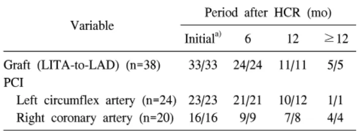
Table 4. Patency of graft and PCI (no. of patents/total) Variable Period after HCR (mo)
Initial
a)6 12 ≥12
Graft (LITA-to-LAD) (n=38) PCI
Left circumflex artery (n=24) Right coronary artery (n=20)
33/33
23/23 16/16
24/24
21/21 9/9
11/11
10/12 7/8
5/5
1/1 4/4 PCI, percutaneous coronary intervention; HCR, hybrid coronary revascularization; LITA-to-LAD, left internal thoracic artery-to- left anterior descending artery.
a)