조선대학교 치의학전문대학원 치과재료학교실
18
0
0
전체 글
(2)
(3)
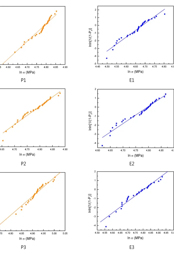
P2 Polyglass polishing with 6 μm diamond paste subsequent to grinding
(5)
(6)
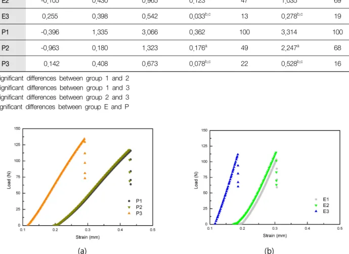
E2 -0.105 0.430 0.965 0.123a
E3 0.255 0.398 0.542 0.033b,c
P2 -0.963 0.180 1.323 0.176a
P3 0.142 0.408 0.673 0.078b,c
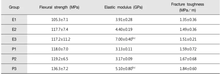
E3 117.2±11.2 7.00±0.40b,c
P3 136.3±7.2 5.10±0.80b,c
(9)
E3 12.72±0.52b,c
P2 22.10±0.67a,*
P3 22.46±0.89b,c,*
(11)
(12)
(13)
(14)
(15)
(16)
(17)
(18)
수치

+3
관련 문서
Influence of agitation or non-agitation on bond strength of Adper TM Easy Bond to pre-etched
The influence of surface conditioning on the shear bond strength of self-adhesive resin cement to zirconia ceramics.. Zirconia as a
The influence of normalized middle school physical education and after school sports club on personality. Kim
Influence of time and rate of nitrogen application on production and botanical composition of forage.. Sudangrass and sorghum-sudangrass hybrids
2001.. to determine influence of planting space and the number of plants per hill on agronomic characters, forage yield and quality of Jeju Italian millet..
Changes in body, flexibility, and physical strength levels of women participating in Pilates have a significant influence on psychological chan ges.. Summarizing
The major contributions of this study are identifying the influence on the performance of venture business by means of structural equation model and the
The average tensile strength value before heat treatment of PLA output was 9.67 N/㎟, and the average tensile strength value after heat treatment was 24.17 N/㎟, which
