618
FOREST SOCIETY
Impact of Transplanting on Tree Growth and Compartmentalization of Pruning Wounds in Acer palmatum Thunb.
Kyu Hwa Lee1*, Kyung Joon Lee1, Ki-Seob Gwak1 and In-Gyu Choi1, 2
1Department of Forest Sciences, Seoul National University, Seoul 151-921, Korea
2Research Institute for Agriculture and Life Sciences, Seoul National University, Seoul 151-921, Korea
Abstract :The objective of this study was to examine the impact of pruning (P treatment) and transplanting (T treatment) of Acer palmatum on cambial growth and compartmentalization of pruning wounds for one year after treatments. Changes of cambial electrical resistance (CER), sizes of pruning wounds, cambial growth of trunks and stems near the wounds, and total phenols at branch unions during the period were examined using a total of 49 trees. After harvesting, areas of discolored wood behind the wounds, relative proportions of extractives, holocellulose and lignin at branch unions were also determined. CER and the cambial growth of trunk at 30 cm above the ground (TGR) were inversely correlated, and differences of CER and TGR among three treatments were significant. TGRs of control, P treatment and P+T treatment after the treatments were 112.2%, 72.4% and 52.5% of the annual growth for the year before the treatments, respectively. The cambial growth rate of stem (SGR) at 1.5 cm above the branch bark ridge and the closure rate of pruning wound (WCR) for one year after treatments were positively correlated, and WCR of P treatment of 39.8% was significantly higher than that of P+T treatment of 31.8%. Wounds of P+T treatment formed greater discolored area per unit area of pruning wound (D/W Ratio) than those of P treatment significantly.
Lower WCR and higher D/W Ratio of P+T treatment suggested less ability of compartmentalizing the wounds than P treatment. Total phenols at branch core of pruning wound for both treatments heightened a month after treatment, and then lowered. The contents at below core of the wound were higher than those at control ones continuously, while they became similar each other at above core. Relatively high phenol contents of the extractives at P+T treatment implied that trees with P+T treatment allocated more energy to compartmentalize their wounds. Holocellulose and lignin contents at the branch core of treated branch unions of both treatments were lower and higher, respectively, than at the same part of the union with living branch, as results of the tree reaction to protection from wounding and microbial invasion.
Key words :cambial growth, discolored wood, total phenols, extractives, holocellulose, lignin
Introduction
Transplanting of large trees is an essential practice these days to achieve intended landscape effects or to produce desired yield without the long wait associated with direct seeding or small transplant technology (Arnold, 2005).
Digging a tree from the nursery for transplanting can result in the loss of 82% to 96% of the tree roots (Gil- man and Beeson, 1996; Hartman et al., 2000; Watson and Sydnor, 1987), which imposes severe physiological stress. It also causes reduction of top growth in trees by limited water absorption which may induce water stress, reduce mineral absorption and assimilation, and reduce
hormone synthesis, especially cytokinin, from the roots (Randolph and Wiest, 1981; Richards and Rowe, 1977).
In addition, root pruning may stimulate production of new roots by directing more assimilates to the root sys- tem. Thus, the growth is redistributed in favor of the roots and the relative shoot growth is reduced again (Geisler and Ferree, 1984).
There have been arguments over the branch pruning at the time of transplanting to compensate for root loss.
Pruning back 15~40% of the top prior to transplanting was a standard recommendation to landscape contractors earlier (Evans and Klett, 1984; Kozlowski and Davies, 1975). But subsequent studies indicated that this practice might not be beneficial in preventing transplant shock and be detrimental by reducing the capacity for photo- synthesis and for hormone production (Ranney et al., 1989; Shoup et al., 1981). So additional pruning beyond
*Corresponding author E-mail: [email protected]
broken, weak, diseased, or interfering branches is rec- ommended to be delayed until after the first growing season (Harris et al., 2004; Hartman et al., 2000).
In spite of the latest recommendation, it is inevitable for trees to get injuries in the process of transplanting, and those injuries including pruning wounds are to be compartmentalized by the trees. According to CODIT model (Shigo and Marx, 1977), lesions in functional sapwood are bounded by four walls laid down in the wood, envisaged as essentially static barriers preventing the spread of infection. Walls 1 to 3, formed in wood present at the time of wounding are equivalent to reac- tion zones, but wall 4 is distinct, comprising a tissue laid down de novo by the cambium in the vicinity of wounds, and is the most durable of the four kinds of compartmentalization walls.
Pearce (2000) postulated that water plays key roles in developing reaction zone (wall 1 to 3) by carrying sec- ondary metabolites produced by elicited xylem paren- chyma cells to the zone. Further, increased water itself in the zone might confer protection against fungal attack by creating a microenvironment inimical to fungal growth, by restricting oxygen availability in these tissues. Trees under drought conditions as transplanted ones may be predisposed to infection or colonization by the same rea- sons mentioned above (Ayres, 1991), and bark diseases and stem cankers commonly developed more rapidly and extensively under water stress (Bier, 1964; Schoene- weiss, 1981; 1986).
Successful transplanting of large trees is important to establish urban landscape. However, little attention has been given to the importance of transplanting stress in predisposing plants to disease since H.M. Ward postu- lated it in 1901, because most plant pathologists worked on diseases of annual crop plants and natural vegetation in forest (Schoeneweiss, 1975). Most of the researches, even limited, were focused mainly on the transplanting stress of small bare-root trees and the stress of root prun- ing in the nursery before transplanting, which are diffi- cult to apply to the transplanting practice of large trees in Korea.
This study was conducted to examine the impact of transplanting on the cambial growth and the ability of transplanted large trees to compartmentalize the pruning
wounds, which is helpful to establish the proper trans- planting practice. The trees selected for this experiment was Acer palmatum Thunb. which is broadly planted in the urban forest of Korea.
Materials and Methods
1. General description of experimental species and experimental conditions
Forty-nine field-grown Acer palmatum Thunb. trees were used for this experiment. Of the forty-nine trees, seventeen trees were chosen for pruning treatment (P treatment), and another seventeen trees for the treatment of pruning together with transplanting (P+T treatment), while remaining fifteen trees were left untouched for a control treatment to observe changes of vigor and cam- bial growth of the species. A. palmatum growing in Yeoju, Gyeonggi-do, Korea (37.10.841N, 127.38.900E) were thirteen years old, 11.1 cm in diameter at 30 cm above the ground and 370 cm tall in average (Table 1).
The trees were growing on well-drained brown forest soils, which was the prevailing forest soil in Korea (Jin et al., 1994).
The forty-nine trees were assigned to each group so that average tree vigor of the group might not be sig- nificantly different using cambial electrical resistance (CER) of the trees (Table 1). The CER of each tree determined by averaging two readings per tree on oppo- site sides of the stem (Shigo and Shortle, 1985) was measured on around the tenth of every month from July to November 2007 for five months with a Shigometer (Model: OZ-93).
2. Terminology
Specific definitions for trunk, stem and branch for this study were introduced to avoid confusion with the understanding of general biological terms. A trunk means a portion of a tree between the root collar and the first branch developed on the main stem of the tree. A stem is a relative concept to the branch, that is, the stem means a portion of a tree bearing a branch or branches which may be removed when a pruning cut is made. So a branch can be called a stem if the particular branch has a developed branch or branches which are cut off by Table 1. Average cambial electrical resistance (CER) for five months from July to November 2007 before pruning treatments, average diameter at the time of the treatments, and average intensity of branch removal of the trees allocated to each treatment. (P treatment: pruning treatment; P+T treatment: pruning and transplanting treatment; SE: standard error; Means with the same letter are not significantly different at P≤0.05.)
Treatment Cambial electrical resistance
(mean±SE, kΩ) Diameter at 30 cm above
the ground (mean±SE, cm) Imposed intensity of branch removal (mean±SE, %)
P treatment 12.5±0.7 a 10.9±0.2 a 45.1±2.2 a
P+T treatment 12.2±0.5 a 11.2±0.1 a 50.0±1.6 a
pruning. A branch, which is used as a relative concept to the stem above mentioned, means a portion of a tree that grows out from the main stem or trunk and has leaves, fruit, or smaller branches growing from it. In this study, the size of the branches for pruning was between 1 cm and 4 cm in diameter at 1.5 cm above the branch bark ridge (BBR).
3. Treatments and experimental period
Pruning cuts were confirmable to the American National Standard (American National Standard Insti- tute, 2001). A pruning cut was made close to the trunk or parent limb, without cutting into the BBR or collar, or leaving a stub (Figure 1). In case where branch collar was not readily apparent, the cut bisected the angle between its BBR and an imaginary line perpendicular to the branch. A branch that was too large to support with one hand was precut to avoid splitting of the wood or tearing of the bark. The number of the cuts per tree was
minimum four for the tree with a few branches up to sixteen for the tree with enough ones. Pruning intensity was estimated by the proportion of number of removed shoots about 1 cm thick. The number of shoots of each tree was counted before pruning. Imposed average inten- sities after pruning were 45.1%±2.2% for P treatment and 50.0%±1.6% for P+T treatment (Table 1).
Transplanting operations were performed in accor- dance with the way prescribed in the American National Standard for nursery stock (American Nursery and Land- scape Association, 2004) and transplanting (American National Standard Institute, 2005). Lifting and transport- ing to other place in transplanting treatments were not performed in this experiment to avoid potential distor- tions of the results caused by any damages of root balls and different environments from the control groups.
Sizes of root balls were based on the minimum root ball diameter and depth of soil balls for Type 4 with small spreading trees mentioned in the standard. For example, a tree with 4 inches (about 10 cm) of diameter at 6 inches (about 15.2 cm) above the ground requires a root ball with 42 inches (about 107 cm) wide and not less than 27.3 inches (about 69 cm) deep (American Nursery and Landscape Association, 2004). The operation started with lining the area of the intended root ball, and dug a trench outside the line (Figure 2). Large roots except tap roots were cut cleanly with hand shears. The trench was backfilled with the soil removed from the trench without amendment. Artificial supports were installed to prevent excessive swaying in the wind, and the root ball and backfill were watered to bring the root ball to field capacity. No more water was supplied thereafter because there was enough rain five days after the transplanting works.
Treatments for pruning and transplanting were made in early March 2008, the end of dormancy in Korea, which was generally recommended as a proper time for pruning and transplanting in temperate zone with a cold winter (Hartman et al., 2000; Lee and Lee, 2001). Two Figure 1. A pruning cut was made close to the stem
without cutting into the branch bark ridge (BBR), or leaving a stub. A Branch too large to support with one hand was precut to avoid splitting of the wood or tearing of the bark (American National Standard Institute, 2001).
Figure 2. The transplanting operation started with lining the area of the intended root ball (1), and dug a trench outside the line of intended ball size (2).
trees from each treated group were harvested earlier to examine the changes of phenolic compounds after treat- ments. Of the two of each group, one was harvested one month after treatments and the other one six months after treatments. The rest of the treated trees were har- vested in early March 2009, one year after the treat- ments. Three trees in the control group were harvested simultaneously to compare their cambial growth with those of treated groups. The harvested trees from the control group after one year and from the treated group earlier were the ones whose CERs were the closest to the arithmetic mean of the group.
4. Measurements
Prior to pruning treatment, diameters of branches and stems at 1.5 cm above the BBR were gauged with a cal- iper. Immediately after each cut, horizontal width and vertical length of the pruning wounds were measured to calculate the wound area. CER was checked every month from March to November during the growing season to monitor the impact of the treatments on the tree vigor with the Shigometer as the same manner men- tioned earlier.
Disks of the trunks and samples with pruning wounds were secured when the trees were harvested. The disks were collected from treated trees and three trees in con- trol group at 30 cm above the ground to examine the impact of branch removal and transplanting on cambial growth. After harvesting, the width of annual rings for the year immediately before and after treatments on two radii at right angles was measured from the disks, and the averaged widths of the each ring was considered annual cambial growth of the tree (Husch et al., 2003).
Stem diameters at 1.5 cm above the BBR, and both hor- izontal width and vertical length of the unenclosed prun- ing wounds were gauged to determine cambial growth of the stem adjacent to the pruning wound, and the enclo- sure of the pruning wound, respectively.
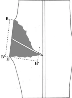
The harvested samples were dissected with a cleaver along the medial longitudinal plane and shaved with a sharpened knife to measure discolored areas formed after branch removal. The area of discolored stem tissue on the medial longitudinal surface was estimated by the length of pruning wound (B-B’) and the deepest pene- tration of discolored wood into the trunk (H-H’) with the formula squaring the area of a triangle supposing the shape of discolored wood as a triangle (Figure 3). The length of H-H’ was measured along the imaginary line perpendicular to the pruning wound.
5. Determination of total phenols
Total phenols were determined three times, one month,
six months and twelve months after treatments. Eight branch union samples were obtained from one tree in each treatment at each point of time after treatments, Figure 3. A diagram of a branch union sample split along the medial longitudinal plane one year after pruning cut.
Length of pruning wound (B to B’) and depth of the discolored wood (H to H’) were measured to square the area of discolored wood interior to the pruning wound.
Figure 4. A diagram of a branch union sample dissected along the medial longitudinal plane. The parts beyond an imaginary line perpendicular to the stem pith at 1.5 cm above the branch bark ridge and an imaginary line perpendicular to the stem pith at 1.5 cm below the end of branch pith were trimmed off. The trimmed sample was divided into three parts, the core of branch (C), above core (AC) and below core (BC) for chemical analyses of those tissues.
whose CER was the closest to the arithmetic mean of the group. The eight samples consisted of four with pruning wounds and the other four with living branches. The 0.06 g Fresh Weight (FW) of wood tissues was collected with a chisel from three separated parts of each sample, which were core of the branch, above core and below core (Figure 4). A total of 0.24 g FW shavings for each part collected from four samples was extracted in 4 mL of 100% methanol for 24 hours at 4oC. After extraction, the wood tissues were dried at 105±1oC and then weighed.
The methanol solution was filtered with 0.50 μm pore hydrophobic syringe filter. Total phenols were estimated by the Folin-Ciocalteu method as described by Bonello and Pearce (1993). For each sample, 15 μL of methanol extract was diluted to 1500 μL water. To this solution, 750 μL of Folin and Ciocalteu’s reagent (Sigma Chemical Co.) diluted two-fold with water was added and left for 3 minutes, followed by the addition of 750 μL of 1 M aqueous Na2CO3. The solution was shaken and left for 1 hour before the absorbance was measured at 725 nm with a spectrophotometer. Concentrations of total phe- nols were calculated with reference to a gallic acid stan- dard curve (10-200 μg/mL) by applying regression analysis and results were expressed as gallic acid equiv- alent (mg) per g Dry Weight (DW).
6. Determinations of extractives, holocellulose and lignin Chemical analyses for the tissues of branch core, and above and below core were performed to examine the
changes of their chemical properties one year after treat- ments. Two trees from each treatment whose CERs were the closest to the arithmetic mean of the group were selected for the analysis. A total of sixteen branch union samples, eight from each tree, were collected from each treatment, and eight samples of each tree consisted of four with pruning wounds and the other four with living branches.
The samples were dissected with a cleaver along the medial longitudinal plane, and the parts beyond an imaginary line perpendicular to the stem pith at 1.5 cm above the BBR and an imaginary line perpendicular to the stem pith at 1.5 cm below the end of branch pith were trimmed off (Figure 4). The trimmed samples were air-dried, split into three parts such as core of branch, above and below core, and then ground through a 40 mesh screen.
The content of extractive was determined by the soxhlet method. Two grams of sample in a thimble filter was extracted by 150 mL of ethanol/benzene mixture (1:2 v/v) using a soxhlet extractor with reflux condenser at 80oC for 6 hours. The extracted solution was evapo- rated under reduced pressure. The extractive was dried at 105±1oC and then weighed. The extractive-free sample left in the thimble filter was used for analyses of holo- cellulose and Klason lignin.
The content of holocellulose was determined as the delignified residue by NaClO2. The 1.25 g of extractive- free sample in 250 mL flask was treated with 75 mL of Table 2. Indicators to analyze the changes of trunks, stems and areas of pruning wound, and development of discolored wood on the medial longitudinal surface after treatments.
Indicators Formulae
Trunk Growth Rate (TGR, %)
: width of annual ring at 30 cm above the ground for the year immediately after pruning : width of annual ring at 30 cm above the ground for the year immediately before pruning Stem Growth Rate (SGR, %)
: stem diameter at 1.5 cm above the BBR adjacent to the pruning wound at the time of pruning : stem diameter at 1.5 cm above the BBR adjacent to the pruning wound one year after pruning Wound Closure Rate (WCR, %)
: area of pruning wound at the time of pruning : area of pruning wound 1 year after pruning Discolored/Wound Area Ratio (D/W Ratio)
: area of discolored stem tissue on the medial longitudinal surface one year after pruning : area of pruning wound at the time of pruning
TGR WARy 1+ WARy 1– --- 100×
= WARy 1+
WARy 1–
SGR SDy 1+ –SDy0 SDy0
--- 100×
= SDy0
SDy 1+
WCR PWAy0–PWAy 1+ PWAy0
--- 100×
= PWAy0
PWAy 1+
D W Ratio⁄ DAy 1+ PWAy0 ---
= DAy 1+
PWAy0
distilled water, 0.5 g of NaClO2 and 0.1 mL of CH3COOH at 80oC for 1 hr. This procedure was repeated 2 more times. The solution was filtered by using a glass filter (1G3, Iwaki, Japan), and then washed with 250 mL of cold distilled water and 25 mL of acetone, successively.
The filtrated residue was dried at 105±1oC and then weighed.
Klason lignin was analyzed according to the standard NREL procedures (NREL, 2005). The 0.3 g of extrac- tive-free sample in 50 mL flasks was hydrolyzed with 3 ml of 72% H2SO4 at 30oC for 1 hr. The hydrolysate was then transferred to 100 mL flask and diluted to 4%
H2SO4 by adding 84 mL of distilled water. The flasks were sealed and autoclaved for 1 hr at 121oC. The solu- tion was then filtered by using a glass filter (1G4, Iwaki, Japan). The filtrated residue was dried at 105±1oC and then weighed.
7. Data analysis
Some indicators like the CER for tree vigor were intro- duced for this study (Table 2). Trunk growth rate (TGR) for the relative cambial growth at 30 cm above the ground was calculated by comparing the width of annual rings of the year immediately after pruning to the width of the year immediately before pruning. On the other hand, stem growth rate (SGR) for the cambial growth at 1.5 cm above the BBR was estimated by comparing the diameter of the stem at 1.5 cm above the BBR one year after prun- ing to the diameter at the time of pruning.
Wound closure rate (WCR) and discolored/wound area ratio (D/W Ratio) were introduced to examine the changes of abilities to compartmentalize the pruning wounds after pruning. The WCR represented the enclos- ing extent of the pruning wound during the year after pruning, and D/W Ratio was computed by comparing the area of discolored stem tissue on the medial longi- tudinal surface one year after pruning with the area of pruning wound at the time of pruning. In CODIT model,
the WCR indicated vitality of the cambium around the wound which formed wall 4 after the tree was wounded, while D/W Ratio expressed the ability of a tree to limit spread of wood discolorations and decays with wall 1, 2 and 3 which were already present in the wood at the time of the pruning.
The statistical differences between the means of the treatments by indicator were determined by the analysis of variance (ANOVA). In all statistical analyses, the signif- icance level of α-value was 0.05 unless otherwise speci- fied.
Results 1. CER and cambial growth of trunk
Figure 5 shows the trends of CER for three different treatments which are control, P treatment and P+T treat- ment. There were no differences among the CERs of each group in early spring when the trees just started to grow, but the differences became larger from May when new leaves and shoots were developed, and maintained up to the end of growing season. The differences among the treatments were significant, while the difference between P treatment and P+T treatment were more sig-
Figure 5. Trends of the cambial electrical resistance (CER) by treatment for the year 2008 after pruning and transplanting treatments were made in March to Acer palmatum. (P treatment: pruning treatment; P+T treatment:
pruning and transplanting treatment)
Figure 6. Comparison between cambial electrical resist.ances (CER) for five-month averages from May to September, period in the midst of growth, and the trunk growth rate (TGR) at 30 cm above the ground for one year after pruning and transplanting treatments. (P treatment: pruning treatment; P+T treatment: pruning and transplanting treatment;
Means (±standard error) with the same letter are not significantly different at P ≤ 0.05.)
nificant (P=0.004) than that between the control and P treatment (P=0.032). This implies that transplanting gave significant impact on the tree vigor remarkably.
Figure 6 shows the correlations between the CERs which were averages for five months from May to Sep- tember, the period of the midst of growth, and TGR at 30 cm above the ground for one year after the treat- ments. The CER and TGR were inversely correlated, which meant the TGR was positively correlated with the tree vigor. Differences of the CERs were significant among the three treatments as mentioned above, and so were the differences of the TGRs. TGR of the control treatment was 112.2%, which meant that the annual cambial growth increased by 12% during the experimen- tal period of one year, while TGRs of P treatment and P+T treatment were reduced to 72.4% and 52.5%, respectively, which were significantly lower than that of the control (P=0.025 and 0.009, respectively). Branch removal of about 50% influenced the cambial growth of trees significantly, and the impact of transplanting on top of the pruning was also significant (P=0.015).
2. Closure of pruning wound and growth rate of stem Figure 7 shows the wound closure rate (WCR) and the stem growth rate (SGR) at 1.5 cm above the BBR for one year after treatments. The higher SGR was the higher WCR was. WCR of P treatment with 39.8% was significantly higher than that of P+T treatment with 31.8% (P=0.017), and the difference of SGR between two treatments (18.1% and 14.1%, respectively) was also significant (P<0.01). This result was consistent with that of TGR showing adverse effects of transplanting.
3. Extent of discolored area after pruning
Figure 8 shows variation of discolored/wound area (D/
W) ratio which means discolored area of the stem tissue on the medial longitudinal surface one year after treat- ment relative to the area of pruning wound at the time
of pruning. The wounds of P+T treatment formed more discolored area per unit area of the pruning wound (D/
W ratio 50.2) than P treatment (D/W ratio 44.8) and the difference between the two ratios was significant (P=0.002).
This result meant trees with P+T treatment were weaker in the ability of compartmentalizing the wounds than those with P treatment.
4. Total phenols and extractives
Table 3 shows differences of total phenols between the treatments of P and P+T, and between the branch unions with pruning wound and living branch, and changes of the compounds one month, six months and 12 months after treatments. Changes of phenol contents at the three parts of the treated branch unions were different from each other. The contents at the branch core increased one month after treatments, and sharply decreased after six months, then recovered up to the level of same part of the control union as time went by. But the contents at below core were continuously higher than those at the same part of the control union, while the contents at
Figure 7. The closure rate of pruning wound (WCR) and the growth rate of stem (SGR) at 1.5 cm above the branch bark ridge (BBR) bearing the pruning wound for one year after treatments. (P treatment: pruning treatment; P+T treatment:
pruning and transplanting treatment; Means (±standard error) with the same letter are not significantly different at P ≤
0.05.).
Figure 8. Variation of D/W ratios which means discolored area of stem tissue on the medial longitudinal surface one year after treatments relative to the area of pruning wound at the time of pruning. (P treatment: pruning treatment; P+T treatment: pruning and transplanting treatment; Means (±standard error) with the same letter are not significantly different at P ≤ 0.05.).
above core maintained similar levels with the same part of the control one.
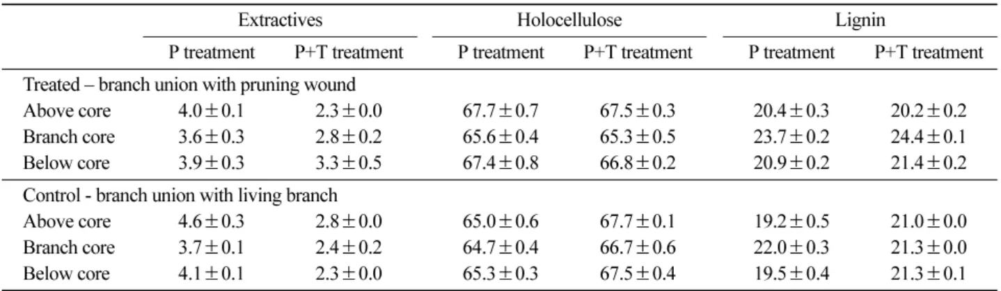
Table 4 shows contents of extractives at the same parts of the treatments twelve months after treatments. Extrac- tives, which includes phenolic compounds, at P+T treat- ment was lower than those at P treatment, but there were no differences between two treatments in the contents of total phenols at treated branch union (Table 3).
5. Variation of holocellulose and lignin
Table 4 shows relative contents per unit weight of the parts among extractives, holocellulose and lignin of three parts around the branch union with the pruning wound and the living branch, and their changes by treat- ment. In holocellulose, the relative contents of branch core at the treated union of both treatments were lower than those at the control union with living branch. On the contrary, lignin contents at the branch core of the two treatments (23.7%±0.2%, 24.4%±0.1%, respec- tively) were remarkably higher than those at the control union (22.0%±0.3%, 21.3%±0.0%, respectively). P+T treatment showed relatively higher lignin contents and lower holocellulose contents than P treatment.
Discussion
Changes of tree vigor caused by pruning and trans- planting practices can be examined by measuring cam- bial electrical resistances (CERs) of the trees, because thicker cambial zone of vigorous tree holds more mois- ture and ions which are major factors affecting electrical resistance (Shigo and Shortle, 1985). Evaluation of the results of the present study is not easy because of limited previous researches on effects of arboricultural practices on the CER. However, but changes of the CERs caused by the treatments can be inductive when comparing the cambial growth among the treatments later. In case of P+T treatment, significantly higher CER would be caused by sharply decreased absorption of water and minerals after removal of major root systems.
Many researches on the correlations between the CER and tree growth were reported. Shortle et al. (1977) reported the resistance to a pulsed electric current in the cambial zone of sprout red maple and hybrid poplar was inversely proportional to the rate of growth. Similar results about correlations between CER and the tree growth were obtained from the studies on mechanism Table 3. Differences of total phenols between the treatments of pruning only (P treatment) and pruning together with transplanting (P+T treatment), and between branch union with pruning wound and with living branch, and changes of the phenols one month, six months and 12 months after treatments. unit: gallic acid equivalent (mg) per g Dry Weight
P treatment P+T treatment
Period after treatment 1 month 6 months 12 months 1 month 6 months 12 months Treated-branch union with pruning wound
Above core 65 55 59 63 51 53
Branch core 72 34 58 75 34 59
Below core 72 76 67 71 66 74
Control -branch union with living branch
Above core 67 53 53 59 45 55
Branch core 68 50 57 60 51 51
Below core 72 54 65 65 50 52
Table 4. Contents of extractives, holocellulose and lignin of the three parts (core of the branch, above core and below core) of branch unions with pruning wound and with living branch, and their changes by treatment. (P treatment: pruning treatment; P+T treatment: pruning and transplanting treatment; mean ± Standard Error) unit: %
Extractives Holocellulose Lignin
P treatment P+T treatment P treatment P+T treatment P treatment P+T treatment Treated – branch union with pruning wound
Above core 4.0±0.1 2.3±0.0 67.7±0.7 67.5±0.3 20.4±0.3 20.2±0.2 Branch core 3.6±0.3 2.8±0.2 65.6±0.4 65.3±0.5 23.7±0.2 24.4±0.1 Below core 3.9±0.3 3.3±0.5 67.4±0.8 66.8±0.2 20.9±0.2 21.4±0.2 Control - branch union with living branch
Above core 4.6±0.3 2.8±0.0 65.0±0.6 67.7±0.1 19.2±0.5 21.0±0.0 Branch core 3.7±0.1 2.4±0.2 64.7±0.4 66.7±0.6 22.0±0.3 21.3±0.0 Below core 4.1±0.1 2.3±0.0 65.3±0.3 67.5±0.4 19.5±0.4 21.3±0.1
relating CER to periodic growth rate of balsam fir (Abies balsamea L.) with varying in level of spruce budworm defoliation (Blanchard et al., 1983). In addition, a sig- nificant correlation between CER and growth of peach cultivar (Prunus persica) was demonstrated (Wisniewski et al., 1985), and Lindberg and Johansson (1989) reported a fairly good correlation between the growth rate and CER of both dominant and suppressed Norway spruce (Picea abies Karst.) trees.
Regarding the effects of top pruning on the tree growth, Layne and Flore (1992) reported that dry weight increase was reduced at each of three levels of leaf area removal (10, 20, or 30%), but a disproportionally large decrease in dry weight increment occurred when the amount of leaf area removed increased from 20 to 30%, that is, the threshold level of leaf area removal, based on photosynthesis of individual leaves, was 20%. Many authors (Bennett, 1955; Lehtpere, 1957; Stein, 1955) reported similar results that removal of one-third of the vertical length of the live crown reduced cambial incre- ment. About 45% of pruning intensities in the present study would be enough to influence the cambial growth of treated trees.
Researches on the effect of root removal on the tree growth were concentrated on root pruning and water stress. Removing half of the root system of young apple trees in midsummer reduced top growth by 30%
(Maggs, 1965). Richards and Rowe (1977) reported that the immediate response of peach seedlings to root prun- ing was rapid decrease in root growth and a depression in shoot extension and leaf emergence. Severe water stress on Scots pine and Norway spruce also had about 79% and 43% lower biomass of current shoot, respec- tively (Turtola et al., 2003). Studies on combined treat- ments of top pruning together with transplanting were very few. But considering the aforesaid effects of top and root pruning on the top growth, a quite low TGR of 52.5% at P+T treatment of the present study might be regarded as a feasible result.
Few researches were made about the effect of trans- planting on the closure of pruning wound. Past experi- ments on wound closure were conducted on the artificial wounds made on stems. The results of the experiments showed high correlations between the wound closure and cambial growth of the stem. The pruning wounds and the growth of stems bearing the pruning wounds of the present study could be substituted for the artificial wounds and the cambial growth of the stem, respec- tively. Neely (1973; 1983) reported the results of several researches about the correlation between wound closure and cambial growth. Three-year experiment on white ash, honey locust and pin oak revealed that the healing rate of the wounds made 60 to 180 cm above the ground
was directly correlated with cambial growth at 100 cm above the ground of the trees. Shortle (1979) also reported that wounds closed more quickly in hybrid pop- lar trees of high vigor. These researches are consistent with the present study which indicated that less vigorous trees caused by transplanting with higher CERs showed slower cambial growth, which led to lower rates of wound closure.
Many studies were conducted on the correlations between water stress and host susceptibility. The thresh- old stem water potentials to be a critical factor in pre- disposition was -12 bars (Schoeneweiss, 1975), and the rate of penetration by Fomes annosus in roots of 12- year-old Pinus taeda was significantly enhanced by induced drought conditions (Towers and Stambaugh, 1968). Increased susceptibility by exposing host plants to controlled water stress was reported in crab apples in response to Physolospora obtusa (Landis and Hart, 1967)), in loblolly pines to Fomes annosus (Towers and Stambaugh, 1968)), and in aspens to Hypoxylon pruin- atum (Bagga and Smalley, 1974). Greater discolored area per unit area of the pruning wound in P+T treat- ment might be caused by water stresses due to root removal for transplanting.
A tree begins to form a protective chemical shield, a reac- tion zone with mostly phenolic compounds in angiosperms, around and immediately behind the wound, and the barrier could eventually be eroded and retreated by fungal activ- ities (Shigo, 1979). Lower contents of total phenols and extractives at the branch union with living branch in P+T treatment might be caused by the root removal which led to water stress and weakened tree vigor even- tually. Relatively high phenol contents at the treated branch union in spite of lower extractives contents in P+T treatment implied that trees with P+T treatment allocated more energy in compartmentalizing the wounds.
Levels of the phenolic contents at treated union changed in accordance with seasonal variation in the production of phenolic compounds (Jalal et al., 1982; Mireku and Wilkes, 1989) as the case of above core, while more dis- cussion is necessary for the changes of the contents at core and below core of the both treatments.
The vascular cambium and the growth rings it pro- duces are continuous from trunk to branch, but cells formed by the cambium in the upper junction of branch and trunk are oriented right angle to the normal orien- tation in the trunk and branch, while the branch xylem is oriented downward at the branch base and encircles it to form a collar which meets on the trunk below the branch. Thus conduction into and out of the branch fol- lowed the pathway of the branch collar and there was no local direct conduction between trunk xylem above a branch and within a branch (Shigo, 1985). Consequently,
the phenol contents at above core were not affected by the treatments during a year in the present study, and the contents at core and below core changed in accordance with the extension of discolored sapwood, which pro- ceeded further in the axial rather than radial or tangential wood alignments (Deflorio et al., 2007).
In case of branch core, total phenols at the wounds increased slowly reaching a maximum after 21 days (Barry et al., 2001), then would decrease with the exten- sion of the discolored area and the retreat of reaction zones rich in phenols, as time went by. Higher total phe- nols at below core in P+T treatment in the present study would be caused by faster discoloration of the pruning wounds in P+T treatment than P treatment, as the dis- coloration area extended downward along the conduction pathway. Further researches would be necessary to examine both the sharp decreases of the contents at branch core of treated branch union in both treatments, and increase of the contents at below core in P treatment after six months. But it might be a seasonal phenomenon considering the lower contents at the parts in the control at the same point of time.
Formation of lignin is one of defense mechanisms in the wood tissues to protect trees from wounding and microbial invasion (Yamada, 2001). There are numerous reports on lignification of conifers in response to wound- ing or infection are numerous (Biggs et al., 1984; Rit- tinger et al., 1987)) and Geiger et al. (1986) reported an increase of 25-30% in lignin-like material in the wood of
Hevea brasiliensis taproots close to the infection front of
Rigidoporus lignosus. Increases of lignin contents at the core of the treated union by about 8~15% comparing with the core of the control in the present study would result from lignification of exposed xylem tissues as a defense mechanism of the trees. The content of holocel- lulose is a relative proportion per unit weight, and the higher lignin content is accompanied by the lower rela- tive content of holocellulose.
Transplanting had adverse effects on tree growth and compartmentalization of pruning wounds through root loss and following water stress. Therefore, transplanting a large tree, which is inevitably accompanied by lots of big pruning wounds and loss of most root systems, should be restricted within a few limited purposes. When it is necessary, the trees should be cared to get less wounds and to keep more root systems. Once trans- planted, the soil around the tree must be watered prop- erly until the tree is firmly established.
Literature Cited
1. American National Standard Institute. 2001. American National Standard for Tree Care Operations-Tree,
Shrub, and Other Woody Plant Maintenance - Standard Practices (Pruning). ANSI A300 (Part 1)-2001 Pruning.
American National Standard Institute, Washington, DC.
2. American National Standard Institute. 2005. American National Standard for Tree Care Operations-Tree, Shrub, and Other Woody Plant Maintenance - Standard Prac- tices (Transplanting) ANSI A300 (Part 6)-2005 Trans- planting. American National Standard Institute, Washington, 3. American Nursery and Landscape Association. 2004.DC.
American Standard for Nursery Stock. ANSI Z60.1- 2004. Washington, DC.
4. Arnold, M.A. 2005. Challenges and benefits of trans- planting large trees: an introduction to the workshop.
HortTechnology 15: 115-117.
5. Ayres, P.G. 1991. Growth responses induced by patho- gens and other stresses. pp. 227-248. In: Mooney, H.A., Winner, W.E., Pell, E.J. and Chu, E. (Eds). Response of Plants to Multiple Stresses. Academic Press, San Diego, CA.
6. Bagga, D.K. and Smalley, E.B. 1974. The development of Hypoxylon canker of Populus tremuloides: role of interacting environmental factors. Phytopathology 64:
658-662.
7. Barry, K.M., Pearce, R.B., Evans, S.D., Hall, L.D. and Mohammed, C.M. 2001. Initial defence responses in sapwood of Eucalyptus nitens (Maiden) following wounding and fungal inoculation. Physiological and Molecular Plant Pathology 58: 63-72.
8. Bennett, F.A. 1955. The effect of pruning on the height and diameter growth of planted slash pine. Journal of Forestry 53: 636-638.
9. Bier, J.E. 1964. The relation of some bark factors to canker susceptibility. Phytopathology 54: 272-275.
10. Biggs, A.R., Merrill, W. and Davis, D.D.. 1984. Dis- cussion: Response of bark tissues to injury and infec- tion. Canadian Journal of Forest Research 14: 351-356.
11. Blanchard, R.O., Shortle, W.S. and Davis, W. 1983.
Mechanism relating cambial electrical resistance to periodic growth rate of balsam fir. Canadian Journal of Forest Research 13: 472-480.
12. Bonello, P. and Pearce, A.D.M. 1993. Biochemical defence responses in primary roots of Scots pine chal- lenged in vitro with Cylindrocarpon destructans. Plant Pathology 42: 203-211.
13. Deflorio, G., Barry, K.M., Johnson, C. and Mohammed, C.L. 2007. The influence of wound location on decay extent in plantation-grown Eucalyptus globulus and Eucalyptus nitens. Forest Ecology and Management 242: 353-362.
14. Evans, P.S. and Klett, J.E. 1984. The effects of dormant pruning treatments on leaf, shoot and root production from bare-root Malus sargentii. Journal of Arboricul- ture 10: 298-302.
15. Geiger, J.P., Rio, B., Nicole, M. and Nandris, D. 1986.
Biodegradation of Hevea brasiliensis wood by Rigi- doporus lignosus and Phellimus noxius. European Jour- nal of Forest Pathology 16: 147-159.
16. Geisler, D. and Ferree, D.C. 1984. Response of plants to root pruning. Hoticultural Reviews 6: 155-188.
17. Gilman, E.F. and Beeson, R.C. 1996. Production method affects tree establishment in the landscape. Journal of Environmental Horticulture 14: 81-87.
18. Harris, R.W., Clark, J.R. and Matheny, N.P. 2004.
Arboriculture (forth edition). Prentice Hall, Upper Sad- dle River, NJ.
19. Hartman, J.R., Pirone, T.P. and Sall, M.A. 2000.
Pirone's Tree Maintenance (7th ed.). Oxford University Press, Inc., New York.
20. Husch, B., Beers, T.W. and Kershaw, J.A., Jr. 2003.
Forest Mensuration (4th ed.). John Wiley & Son, Inc., Hoboken, NJ.
21. Jalal, M.A.F., Read, D.J. and Haslam, E. 1982. Phe- nolic composition and its seasonal variation in Calluna vulgaris. Phytochemistry 21: 1397-1401.
22. Jin, H.O., Yi, M.J., Shin, Y.O., Kim, J.J. and Chon, S.K.
1994. Forest Pedology. Hyangmunsa, Seoul, Korea.
23. Kozlowski, T.T and Davies, W.J. 1975. Control of water balance in transplanted trees. Journal of Arbori- culture 1: 1-10.
24. Landis, W.R. and Hart, J.H. 1967. Cankers of orna- mental crabapples associated with Physalospora obtusa and other microorganisms. Plant Disease Reporter 51:
230-234.
25. Layne, D.R. and Flore, J.A. 1992. Photosynthetic com- pensation to partial leaf area reduction in sour cherry.
Journal of the American Society for Horticultural Sci- ence 117: 279-286.
26. Lee, K.J. and Lee, S.J. 2001. Arboriculture (1st ed.).
SNU Press, Seoul.
27. Lehtpere, R. 1957. The influence of high pruning on the growth of Douglas fir. Forestry 30: 9-20.
28. Lindberg, M. and Johansson, M. 1989. The use of elec- trical resistance of cambium and phloem as a measure of tree vigor. Scandinavian Journal of Forest Research 4: 175-185.
29. Maggs, D.H. 1965. Growth rates in relation to assim- ilate supply and demand. II. The effect of particular leaves and growing regions in determining the dry mat- ter distribution in young apple trees. Journal of Exper- imental Botany 16: 387-404.
30. Mireku, E. and Wilkes, J. 1989. Seasonal variation in the ability of the sapwood of Eucalyptus maculata to compartmentalize discolouration and decay. Forest Ecology and Management 28: 131-140.
31. Neely, D. 1973. Tree wound healing and radial growth correlations. HortScience 8: 384-385.
32. Neely, D. 1983. Tree trunk growth and wound closure.
HortScience 18: 99-100.
33. NREL. 2005. Biomass Analysis Technology Team Lab- oratory Analytical Procedure. National Renewable Energy Laboratory, US Department of Energy.
34. Pearce, R.B. 2000. Decay development and its restric- tion in trees. Journal of Arboriculture 26: 1-11.
35. Randolph, W.S. and Wiest, C. 1981. Relative impor- tance of tractable factors affecting the establishment of transplanted holly (Ilex crenata). Journal of the Amer- ican Society for Horticultural Science 106: 207-210.
36. Ranney, T.G., Bassuk, N.L. and Whitlow, T.H. 1989.
Effect of transplanting practice on growth and water relations of ‘Colt’ cherry trees during reestablishment.
Journal of Environmental Horticulture 7: 41-45.
37. Richards, D. and Rowe, R.N. 1977. Root-shoot inter- actions in peach: the function of the root. Annals of Botany 41: 1211-1216.
38. Rittinger, P.A., Biggs, A.R. and Peirson, D.R. 1987.
Histochemistry of lignin and suberin deposition in boundary layers formed after wounding in various plant species and organs. Canadian Journal of Botany 65:
1886-1892.
39. Schoeneweiss, D.F. 1975. Predisposition, stress, and plant disease. Annual Review of Phytopathology 13:
193-211.
40. Schoeneweiss, D.F. 1981. The role of environmental stress in diseases of woody plants. Plant Disease 65:
308-314.
41. Schoeneweiss, D.F. 1986. Water stress predisposition to disease - an overview. pp. 157-174. In: Ayres, P.G. and Boddy, L. (eds.). Water, fungi and plants. Cambridge University Press, Cambridge, UK.
42. Shigo, A.L. 1979. Tree decay - an expanded concept.
Forest Service, U.S.D.A., Durham, NH.
43. Shigo, A.L. 1985. How tree branches are attached to trunks. Canadian Journal of Botany 63: 1391-1401.
44. Shigo, A.L. and Marx, H.G. 1977. Compartmentaliza- tion of Decay In Trees. Forest Service, USDA.
45. Shigo, A.L. and W.C. Shortle. 1985. Shigometry: A reference guide. Agriculture Handbook No. 646. Forest Service U.S.D.A.
46. Shortle, W.C. 1979. Compartmentalization of decay in red maple and hybrid poplar trees. Phytopathology 69:
410-413.
47. Shortle, W.C., Shigo, A.L., Berry, P. and Abusamra, J.
1977. Electrical resistance in tree cambium zone: rela- tionship to rates of growth and wound closure. Forest Science 23: 326-329.
48. Shoup, S., Reavis, R. and Whitcomb, C.E. 1981. Effects of pruning and fertilizers on establishment of bareroot deciduous trees. Journal of Arboriculture 7: 155-157.
49. Stein, W.J. 1955. Pruning to different heights in young Douglas fir. Journal of Forestry: 352-355.
50. Towers, B. and Stambaugh, W.J. 1968. The influence
of induced soil moisture stress upon Fomes annosus root rot of loblolly pine. Phytopathology 58: 269-272.
51. Turtola, S., Manninen, A.M., Rikala, R. and Kainulainen, P. 2003. Drought stress alters the concentration of wood terpenoids in Scots pine and Norway spruce seedlings. Journal of Chemical Ecology 29: 1981-1995.
52. Watson, G.W. and Sydnor, T.D. 1987. The effect of root pruning on the root system of nursery trees. Journal of Arboriculture 13: 126-130.
53. Wisniewski, M., Bogle, A.L. and Wilson, C.L. 1985.
Seasonal variation in cambial electrical resistance and its relation to growth in two cultivars of peach. Cana- dian Journal of Plant Science 65: 345-350.
54. Yamada, T. 2001. Defense mechanisms in the sapwood of living trees against microbial infection. Journal of Forest Research 6: 127-137.
(Received October 1, 2009; Accepted October 22, 2009)