Introduction
The bitterling (subfamily Acheilognathinae), is a small freshwater cyprinid fishes with deep body and a semi-inferior mouth and distributed in temperate regions of Europe and Asia, includ- ing Korea, Japan, Taiwan, and China (Nelson, 1984; Berra, 2001; Kim and Park, 2002; Kim et al., 2005). It was widely known that the female possess a long ovipositor behind its anal opening that they use to place their eggs onto the gills of a mussel. The male fertilizes the eggs by releas-
ing sperm into the inhalant siphon of the mussel, and the fertilized eggs develop inside the mussel gill cavity and leave the mussel as actively swim- ming larvae (Nakamura, 1969; Arai and Akai, 1988; Heschl, 1989; Suzuki and Jeon, 1990;
Reynolds and Guillaume, 1998; Aldridge, 1999;
Candolin and Reynolds, 2001; Kim and Park, 2002; Mills and Reynolds, 2003; Smith et al., 2004; Kawamura and Uehara, 2005; Kim et al., 2005).
During the larvae are lodged in the mussel just before leaving the gill cavity, minute tubercles on the skin surface of the larvae develops.
Among the known Korean bitterling, 3 genera and 14 species including 9 endemic species (Kim
Histological Study of the Minute Tubercles on Larval Skin Surface of a Korean Endemic Bitterling, Acheilognathus
koreensis (Pisces, Cyprinidae), with Its Larval Growth
Chi-Hong Kim, Jong-Young Park
1*, Min-Kyong Park
1, Eon-Jong Kang
2and Jong-Hwa Kim
Inland Fisheries Ecological Research Institute, NFRDI.
Cheongpyeong-ri Cheongpyeong-myeon Kapyeong-gun, Kyunggi-do 477-815, Korea
1Faculty of Biological Sciences and Institute for Biodiversity Research, Chonbuk National University, Jeonju 561-756, Korea
2Inland Fisheries Aquaculture Research Institute, Jinhae 645-806, Korea
Morphology and distribution of the minute tubercles projected on the skin surface of larvae with its development was observed in the Korean bitterling, Acheilognathus koreensis, known as an endemic freshwater fish. The epidermis of the larvae consisted of a thin single layer, having smaller basophilic flat or round-flattened basal cells. In between the single cell layer, two or three layers were added and they consisted mainly of large epidermal cells just above basal cells. These large unicellular epidermal cells were mainly scale-shaped and rarely cone-shaped, and do not give any histochemical tests for mucosubstances. They were present in anterior region and most region of yolk sac. Whereas, vestigial epidermal cells were distributed in the body region and the caudal fin-fold region. These two kinds of epidermal cells, called minute tubercles, increased in number and height from Just to 8 days after hatching, but as the larvae develop gradually, they became to reduce.
At 31days after hatching of free swimming stage and absolute absorption stage of the yolk sac, the minute tubercles did not exist on the whole skin of the larvae.
Key words : Korean bitterling, Acheilognathus koreensis, larva, minute tubercle, skin
*Corresponding author: [email protected]
─
─ 170 ──
et al., 2005), the morphology and distribution on the minute tubercles in several bitterling fishes was reported and sometimes they have been treated as an useful character for phylogenetic studies of Rhodeus (Suzuki and Hibia, 1984;
Suzuki and Jeon, 1987; Suzuki and Jeon, 1988a, b, c, d, 1989, 1990; Arai et al., 2001). However, these studies on the minute tubercles were just described with the development of the eggs and larvae, and it was little known on detailed histo- logical information.
Therefore, we studied on the development, basic structure and nature of the minute tubercles by comparing its larval stage in Acheilognatus koreensis.
Materials and Method
Parental fishes of Acheilognatus koreensis were collected from Gwanchon-myeon, Imsil-gun, Jeollabuk-do, Somjin River, Korea, on April 18, 2006. Artificial insemination was carried out several times from April 19 to March 25 using
six pairs. Method of artificial insemination and rearing of eggs and larvae followed those of Suzuki and Hibia (1984).
For light microscope observations of the minute tubercles on the skin surface of larvae, the entire larvae by stage were fixed in 10% neutral buffered formaldehyde. These larvae were dehydrated through a standard ethanol series to 100%, clear- ed in xylene and then embedded in wax (Para- plast, Oxford). Five µ m sections were deparaf- finized and stained with Ehrlich haematoxylin, counter-stained with eosin for general histology.
For histochemical test, it was alcian blue solu- tion (AB) at pH 1.0 and 2.5 (Steedman, 1950; Lev and Spicer, 1964), and periodic acid-Schiff (PAS) method (Lilllie and Greco, 1947). For scanning electron microscope observations of the tuber- cles, three specimens were fixed in each stage of larval development for 24 hours under 4� C in cacodylate-buffered 2.5% glutaraldehyde, dehy- drated by a graded series of ethanol, dried to a critical point with liquid CO
2. The dried mate- rials were coated with gold by an ion sputtering and then examined with a Hitach S-450 scann-
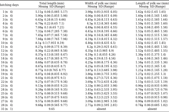
Table 1. Total length of the larva, width and length of the yolk by larval growth in Acheilognthsus koreensis
Hatching days Total length (mm) Width of yolk sac (mm) Length of yolk sac (mm)
Mean±SD (Range) Mean±SD (Range) Mean±SD (Range)
1 (n==4) 5.11±0.04 (5.08~5.17) 3.98±0.05 (3.93~4.05) 1.62±0.02 (1.6~1.65) 2 (n==4) 5.90±0.06 (5.85~5.98) 4.06±0.04 (4.01~4.09) 1.60±0.04 (1.54~1.64) 3 (n==4) 6.43±0.24 (6.1~6.66) 4.45±0.24 (4.11~4.63) 1.61±0.02 (1.58~1.64)
4 (n==4) 6.79±0.22 (6.6~7.1) 4.5±0.12 (4.36~4.66) 1.59±0.01 (1.58~1.60)
5 (n==4) 7.08±0.1 (6.6~7.21) 4.49±0.04 (4.45~4.55) 1.53±0.04 (1.49~1.58)
6 (n==4) 7.32±0.04 (7.28~7.38) 4.41±0.19 (4.19~4.66) 1.52±0.05 (1.46~1.58) 7 (n==4) 7.45±0.07 (7.36~7.54) 4.50±0.14 (4.34~4.68) 1.53±0.02 (1.51~1.56) 9 (n==4) 7.84±0.08 (7.76~7.94) 4.19±0.13 (4.01~4.32) 1.54±0.02 (1.51~1.57)
10 (n==4) 8.1±0.1 (7.95~8.19) 4.46±0.03 (4.43~4.5) 1.43±0.02 (1.4~1.45)
11 (n==4) 8.27±0.09 (8.17~8.39) 4.2±0.29 (3.92~4.61) 1.50±0.04 (1.44~1.54)
12 (n==4) 8.36±0.22 (8.06~8.58) 4.35±0.4 (3.98~4.9) 1.52±0.03 (1.48~1.55)
13 (n==4) 8.37±0.13 (8.18~8.47) 4.19±0.1 (4.05~4.26) 1.38±0.01 (1.37~1.4)
14 (n==4) 8.62±0.17 (8.38~8.77) 4.29±0.19 (4.1~4.54) 1.4±0.04 (1.36~1.46)
16 (n==4) 8.68±0.07 (8.63~8.78) 4.25±0.08 (4.17~4.36) 1.34±0.01 (1.33~1.36) 17 (n==4) 8.67±0.03 (8.63~8.7) 4.25±0.05 (4.19~4.32) 1.37±0.02 (1.34~1.4) 18 (n==4) 8.77±0.0 (8.76~8.77) 3.87±0.13 (3.68~3.98) 1.21±0.06 (1.14~1.28) 19 (n==4) 8.87±0.04 (8.83~8.92) 3.86±0.08 (3.75~3.95) 1.27±0.02 (1.25~1.3) 21 (n==4) 9.03±0.05 (8.97~9.1) 4.08±0.27 (3.72~4.36) 1.13±0.05 (1.07~1.18) 22 (n==4) 9.27±0.07 (9.17~9.34) 3.69±0.75 (2.63~4.24) 1.15±0.03 (1.12~1.19) 23 (n==4) 9.13±0.08 (9.05~9.23) 3.33±0.90 (2.05~3.98) 0.96±0.02 (0.93~0.99) 24 (n==4) 9.28±0.08 (9.16~9.35) 3.41±0.63 (2.53~3.95) 0.76±0.03 (0.72~0.79) 25 (n==4) 9.57±0.08 (9.51~9.68) 3.49±0.05 (3.42~3.55) 1.01±0.07 (0.92~1.07) 26 (n==4) 9.57±0.07 (9.47~9.63) 3.38±0.12 (3.22~3.52) 0.95±0.03 (0.91~0.98) 27 (n==4) 9.57±0.08 (9.49~9.68) 3.09±0.08 (2.98~3.18) 0.90±0.09 (0.83~1.02) 28 (n==4) 9.68±0.09 (9.56~9.77) 2.71±0.09 (2.59~2.81) 0.74±0.06 (0.68~1.82)
31 (n==1) 9.65 absent absent
n== Number of the observed larvae
ing electron microscope.
For evaluations of the minute tubercles, we used Carl Zeiss vision (LE REL. 4.4) on scanning electron microscopy. The measurements of the length and yolk size were carried out under the anatomic microscope using 1/10 mm digital cali- pers.
To facilitate description of the distributional patterns of minute tubercles, the skin surface of the larvae was divided into four regions by the method of Suzuki and Jeon (1988c).
Results
1. General features of the minute tubercles
The larvae from larval stage after hatching to nearly free-swimming stage had a pair of scaly yolk projection, and their whole body skin sur- faces were covered with minute tubercles althou- gh there are differences in height and density with larval growth.
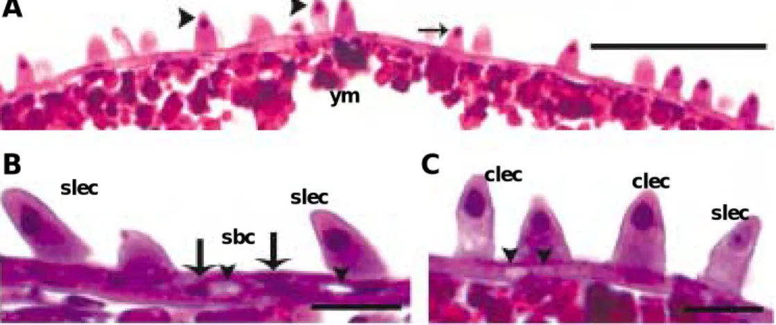
Just after hatching, the epidermis of the larvae consisted of a thin single layer, about 2.5 to 3.8 µ m, having smaller basophilic flat or round- flattened basal cells (Fig. 1, A to C). These basal cells were deeply stained nuclei and surrounded by small amounts of faintly stained cytoplasm (Fig. 1, B and C). However, some regions of the single layer became to formed two layers by the
addition of one layer consisted of large cells, meaning scale-shaped cell, just above basal cells (Fig. 1, A to C). These epidermal layers became thick, about 14 to 30 µ m.
These large cells were an unicellular cell with a scale-shaped body, reaching 12.5 to 25 µm in height. They had a top, spherical or oval nucleus which stained darkly with H&E (Fig. 1, A to C).
Their cytoplasms were basophilic and more or less homogeneous, sometimes vacuolated (Fig. 1, B and C). However, they did not give any histo- chemical tests for mucosubstances as AB (pH 1.0 and 2.5), PAS reaction and AB (pH 2.5)-PAS reaction. There were small and flat vacuole-like structures in the epidermal layer (Fig. 1, B and C). As the larvae grow, these scaly large cells reduced. They lost their nucleus, and their cyto- plasms were vacuolated and shrunk, causing cell boundary unclear. They were distributed on the anterior region of the yolk sac protruded, and most part of the yolk sac, the mid-yolk sac and the mid-body regions.
Interestingly, besides these scale-shaped large cells, vestigial epidermal cells existed rarely on the most posterior region of the yolk (body) and the caudal fin-fold region. Their cell bodies were shrunk and cell boundaries were unclear.
2. Development of the minute tubercles with larval growth
The presence of the minute tubercles on the
Fig. 1. Transverse sections of the skin surface of larval Acheilognathus koreensis, stained with Ehrlich haematoxylin and eosin (bars indicate 20µm). A, Two kinds of the minute tubercles cover the anterior epidermal region of the yolk (region A), 1 day after hatching. ym, yolk mass; arrowheads, circular cone-shaped tubercles; arrows, scale-shaped tubercles. B, The epidermis having 2 or 3 epidermal cell layers consists of small basal cells (sbc) and scaly large cells (slec), the anterior epidermis in 4 days after hatching (region A). There are some vacuoles (arrowheads) in the epidermis. C, In the same regions with the above (B), the epidermis has two kinds of large cells, large and circular cone-shaped cells (clec) and large scale-shaped cells (slec). Note some vacuoles (arrowheads).
A
B C
slec
sbc
slec ym
clec clec
slec
skin of the larvae was closely related to its yolk sac with larval growth (Table 1). The develop- ment of the minute tubercles may be classified into four stages: formation, growth, reduction and disappearance stage.
1) Formation stage
(1) Minute tubercles
At the this stage, the scale-shaped minute tu- bercles covered the whole body skin of the larvae which range just after hatching to 3 days after hatching. At the this stage, the minute tubercles are seems to be similar to each stage in its num- ber, 3 to 7 per 100 µm and height, 12.5 to 30 µm (Fig. 2, A to D).
(2) Larval stage (just after hatching to 3 days after hatching)
The total length ranges from 5.08 to 6.66 mm.
At the 3 days after hatching, the tail elongated backward and caudal fin-fold slightly developed.
The anteriormost part of the yolk sac elongated slightly forward. The dorsal part of head rose slightly and the brain had undergone further development. The yolk sac length and projection height in this stage became more developed with 4.1×1.9 mm respectively (Fig. 3).
2) Growth stage
(1) Minute tubercles
At the growth stage, the scale-shaped minute
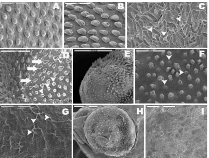
Fig. 2. Minute tubercles distributed on the skin surface of larvae of Acheilognathus koreensis, A, Hemispheric minute tubercles cover at the anterior epidermal region of the yolk (region A), 1 day after hatching. B, Scale-shaped minute tubercles at the region (B), 1 day after hatching. C, Vestigial minute tubercles (arrowheads) at the region (C), 1 day after hatching. D, Scaly (arrows) and hemispheric minute tubercles (arrowheads) at the region (A), 2 days after hatching. They incline to tilt posteriorly. E, The minute tubercles around the eye cup of the region (A) become to reduce, 9 days after hatching. F, The minute tubercles of the region (B) become to reduce, 9 days after hatching.
Note a reduction of the minute tubercles (arrowheads). G, The vestigial minute tubercles (arrowheads) reduce at the regions (C), 9 days after hatching. H, In 25 days after hatching, the minute tubercles of the eye cup still remain.
I, In 25 days after hatching, there are no the minute tubercles around caudal region.
tubercles on the whole body skin of the larvae increased in number and height. The whole body skin was densely covered with the developed tubercles. These tubercles ranged about 5 to 10 per 100 µ m in number and about 16.5 to 40.3 µ m in height and these values peaked at 6 to 8 days after hatching. This stage ranged approximately 4 to 8 days after hatching.
(2) Larval stage (4 to 8 days after hatching) The total length ranges from 6.6 to 7.64 mm. At the 6 to 8 days after hatching, the height of dor- sal and ventral yolk projection in this stage be- came creased slightly but yolk sac gradually elongated posterior. The number of myotome ranged from 30 to 33. The circulatory system was already established and blood cells became red- dish to increase in number (Fig. 3).
3) Reduction stage
(1) Minute tubercles
These tubercles become to reduce in number and height. This stage ranges approximately from 9 to 30 days after hatching (Fig. 3, E to I).
At 9 to 10 days after hatching, the tubercles started to reduce (Fig. 3, E to G). As the larvae grow, they was randomly distributed, and more less than half of the tubercles reduced at 18 to 20 days after hatching. At 25 to 30 days after hatch- ing, they reduced rapidly remained on the eye cups at 30 days after hatching (Fig. 2, H and I).
These tubercles ranged about 0 to 1 per 100 µ m in number and about 6.5 to 13.5 µ m in height.
(2) Larval stage (9 to 30 days after hatching) The total length ranges from 7.76 to 9.63 mm.
At the 20 to 25 days after hatching, the gas bladder completely divided into front and hind lobes. This means that the larvae are able to swim actively with good balanced orientation for several minutes. The yolk projection on the breast was so reduced that it was difficult to find. The mouth and anus cannel was formed completely. Although the yolk still remains, the larvae at this stage began to feed (Fig. 3).
4) Disappearance stage
(1) Minute tubercles
At this stage, the minute tubercles did not exist on the whole body skin any more.
(2) Larval stage (31 days after hatching) The total length was 9.65 mm. At 31 days after hatching, free swimming stage starts. The cau- dal fin was forked into branches. Larvae can cat- ch and eat small planktons. Yolk sac was com- pletely absorbed and digestive system was com- pleted.
3. Distribution pattern of the minute tubercles by region
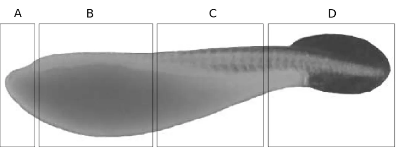
To observe the distribution of the minute tu- bercles, the skin surface of the larvae was divid- ed into four regions as follows (Fig. 4): (A) an- terior region of the yolk sac protruded, (B) most region of the yolk sac, the mid-yolk sac and the mid-body regions, (C) posterior region of the yolk sac and the body, between regions (b) and (d); (D) the caudal fin-fold region.
At immediately after hatching, the minute tu- bercles of regions (A) were hemispheric, about 7.0 to 11.2 µ m (Fig. 2A). However, these small hemispheric tubercles began to change to scale- like shape between 1 and 2 days after hatching (Fig. 2D). Finally, the region (A) had lots of scaly and a few circle cone-shaped minute tubercles (Fig. 2B). During the larvae grow, they increased in height and number, and particularly reached
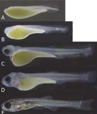
Fig. 3. Larvae of Acheilognathus koreensis from Somjin River in Korea. A, 1 day after hatching, 5.13 m in TL. B, 3 days after hatching, 6.52 m in TL. C, 6 days after hatching, 7.38 m in TL. D, 12 days after hatching, 8.41 m in TL. E, 26 days after hatching, 9.52 m in TL.
A
B
C
D
E
maximum size at 6 to 8 days after hatching.
Thereafter, the minute tubercles became to re- duce (Fig. 2F). Region (B) had skin surface sur- rounded with scale-shaped minute tubercles, and these minute tubercles were inclined to tilt pos- teriorly (Fig. 2, B and D). Gradually, they reduc- ed in height and number with larval develop- ment. The region (C), unlike those of regions (A) and (B), had many vestigial minute tubercles of shrunk cell bodies and unclear cell boundaries (Fig. 2, C and G). They were reduced during the larvae develop as in the scaly minute tubercles.
The region (D), had a few minute tubercles dis- tributed rarely on its skin surface.
Discussion
Bitterling larvae display unique minute tuber- cles on the skin surface that enable them to sur- vive in a mussel gill chamber and which vary among genera. Bitterling embryos of the genus Rhodeus have 2 wing-like yolk projections (Suzuki et al., 1986; Suzuki and Jeon, 1987, 1988b; Aldridge, 1999), while species of Acheilog- nathus and Tanakia have none. Also, all species of Rhodeus, and some of Acheilognathus and Tanakia possess scaly tubercles on their yolk-sac (Fukuhara et al., 1982; Suzuki and Jeon, 1988a, c, d), which may play a role in helping the em- bryo remain lodged in the gills of its mussel host.
For establishment of taxonomic position of Acheilognathus limbata distributed over Japan and Korea, Suzuki and Jeon (1988c) compared
Korean and Japan bitterling on the basis of the egg and larvae development, and they docu- mented the minute tubercles of Korean A. lim- bata, sampled from two localities of Kum River and Somjin River, Korea. However they regarded Korean and Japanese population as one species, A. limbata, not is separate species. Since then, Korean A. limbata redescribed as a new Korean endemic species, A. koreenis, by Kim and Kim (1990).
The minute tubercles of the Korean A. limbata reported by Suzuki and Jeon (1988c) were sim- ilar to those of A. koreensis studied in the pre- sent study: scaly minute tubercles on the skin surface of the larvae without 2 wing-like yolk projections. However, there were some differ- ences between Korean A. limbata and A. koreen- sis as follows: 1) at the posterior region of the yolk sac and the body region of the larvae, Korean A. limbata had hemispheric minute tubercles but A. koreensis vestigial minute tubercles, and 2) In the caudal fin-fold region, Korean A. limbata had lots of vestigial one but A. koreensis a few ves- tigial one.
Although various minute tubercles on the skin surface of bitterling larvae was known by several researchers, it was almost focused on its overall shape (Suzuki and Hibia, 1984; Suzuki and Jeon, 1987, 1988a, b, c, d, 1989, 1990; Arai et al., 1991).
Through our observation, we could confirm that the minute tubercles were formed by large uni- cellular epidermal cells with scale-shaped body in a thin single layer having basophilic flat or round-flattened basal cells. Their cytoplasms
A B C D
Fig. 4. Diagram showing regions of the skin surface distributed larval minute tubercles introduced by Suzuki and Jeon (1988c). A, anterior region of the yolk sac protruded. B, most part of the yolk sac, the mid- yolk sac and the mid- body regions. C, posterior region of the yolk sac and the body, between regions (b) and (d). D, the caudal fin-fold region.
were basophilic and more or less homogeneous, sometimes vacuolated. However, they did not give any histochemical tests for mucosubstances as AB at pH 1.0 and 2.5, PAS reaction and AB (pH 2.5)-PAS reaction. However, these histolog- ical results are little known in the previous pa- pers.
The minute tubercles function as an attach- ment that enables them to survive in a mussel gill chamber (Aldridge, 1999). In general, the cells related to attachment in fish egg were known as secreted materials as mucus, mucin, and mucil- age, or gelatin, (Laale, 1980). However, because these minute tubercles in A. koreensis are un- certain whether they are secreted from a large epidermal cell or are a simple growth of epider- mal cell, detailed researches on its nature will be needed in future.
Acknowledgments
The project was supported by RP-2006-RE-003, NFRDI, Korea.
References
Aldridge, D.C. 1999. Development of European bitterling in the gills of freshwater mussels. J. Fish Biol., 54 : 138~151.
Arai, R., S.R. Jeon and T. Ueda. 2001. Rhodeus pseudo- sericeus sp. nov., a new bitterling from South Korea (Cyprinidae, Acheilognathinae). Ichthyol. Res., 48 : 275~282.
Arai, R. and Y. Akai. 1988. Acheilognathus melanogaster, a senior synonym of A. moriokae, with a revision of the genera of the subfamily Acheilognathinae (Cyprini- formes, Cyprinidae). Bulletin of the National Science Museum, Series A, 14 : 199~213.
Berra, T.M. 2001. Freshwater fish distribution. Academic press. New York, 604 pp.
Candolin, U. and J.D. Reynolds. 2001. Sexual signaling in the European bitterling: females learn the truth by direct inspection of the resource. Behav. Ecol., 12 : 407~411.
Fukuhara, S., Y. Nagata and W. Maekawa, 1982. Minute scaly tubercles on the yolksac of rhodeine cyprinid fishes in prolarval stages. Japan. J. Ichthyol., 29 : 232
~236. (in Japanese)
Heschl, A. 1989. Integration of “innate” and “learned”
components within the IRME for mussel recognition in the European bitterling Rhodeus amarus (Bloch).
Ethology, 81 : 193~208.
Kawamura, K. and K. Uehara. 2005. Effects of temper- ature on free embryonic diapause in the autumn spawn-
ing bitterling Acheilognathus rhombeus (Teleostei:
Cyprinidae). J. Fish Biol., 67 : 684~695.
Kim I.S. and C.H. Kim. 1990. A new acheilognathine fish Acheilognathus koreensis (Pisces: Cyprinidae) from Korea. Korean J. Ichthyol., 7 : 47~52.
Kim, I.S., Y. Choi, C.L. Lee, Y.J. Lee, B.Y. Kim and J.H.
Kim. 2005. Illustrated book of Korean fishes. Kyo-Hak Publishing Co. Ltd., 613 pp (in Korean).
Kim, I.S. and J.Y. Park. 2002. Freshwater fishes of Korea.
Kyo-Hak Publishing Co. Ltd., 463 pp (in Korean).
Laale, H.W. 1980. The perivitelline space and egg enve- lopes of bony fishes; a reviews. Copeia, 1980 : 210-226.
Lev, R. and S.S. Spicer. 1964. Specific staining of sul- phated groups with alcian blue at low pH. J. Histochem.
Cytochem., 12 : 309.
Lillie, R.D. and J. Greco. 1947. Malt diastase and ptylin in place of saliva in the identification of glycogen. Stain Techn., 22 : 67~70.
Mills, S.C. and J.D. Reynolds. 2003. Operational sex ratio and alternative reproductive behaviours in the European bitterling, Rhodeus sericeus. Behav. Ecol. Sociobiol., 54 : 98~104.
Nakamura, M. 1969. Cyprinid fishes of Japan. Special publications of the Research Institute for Natural Resources, No. 4. Tokyo: Research Institute for Natural Resources.
Nelson, J.S. 1984. Fishes of the world. A Willey-Inter- science Publication, New York, 523 pp.
Reynolds, J.D. and H.P. Guillaume. 1998. Effects of phos- phate on the reproductive symbiosis between bitterling and freshwater mussels: implications for conservation.
J. Appl. Ecol., 35 : 575~581.
Smith, C., M. Reichard, P. Jurajda and M. Przybylski.
2004. The reproductive ecology of the European bit- terling (Rhodeus sericeus). J. Zool., 262 : 107~124.
Steedman, H.F. 1950. Alcian blue 8G: a new stain for mucin. Quart. J. Micr. Sci., 91 : 477~479.
Suzuki, N. and T. Hibia. 1984. Development of eggs and larvae of two bitterlings, Rhodeus atremius and R.
suigensis (Cyprinidae). Japan. J. Ichthyol., 31: 287-296.
Suzuki, N. and S.R. Jeon, 1987. Development of the bit- terling, Acheilognathus yamatsutae (Cyprinidae), with notes on minute tubercles on the skin surface and par- yngeal apparatus. Korean. J. Limnol., 20: 229~241. (in Korean with English abstract)
Suzuki, N. and S.R. Jeon, 1988a. Development of the bitterling, Acheilognathus signifer (Cyprinidae), with note on minute tubercles on the skin surface. Korean J.
Limnol., 21 : 165~179.
Suzuki, N. and S.R. Jeon, 1988b. Development of eggs, larvae and juveniles of Rhodeus ocellatus from Ansung- river, Korea (Pisecs: Cyprinidae), with notes on minute tubercles on the skin surface. Korean J. Limnol., 21 : 1~15. (in Korean with English abstract)
Suzuki, N. and S.R. Jeon, 1988c. Development of the bitterling, Acheilognathus limbata (Cyprinidae) from Korea and Japan, with notes on minute tubercles on the skin surface and on the genetic implication in hy- brid embryos. Korean J. Limnol., 21 : 211~229.
Suzuki, N. and S.R. Jeon, 1988d. Development of the
bitterling, Acheilognathus suigensis (Cyprinidae) from Korea, with note on minute tubercles on the skin sur- face. Korean J. Limnol., 21 : 231~242. (in Korean with English abstract)
Suzuki, N. and S.R. Jeon, 1989. Development of the bitter- ling, Acanthorhodeus asmussi (Cyprinidae) with note
on minute tubercles on the skin surface. Korean J.
Ichthyol., 1 : 73~82 (in Korean with English abstract).
Suzuki, N. and S.R. Jeon, 1990. Development of the bitter- ling, Acanthorhodeus geacilis (Cyprinidae), with note on minute tubercles on the skin surface. Korean. J.
Ichthyol., 2 : 169~181.
Received : July 9, 2006 Accepted : August 30, 2006
칼납자루
, Acheilognathus koreensis (
어강,
잉어목)
자어의 표피돌기에 관한 조직학적 연구김치홍∙박종영1,*∙박민경1∙강언종2∙김종화 국립수산과학원 내수면생태연구소,
1전북대학교 생물과학부∙전북대학교 부설 다양성연구소
2국립수산과학원 내수면양식연구소
한국 고유종인 칼납자루의 자어시기 표피에서 돌출되어 나타나는 미세돌기의 형태 및 분포를 자어의 발생단계에 따라 조직학적으로 조사한 결과 자어의 표피는 소형의 호염기성이면서 편평 한 기저세포로 구성된 얇은 한 층의 세포층으로 구성되어 있다. 이러한 표피층은 발생이 진행되 면서 기저세포 위에 커다란 상피세포)로 구성된2층이 형성된다. 이러한 커다란 상피세포는 단세 포로서 비늘모양으로 난황낭 앞부분과 난황 부분에서 출현하여, 탄수화물테스트에 어떠한 반응 을 보이지 않았다. 또한 자어의 몸통과 미병부에서는 흔적적인 상피세포가 존재한다. 이러한 두 종류의 상피세들은 미세돌기로 알려져 있다. 이러한 세포들은 부화 직후부터 부화 8일까지 수와 크기가 증가하지만 자어가 발달함에 따라 점점 감소를 반복하다 유영시기와 난황이 완전 흡수되 는 시기인 부화31일에 더 이상 존재하지 않는다.