Copyright ⓒ 2013, The Microbiological Society of Korea
서양뒤영벌 야외개체군에서 Real-Time PCR을 이용한 Nosema ceranae의 검출
이대원
경성대학교 이과대학 생물학과
Detection of a Microsporidium, Nosema ceranae, from Field Population of the Bumblebee, Bombus terrestris, via Quantitative Real-Time PCR
Dae-Weon Lee
Department of Biology, Kyungsung University, Busan 608-736, Republic of Korea (Received August 16, 2013 / Accepted September 7, 2013)
The bumblebee, Bombus terrestris, has played an important role as one of the alternative pollinators since the outbreak of honeybee collapse disorder. Recently, pathogens and parasites such as viruses, bacteria and mites, which affect the life span and fecundity of their host, have been discovered in B. terristris. In order to detect the microsporidian pathogen, Nosema spp. in the field populations of B. terristris, we collected adults and isolated their genomic DNA for diagnostic PCR. The PCR primers specific for Nosema spp. were newly designed and applied to gene amplification for cloning. Only small subunit ribosomal RNA (SSU rRNA) gene of N. ceranae was successfully amplified among examined genes and sequenced, which indicates that N. ceranae mainly infects the examined field population of B. terristris. To detect of SSU rRNA gene, two regions of SSU rRNA gene were selected by primary PCR analysis and further analyzed in quantitative real-time PCR (qRT-PCR). The qRT-PCR analysis demonstrated that SSU rRNA of N. ceranae was detected at concentration as low as 0.85 ng/μl genomic DNA. This result suggests that the detection via qRT-PCR can be applied for the rapid and sensitive diagnosis of N.
ceranae infection in the field population as well as risk assessment of B. terristris.
Keywords: Bombus terrestris, Nosema ceranae, diagnosis, quantitative real-time PCR, risk assessment
*For correspondence. E-mail: [email protected]; Tel.: +82-51-663-4644;
Fax: +82-51-627-4645
꿀벌은 꿀을 생산하는 경제적 가치 뿐만 아니라 화분매개체 로서 자연생태계 유지에도 큰 역할을 한다. 최근 전세계적으로 봉군 붕괴 증후군(colony collapse disorder)이 만연하면서 꿀벌의 화분매개체로서 역할이 급격히 저하되었고, 농업분야에서는 대체 화분매개곤충으로서 서양뒤영벌이 수입되어 활용되고 있다. 서 양뒤영벌은 꿀벌에 비해 화분채집 능력과 날씨에 따른 활동력 등이 우수하여 수입이 점차 증가되고 있다. 이들 화분매개충의 역할은 환경적인 조건, 다른 곤충과의 먹이경쟁, 병원체 등에 영 향을 받는데, 특히 기생충(응애류)과 병원체(균류, 세균, 바이러스)는 감염충이나 사충에서 쉽게 발견되고(Cox-foster et al., 2007), 이들 병원체들 중에서 Nosema spp.는 미국과 유럽에서 꿀벌의 개체 군 감소 및 봉군 붕괴에 중심적 역할을 하고 있다(Bromenshenk et al., 2010; Higes et al., 2010; Cameron et al., 2011).
꿀벌에서 발생하는 노제마병(Nosema disease)은 미포자충 (Microsporodia; Kingdom: Fungi, Phylum: Miscospridia)에 속
하는 Nosema spp.가 성충의 소화기 및 부속기관을 감염시켜 발 병하며, 꿀의 품질저하 주요 요인으로 양봉산업에 막대한 피해를 주고 있다(Choi et al., 2006). 노제마병은 온대기후에서 일반적 으로 나타나며, 소화장애, 수명단축, 봉군크기 감소로 인한 작물 생산량 저하 등을 유발한다(Fries et al., 1984). 서양 꿀벌(Apis mellifera)과 동양 꿀벌(Apis cerana) 모두에서 Nosema spp.가 발견되었는데, 서양 꿀벌에서 노제마병을 일으키는 병원체, Nosema apis가 처음 발견되었고(Zander, 1909), 동양 꿀벌에서는 N. ceranae에 의한 노제마병이 발견되었으며(Fries et al., 1996), 이들은 전세계 적인 발생 분포를 보인다(Singh, 1975; Matheson, 1996). 서양뒤 영벌은 N. bombi의 감염이 보고되었으며(Shykoff and Schmid-Hempel, 1991), 최근 N. ceranae가 서양뒤영벌에서 발견되었다(Li et al., 2012). 위에 언급된 3종의 Nosema는 형태적, 유전적, 병리적 특 징이 잘 연구되어 있다(Fries et al., 2001; Chen et al., 2009).
Nosema 감염을 진단하기 위해 다양한 방법들이 개발되고 있고, 크게 나누어 현미경진단법과 분자진단법으로 구별할 수 있다.
노제마병의 진단은 일반적으로 현미경을 이용한 관찰을 통해 Nosema 포자를 확인한다(Chen et al., 2009). 그러나 봉군에 심
Primer
name Sequence Gene Encompassing
region
GenBank
Accession No. Experiment NC1-F
NC1-R 5′-CCTGACGTAGACGCTATTCC
5′-CCCTGCTGCAATCCTTAG N. ceranae small subunit
ribosomal RNA 17-284 FJ481912 PCR
NC2-F NC2-R
5′-CCTCGTCATTTAATTGGTGAC 5′-AATAAACTGATGGTTACCATCCATC
N. ceranae large subunit
ribosomal RNA 1641-1972 FJ481911 PCR
NC3-F NC3-R
5′-CAGTAAGAGTGAGACCTATCAGCTAG 5′-CAATCATCATACACACTCTTGGAAC
N. apis 16S ribosomal RNA (N. ceranae small subunit ribosomal RNA)
55-341 (150-438)
FJ789792
(FJ481912) PCR
NA1-F NA1-R
5′-CTACTTGTGAAGGTGAAAATACAC 5′-GTGTGTTAGATCATCTTCTGCTC
N. apis RNA polymerase II
largest subunit 122-441 DQ996230 PCR
qrtNC1-F qrtNC1-R
5′-AGGTAATGGCTTAACAAGGCTG 5′-CCCTGCTGCAATCCTTAGAC
N. ceranae small subunit
ribosomal RNA 182-284 FJ481912 qRT-PCR
qrtNC2-F qrtNC2-R
5′-GGACTGCTCAGTAATACTCACTT 5′-CAGCCTTGTTAAGCCATTACC
N. ceranae small subunit
ribosomal RNA 75-203 FJ481912 qRT-PCR
Actin-F Actin-R
5′-TCCTCAAGCTTGGAAAAGAG
5′-GGTGGACAAAGAAGCAAGAA Apis mellifera β-actin 23-379 AB023025 PCR/
qRT-PCR Table 1. Primer list used for DNA amplification
각한 악영향을 주는 N. apis와 N. ceranae의 포자 형태에 의한 구별은 제한적으로만 사용할 수 있어(Fries et al., 2006), Nosema 감염에 대한 정확한 구별법이 분자수준에서 요구되고 있다. 진 단에서 비용과 시간이 상대적으로 많이 드는 검경에 의한 방법보 다, PCR과 같은 유전자 증폭법은 민감도가 훨씬 높고, 정량적인 분석이 가능하다(Hamiduzzaman et al., 2010). Nosema spp.에 속 하는 종들을 구별하기 위해 PCR 후 염기서열검정(Higes et al., 2006; Chen et al., 2008)과 제한효소를 이용한 PCR 산물분석 (Klee et al., 2007) 등의 방법이 보고되었다. Martin-Hernandez 등 (2007)은 duplex PCR을 이용하여 꿀벌에서 N. apis, N. ceranae 의 16S rRNA 부분을 증폭시켜 두 병원체를 확인하였다. 국내에 서도 꿀벌에서 Nosema 감염의 시기별 발생(Kim et al., 2010), PCR을 이용한 진단(Choi et al., 2003; Yoo et al., 2008) 등의 보 고가 있다.
본 연구는 화분매개충으로서 수입이 증가되고 있는 서양뒤영 벌에서 Nosema 감염여부를 먼저 PCR로 확인하고, real-time PCR을 이용하여 Nosema 검출조건을 분석하여 N. ceranae 진 단과 서양뒤영벌의 위해성평가를 위한 기초자료로 활용하기 위 하여 수행되었다.
재료 및 방법 곤충
서양뒤영벌은 수입업체를 통하여 판매된 경상북도 예천 일대의 농가를 방문하여 포장 주변에서 활동 중인 성충을 채집하였다.
시료들은 도착 즉시 실험실에서 genomic DNA 추출을 시행하 였다.
Genomic DNA의 분리
Genomic DNA는 DokDo-PrepTM genomic DNA purification kit (Elpis Biotech, Korea)를 사용하였고, 일부 방법을 변형하여 추출하였다. 해부용 칼을 이용하여 서양뒤영벌의 소화기관부분을 잘게 자르고, 마쇄기에 시료를 넣었다. 실험용기에 조직분해 완
충액(lysis buffer)을 0.4 ml 첨가하고 유리막대로 잘게 갈았다.
상청액을 새로운 시험용기에 넣고, Proteinase K 20 µl (20 mg/ml)를 넣고 잘 섞어준 다음 70℃, 10분간 처리하였다. 시험 용기에 에탄올을 첨가하고, genomic DNA 흡착 컬럼에 반응용 액을 첨가한 다음 13,000 rpm 1분간 원심분리하였다. 컬럼을 세 척용액을 넣고 13,000 rpm, 1분간 원심분리 후 100 µl 멸균증류 수로 genomic DNA를 추출하여 실험에 사용하였다.
Polymerase chain reaction (PCR)
Nosema spp.를 탐지하기 위해 small subunit ribosomal RNA (SSU rRNA), 16S rRNA, Large subunit rRNA, 및 RNA polymerase II largest subunit 유전자에 특이적인 PCR primer를 유전자 서열을 참고하여 제작하였다(Table 1). 모든 PCR 반응은 HiPi Plus 5X PCR premix (Elpis Biotech)를 사용하였다. PCR 반응에 사용한 조건은 다음과 같다: predenaturation 94℃, 1 분을 수행하고, denaturing 95℃ 30초, annealing 55℃ 30초, polymerization 72℃ 1분으로 40 cycle을 반복하고, post-polymerization은 72℃ 2분을 수행하였다.
유전자 클로닝과 염기서열 분석
PCR 후 증폭된 DNA를 GelStarⓇ nucleic acid stain (BioWhittaker Molecular Applications, USA)로 처리된 1% agarose gel을 이용하여 전기영동하였다. 증폭된 DNA는 AccuPrep gel extraction kit (Bioneer, Korea)를 사용하여 회수하였다. DNA는 pGEMT-easy 벡터(Promega, USA)에 클로닝하고 마크로젠(Korea)에 유전자 의뢰하여 DNA 염기서열 분석을 수행하였다.
유전자 증폭의 최적화
선발된 유전자에 대한 유전자 증폭을 최적화 하기 위해, Primer-BLAST (http://www.ncbi.nlm.nih.gov/tools/primer-blast/)를 이용하여 PCR primer를 제작하였다(Table 1). 주형으로 N. ceranae에 감염된 개체의 genomic DNA를 사용하였다. PCR에 사용된 반응조건은 아래와 같다: predenaturing 94℃ 1분을 수행하고, denaturing 95℃
30초, annealing 55–62℃ 30초, polymerization 72℃ 1분으로 40
Fig. 1. Detection of Nosema-specific DNA fragment by PCR. Small subunit ribosomal RNA (SSU rRNA; lane 3) and internal control (β-actin; lane 5) were amplified by PCR. Other smearing bands (lanes 1 and 4) did not show the expected DNA size. Lanes: M, 100-bp DNA ladder; 1, NC1-F and NC1-R (N. ceranae small subunit ribosomal RNA); 2, NC2-F and NC2-R (N. ceranae large subunit ribosomal RNA); 3, NC3-F and NC3-R (N. apis 16S ribosomal RNA or N. ceranae small subunit ribosomal RNA); 4, NA2-F and NA2-R (N. apis RNA polymerase II largest subunit); 5, Actin-F and Actin-R (β-actin of A. mellifera).
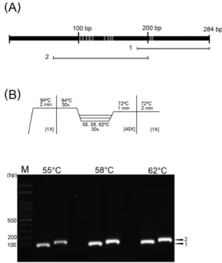
Fig. 2. Amplification of small subunit ribosomal RNA (SSU rRNA) region. (A) Schematic diagram of amplified SSU rRNA region. The gray bars indicate the rRNA region of area 1 and 2 amplified by PCR. The gray areas of SSU rRNA showed the different nucleotides between N. apis and N. ceranae via nucleotide alignment. (B) Reaction procedures of gene amplification for SSU rRNA and gel electrophoresis. Arrow heads indicate amplification of region 1 and 2 of SSU rRNA, respectively. M, 100-bp DNA ladder.
cycle을 반복하고, post-polymerization 72℃ 2분을 수행하였다. 이 반응 중에서 특이적인 유전자 증폭이 확인된 온도를 선택하였다.
2 µl PCR 반응 용액을 전기영동 및 유전자 클로닝과 서열분석을 수행하였다.
Quantitative real-time PCR (qRT-PCR)
서양뒤영벌에서 추출한 genomic DNA를 AquaQuest (Cecil Instruments Ltd., UK)를 이용하여 DNA 농도를 측정하였다.
Nosema 감염개체로부터 genomic DNA의 추출여부를 확인하기 위해 대조구로는 β-actin 유전자를 사용하였다. qRT-PCR은 최적화 반응에서 선택된 조건과 동일하게 수행되었다. 반응에 사용된 primer는 최적화에 이용된 primer를 이용하였다. DNA 증폭과정에는 TOPrealTM qPCR 2X premix (Enzynomics, Korea)를 사용하였다. qRT-PCR 반응은 PrimeQ real-time PCR system (Bibby Scientific Ltd., UK)를 사용하였다. PCR 후 melting curve analysis 수행하여 유전자 증폭 조건에서 유전자 특이적 반응여부를 검정하였다.
결과 및 고찰
서양뒤영벌을 활용하는 농가주변에 야외활동을 하는 약 265개체를 임의로 채집하고 본 연구에 사용하였다. 노제마병은 주로 중장에 있는 상피세포에 영향을 주기 때문에, 중장을 포함하는 부분을 분리하여 genomic DNA를 추출하였으며, 추후 실험 수행 전에 물리적인 분해(shearing) 여부를 DNA 전기영동으로 확인하였다. Nosema spp.를 탐지하기 위해 기존에 염기서열이 보고된 여러 유전자들을 검토하여, small subunit ribosomal RNA (SSU rRNA), 16S rRNA, large subunit rRNA, 및 RNA polymerase II largest subunit 유전자를 선발하고, 이들 유전자에 대한 PCR primer를 제작하여 유전자 증폭에 사용하였다. 이들 유전자 중에서, N. apis 16S rRNA 유전자와 N. ceranae의 SSU rRNA 유전자를 동시에 증폭할 수 있는 NC3-F와 NC3-R primer를 이용한 시험구에서 예상된 크기의 PCR 산물을
얻었다(Fig. 1). PCR 산물을 pGEMT-easy 벡터에 클로닝하고, NCBI에 등록된 서열과 염기서열을 분석한 결과, 증폭된 유전자가 N. ceranae의 SSU rRNA임을 확인하였고, 전체 조사된 265개체 중 18 개체에서 감염을 확인하였다. 이 결과는 조사에 사용된 서양뒤영벌 야외개체군의 주 감염체가 N. ceranae 임을 보여준다.
서양뒤영벌에서 N. ceranae SSU rRNA 유전자 증폭을 최적화하기 위해, 증폭된 SSU rRNA 유전자의 17–438 bp 부분을 영역별로 나누어 PCR 수행하여, 75–284 bp 영역에서 특이적으로 증폭된 DNA를 확인하였다. 증폭된 유전자 영역에 대해서 다시 영역 1과 2로 나누고, 유전자의 증폭에 영향을 미치는 annealing 온도를 55–62℃로 나누어 PCR을 한 결과, DNA 증폭이 모두 특이적으로 나타났다(Figs. 2A and 2B). 영역 1 및 2에서 얻어진 PCR 산물을 클로닝하고 염기서열을 분석하여, 증폭된 유전자가 N. ceranae의 SSU rRNA임을 확인하였다. 영역 1에 비해서 영역 2는 염기서열상의 여러 변이가 발견된 영역으로, 종간의 변이가 심한 유전자 부분으로서 특이적인 유전자 증폭을 이용한 종간 분류 마커로 활용가능한 영역으로 생각된다. 설정된 모든 annealing 온도에서 특이적인 PCR 산물을 얻을 수 있었고, 58℃를 annealing 온도로 선정하여 추후 실험을 진행하였다.
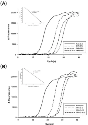
qRT-PCR을 이용하여 대상 유전자를 탐지하기 위해, DNA의 농도를 분광광도계로 정량하고(0.85 ng/µl), 순차적으로 희석하여
Fig. 3. Amplification of SSU rRNA of N. ceranae from Bombus terrestris. (A) Region 1 amplification. (B) Region 2 amplification.
The amplification is the same of optimized PCR procedures in Fig.
2. β-actin gene is used as an internal control. The number in the parenthesis is the dilution ration of the original genomic DNA concentration (0.85 ng/µl). The insets in each panel provide the standard curves which show the PCR efficiency of region 1(A) and region 2(B).
SSU rRNA 유전자의 증폭여부를 검정하였다. 대조구로는 β-actin 유전자를 사용하였다. 대상유전자의 증폭은 DNA 농도가 낮아짐에 따라 증폭이 시작되는 시점이 지연되었다. 주형으로 사용된 genomic DNA의 여러 희석농도와 threshold cycle (Ct)의 상관성은 두 영역 모두 R2 값이 0.96 이상으로 분석되었다(Fig. 3). 영역 1 및 2 모두 대조구로 상용한 β-actin 유전자에 비해 모든 조사농도에서 증폭시간이 10 cycle 이상 차이가 났지만, 극히 낮은 1/100 이하의 희석농도에서도 SSU rRNA 유전자가 증폭되었다(Figs. 3A and 3B).
노제마감염은 미포자충이 성충을 감염시켜 기생하여 발생하는 내부기생충성 전염병으로, 노제마병 감염으로 인해 수명단축, 여왕벌교체, 소화장애, 벌꿀수확량감소, 작물수확량 감소 등이 보고되었다(Fries et al., 1984; Anderson and Giacon, 1992; Malon et al., 1995). 미포자충 중에서 서양뒤영벌에 영향을 주는 것은 Nosema 속의 3종으로, 이들 중에서 N. apis와 N. ceranae는 화분매개충인 꿀벌에도 큰 영향을 준다. 노제마병의 감염은 봉군의 안정적인 관리에 큰 위협이 되고 있으며, 노제마 병 이외에 바이러스나 응애 등의 복합감염을 유발함으로써 봉군붕괴에 영향을
준다.
본 연구는 화분매개충으로 사용되고 있는 서양뒤영벌에서 Nosema 감염여부를 진단하기 위하여, Nosema spp.에서 보고된 유전자들의 서열을 기반으로 PCR을 통한 DNA 증폭을 시도하여 서양뒤영벌 개체에서 N. ceranae의 감염을 확인하였다. 국내에서는 꿀벌에서 Nosema 감염 진단을 위해, 충체에서 Nosema 포자(spore)를 순수분리 후 정량화된 포자수에 따른 PCR(Choi et al., 2003, 2006)을 이용하였다.
Yoo 등(2008)은 real-time PCR 이용한 Nosema 진단 여부를 보고하였으나, 클로닝된 유전자를 주형으로 하여 Nosema 진단법을 제시함으로써 실제 적용에서의 제한을 가지고 있다.
N. ceranae와 N. apis 두 종간의 SSU rRNA 서열은 일부 차이를 보인다는 보고(Fries et al., 1996)가 있었고, 두 종간의 낮은 SSU rRNA 상동성에 관한 연구(Huang et al., 2008)가 진행되었다.
특히 SSU rRNA를 포함한 N. ceranae의 rRNA 유전자들의 위치[5′-5S rRNA-Intergenic spacer (IGS)-SSU rRNA-internal transcribed spacer (ITS)-large subunit (LSU) rRNA-3′; Huang et al., 2008]는 다른 Nosema spp.에서 보여지는 위치(LSU rRNA-ITS-SSU rRNA-IGS-5S rRNA-3′)와는 서로 다르다(Huang et al., 2004; Tsai et al., 2005; Ku et al., 2007). 따라서 SSU rRNA 서열 간의 낮은 상동성과 rRNA 유전자 구조의 특이성은 SSU rRNA 서열의 종간구별 마커로서의 가능성을 높일 것으로 판단된다. 본 연구에서 Nosema 감염여부를 판단할 수 있는 유전자 영역은 선행 연구에서 이용한 유전자영역보다 상대적으로 짧으며, 근연관계에 있는 N. ceranae와 N. apis를 구별할 수 있어, 충체에 감염된 Nosema 종의 진단에 활용가능할 것으로 사료된다.
이상의 결과들을 통해, 본 연구에서 사용된 SSU rRNA 부분은 N. ceranae의 진단에 활용할 수 있을 것으로 판단된다. 또한 real-time PCR을 이용한 Nosema 감염진단은 단순히 감염여부를 판단하는 것이 아니라 화분 매개체로 활용하는 서양뒤영벌 개체에서 N. ceranae의 진단이 가능하고, 수입증가추세에 있는 서양뒤영벌의 위해성 평가에 대한 기초자료로서 활용가능성을 제시하고 있다.
적 요
서양뒤영벌(Bombus terrestris)은 꿀벌의 봉군붕괴증후군(colony collapse disorder)에 대한 대체 화분매개곤충으로서 농업분야에서 중요한 역할을 하고 있다. 최근 서양뒤영벌에서 바이러스, 세균, 응애 등의 여러 병원체와 기생체가 발견되었고, 이들은 서양뒤영벌의 수명과 생식력 등에 영향을 주는 것이 알려져 있다. 서양뒤영벌 야외개체군에서 Nosema spp.를 탐지하기 위해, 서양뒤영벌 성충으로부터 genomic DNA를 추출하여 Nosema spp. 유전자들에 대해 polymerase chain reaction (PCR)을 수행하였다. 이들 유전자 중에서 small subunit ribosomal RNA (SSU rRNA) 유전자만이 증폭되었고, 염기서열분석을 통해 N. ceranae로 확인된 것은 조사된 야외개체군에서 N. ceranae가 서양뒤영벌의 주된 감염체임을 보여준다. Quantitative real-time PCR (qRT-PCR)을 이용하여 SSU rRNA 유전자를 탐지하기 위해, 먼저 PCR을 통해 SSU rRNA 유전자의 2개 영역에 대한 유전자 특이적 증폭을 확인하였다. qRT-PCR을 이용하여 각 개체에서 얻은 genomic DNA의 순차적인 농도희석를 통해
0.85 ng/µl 이하의 genomic DNA 농도에서도 SSU rRNA 유전자가 성공적으로 증폭되는 것이 확인되었다. 이러한 실험 결과, qRT-PCR를 이용한 N. ceranae 특이 유전자 증폭은 서양뒤영벌의 병원체 감염 진단 뿐만 아니라 생태계 위해성 평가에도 활용될 수 있을 것으로 사료된다.
감사의 말
이 논문은 2012학년도 경성대학교 신임교수정착연구비에 의 하여 연구되었음.
참고문헌
Anderson, D.L. and Giacon, H. 1992. Reduced pollen collection by honey bee (Hymenoptera: Apidae) colonies infected with Nosema apis and sacbrood virus. J. Econ. Entomol. 85, 47–51.
Bromenshenk, J.J., Henderson, C.B., Wick, C.H., Stanford, M.F., Zulich, A.W., Jabbour, R.E., Deshpande, S.V., McCubbin, P.E., Seccomb, R.A., Welch, P.M., and et al. 2010. Iridovirus and microsporidian linked to honey bee colony decline. PLoS One 5, e13181.
Cameron, S.A., Lozier, J.D., Strange, J.P., Koch, J.B., Cordes, N., Solter, L.F., and Griswold, T.L. 2011. Patterns of widespread decline in North American bumble bees. Proc. Natl. Acad. Sci. USA 108, 662–667.
Chen, Y.P., Evans, J.D., Murphy, C., Gutell, R., Zuker, M., Gundensen-Rindal, D., and Pettis, J.S. 2009. Morphological, molecular, and phylogenetic characterization of Nosema ceranae, a microsporidian parasite isolated from the European honey bee, Apis mellifera. J. Eukaryot.
Microbiol. 56, 142–147.
Chen, Y., Evans, J.D., Smith, I.B., and Pettis, J.S. 2008. Nosema ceranae is a long-present and wide-spread microsporidian infection of the European honey bee (Apis mellifera) in the United States. J.
Invertebr. Pathol. 97, 186–188.
Choi, J.Y., Lee, M.Y., Je, Y.H., Kim, J.G., Choi, Y.C., and Kim, K.Y. 2003.
Diagnosis of Nosema disease in Honeybee by polymerase chain reaction. Kor. J. Apicult. 18, 131–138.
Choi, J.Y., Lee, M.-Y., Nam, S.-H., Kim, J.-G., Choi, Y.-C., Kim, W.-T., Sim, H.-S., and Kim, K.-Y. 2006. Multiplex polymerase chain reaction assay for simultaneous detection of Nosema ceranae, Ascosphaea apis and Paenibacillus larvae in honeybee. Kor. J.
Apicult. 21, 93–100.
Cox-Foster, D.L., Conlan, S., Holmes, E.C., Palacios, G., Evans, J.D., Moran, N.A., Quan, P.-L., Briese, T., Hornig, M., Geiser, D.M., and et al. 2007. A metagenomic survey of microbes in honey bee colony collapse disorder. Science 318, 283–287.
Fries, I., de Ruijter, A., Paxton, R.J., da Silva, A.J., Slemenda, S.B., and Pieniazek, N.J. 2001. Molecular characterization of Nosema bombi (Microsporidia: Nosematidae) and a note on its sites of infection in Bombus terrestris (Hymenoptera: Apoidea). J. Apicult. Res. 40, 91–96.
Fries, I., Ekbohm, G., and Villumstad, E. 1984. Nosema apis, sampling techniques and honey yield. J. Apicult. Res. 23, 102–105.
Fries, I., Feng, F., da Silva, A., Slemenda, S.B., and Pieniazek, N.J. 1996.
Nosema ceranae n. sp. (Microspora, Nosematidae), morphological and molecular characterization of a microsporidian parasite of the Asian honey bee Apis cerana (Hymenoptera, Apidae). Europ. J.
Protistol. 32, 356–365.
Fries, I., Martin, R., Meana, A., Garcia-Palencia, P., and Higes, M. 2006.
Natural infections of Nosema ceranae in European honey bees. J.
Apicult. Res. 45, 230–233.
Hamiduzzaman, M., Guzman-Novoa, E., and Goodwin, P.H. 2010. A multiplex PCR assay to diagnose and quantify Nosema infections in honey bees (Apis mellifera). J. Invertebr. Pathol. 105, 151–155.
Higes, M., Martin, R., and Meana, A. 2006. Nosema ceranae, a new microsporidian parasite in honeybees in Europe. J. Invertebr.
Pathol. 92, 93–95.
Higes, M., Martín-Hernández, R., Martínez-Salvador, A., Garrido- Bailón, E., González-Porto, A.V., Meana, A., Bernal, J.L., del Nozal, M.J., and Berna, J. 2010. A preliminary study of the epidemiological factors related to honey bee colony loss in Spain. Environ. Microbiol.
Rep. 2, 243–250.
Huang, W.F., Bocquet, M., Lee, K.C., Sung, I.H., Jiang, J.H., Chen, Y.W., and Wang, C.H. 2008. The comparison of rDNA spacer regions of Nosema ceranae isolates from different hosts and locations. J.
Invertebr. Pathol. 97, 9–13.
Huang, W.F., Tsai, S.J., Lo, C.F., Soichi, Y., and Wang, C.H. 2004. The novel organization and complete sequence of the ribosomal gene of Nosema bombycis. Fungal Genet. Biol. 41, 473–481.
Kim, N.-S., Lee, M.Y., Hong, I.-P., Choi, Y.S., Kim, H.K., Lee, M.Y., and Lee, K.G. 2010. Prevalence of honeybee (Apis mellifera) Nosema disease by season in Korea. Kor. J. Apicult. 25, 25–29.
Klee, J., Besana, A.M., Genersch, E., Gisder, S., Nanetti, A., Tam, D.Q., Chinh, T.X., Puerta, F., Ruz, J.M., Kryger, P., and et al. 2007.
Widespread dispersal of the microsporidian Nosema ceranae, an emergent pathogen of the western honey bee, Apis mellifera. J.
Invertebr. Pathol. 96, 1–10.
Ku, C.C., Wang, C.Y., Tsai, Y.C., Tzeng, C.C., and Wang, C.H. 2007.
Phylogenetic analysis between two putative Nosema isolates from cruciferous lepidopteran pests in Taiwan. J. Invertebr. Pathol. 95, 71–76.
Li, J., Chen, W., Wu, J., Peng, W., An, J., Schmid-Hempel, P., and Schmid-Hempel, R. 2012. Diversity of Nosema associated with bumblebees (Bombus spp.) from China. Int. J. Parasitol. 42, 49–61.
Malone, L.A., Giacon, H.A., and Newton, M.R. 1995. Comparison of the responses of some New Zealand and Australian honey bees (Apis mellifera L.) to Nosema apis Z. Apidologie 26, 495–502.
Martin-Hernandez, R., Meana, A., Prieto, L., Salvador, A.M., Garrido-bailon, E., and Higes, M. 2007. Outcome of colonization of Apis mellifera by Nosema ceranae. Appl. Environ. Microbiol. 73, 6331–6338.
Matheson, A. 1996. World bee health update 1996. Bee World 77, 45–51.
Shykoff, J. and Schmid-Hempel, P. 1991. Incidence and effects of four parasites in natural populations of bumble bees in Switzerland.
Apidologie 22, 117–125.
Singh, Y. 1975. Nosema in Indian honey bee (Apis cerana indica). Amer.
Bee J. 115, 59.
Tsai, S.J., Huang, W.F., and Wang, C.H. 2005. Complete sequence and gene organization of Nosema spodopterae rRNA gene. J. Eukaryot.
Microbiol. 52, 52–54.
Yoo, M.S., Kim, I.W., Kang, M.H., Han, S.H., and Yoon, B.S. 2008.
Development of real-time PCR method for the detection of Nosema.
Kor. J. Apicult. 23, 241–249.
Zander, E. 1909. Tierische parasite als krankenheitserreger bei der biene.
Munchener Bienezeitung 31, 196–204.