서 론
꿀벌의 독낭(毒囊)에 들어있는 봉독(蜂毒, bee venom)은 염증(炎症) 및 알러지 등을 유발할 수도 있지만 진통 및 소, 염의 효능이 탁월한 것으로 알려져 있는 약 40여 가지의 유 효성분으로 구성된 물질이다 오랫동안 봉독의 임상적 적용. 을 위하여 봉침(蜂鍼)을 직접 인체에 자입(刺入)하는 방법을 사용하였으나 최근에는 봉독을 추출 가공한 후 인체의 경, ․ 혈(經穴)에 자입하는 봉독약침요법(蜂毒藥鍼療法, Bee ven- 이 주로 활용되고 있다 즉 봉 om herbal acupuncture) [7,10].
독약침요법은 봉독을 질병과 유관한 부위나 혈위에 주입함 으로써 자침(刺鍼) 효과와 봉독의 생화학적 특이물질이 인체 에 미치는 약리작용을 동시에 이용하는 치료요법의 일종이 다 봉독의 성분은 크게. peptide components, non peptide
등으로 구성되어 있다 그 중 components, enzymes [7]. pep-
는 의 약 를 구성하
tide components freeze-dried venom 50%
고 있으며 주요 성분으로는, melittin, apamin, mast cell de-
등이 있다 봉독에 관한 연구는
granulating peptide [3,17].
년대 이후부터 활발히 진행되고 있으며 항암효과를 포
1990 ,
함한 다양한 생리 및 약리 작용에 관한 기전이 점차 알려지 고 있다.
최근 bee venom의 항암작용 가능성이 대두되면서 다양 한 인체암세포를 대상으로 봉독을 포함한 봉독의 구성성분 의 암세포 증식 억제기전의 연구가 활발히 진행되고 있다 또한 암의 발생과 진행뿐만 아니라 [1,5,10,12,13,15,20-22].
염증 반응의 주요 지표인 cyclooxygenase-2 (COX-2) 활성 저해 효과에 대한 보고가 부분적으로 이루어지고 있으나 봉독 자체에 대한 항암활성의 분자생물학적 기전에 [2,8,9]
관하여 아직까지 정확하게 알려진 바는 없다 따라서 본 연. 구에서는 선행연구들에 의해 관찰된 봉독의 항암작용에 관 한 추가적인 작용 기전을 조사하기 위하여 봉독에 의한 인 체 폐암세포의 생존율 저하와 연관된 COX-2의 발현 조절과 연관된 prostaglandin E2 (PGE2) 생성 및 염색체 말단에 존 재하는 telomere의 조절인자와telomerase의 활성에 미치는 봉독의 영향에 관하여 A549 인체폐암세포를 대상으로 조사 하였다.
인체폐암세포에서 봉독에 의한 prostagladin E
2생성 및 telomerase 활성 저하
김종환 황원덕 김병우․ ․ 2․최영현1,2*
동의대학교 한의과대학 신계내과학교실, 1생화학교실, 2대학원 바이오물질제어학과(BK 21 program)
2블루바이오 소재개발센터 및 자연과학대학 응용생명학과
Received March 2, 2009 /Accepted April 15, 2009
Bee Venom -induced Grow th Inhibition of H um an L ung Cancer C ells w as A ssociated w ith Inhibition of Prostagladin E2 Prod uction and Telom erase A ctivity. Jong-H w an Kim , W on-D euk H w ang, Byung-W oo K im2, and Yung H yun C hoi1,2*. Departments of Internal Medicine and1Biochemistry, College of Oriental Medicine,1Department of Biomaterial Control (BK21 program), Graduate School,2Blue-Bio Industry RIC and 2Department of Life Science and Biotechnology, College of Natural Sciences, Dong-Eui University, Busan 614-052 - In modern oriental medicine, bee venom therapy is being used for aqua-acupuncture to relieve pain and to cure inflammatory diseases such as rheumatoid arthritis, os- teoarthritis, and gout. Bee venom therapy has been processed and reported in many experimental studies, with regard to its effects on pain alleviation, anti-inflammation, removal of fever, anti-con- vulsion, suppression of tumor and immunity strengthening, etc., however, its mechanism of action, molecular targeting on prostaglandin E2(PGE2) production and telomere length regulation in human cancer remains unclear. In this study, we investigated the effect of bee venom on the levels of cyclo- oxygenases (COXs) and telomere regulatory components of A549 human lung cancer cells. Bee ven- om-induced anti-proliferative effects of A549 cells were associated with the inhibition of human telo- merase reverse transcriptase (hTERT) as well as human telomerase RNA (hTR), transcription factor c-myc and the activity of telomerase. In addition, bee venom treatment markedly decreased the lev- els of COX-2 mRNA and protein expression without significant changes in the expression of COX-1, which was correlated with a decrease in PGE2 synthesis. Taken together, these findings provide im- portant new insights into the possible molecular mechanisms of the anti-cancer activity of bee venom.
Key w ords : Bee venom , prostaglandin E2, telom erase
*Corresponding author
*Tel +82-51-850-8649, Fax +82-51-853-4036: :
*E-mail : [email protected]
재료 및 방법 실험재료
본 실험에 사용된 봉독은 Sigma Chemical Co. (St. Luis,
에서 구입하였고 분석을 위하여
MO, USA) mRNA Bioneer
에서 구입한 는
(Taejeon, Korea) primer Table 1에 나타낸 바와 같다 단백질 발현 분석을 위하여 사용된 항체는. CalBiochem (San Diego, CA, USA), 및 Santa Cruz Biotechnology Inc.
에서 구입하였으며
(Santa Cruz, CA, USA) , immunoblotting 을 위해 차 항체로 사용된2 peroxidase-labeled donkey an- ti-rabbit 및 peroxidase-labeled sheep anti-mouse im- munoglobulin은 Amersham Life Science Corp. (Arlington
에서 구입하였다
Heights, IL, USA) . PGE2의 생성양의 측정은
의 를 사용
Amersham Corp. enzyme immunoassay (EIA) kit 하였으며, telomerase 활성의 측정은 polymerase chain re-
에 기초를 둔 면역반응분석법
action (PCR) [PCR-based telo- meric repeat amplification protocol (TRAP) enzyme-linked
을 이용하였고 이를 위한 immunosorbent assay (ELISA)] kit 는Boehringer Mannheim (Mannheim, Germany)에서 구입하 였다.
암세포의 배양
실험에 사용한 A549 폐암세포는 생명공학연구소(KRIBB,
에서 분양 받았으며 의 배지
Taejeon, Korea) 90% RPMI-1640 (Gibco BRL, Grand Island, NY, USA), 10% fetal bovine se-
에 의 및
rum (FBS, Gibco BRL) 1% penicillin streptomycin 이 포함된 배지를 사용하여
(Gibco BRL) 5% CO2, 37oC의 조건 하에서 배양하였다 봉독은 차 증류수에 희석하여. 3 stock용액 으로 제조한 뒤 적정 농도로 배지에 희석하여 처리하였으며,
봉독처리에 따른 암세포의 형태변화는 도립현미경 하에서 관 찰하였다.
를 이용한 세포 성장억제 조사 MTT assay
세포배양용6 well plate에well 당1×10⁵개의 세포를 분주 하고24시간 동안 안정화시킨 다음 봉독을 배지에 희석 처리
시간 후 배지를 제거하고
48 0.5 mg/µl tetrazolium bromide 를 처리하였다 시간 후 를 제거하고 salt (MTT, Sigma) . 3 MTT
를 이용하여 에 생성된 을 녹인 후
DMSO well formazin ELISA
reader (Molecular Devices, Sunnyvale, CA, USA)로540 nm 에서 흡광도를 측정하였다 측정은 모두 회 실시하였으며. 3 , 그에 대한 평균값과 표준 오차를 Microsoft EXCEL program 을 사용하여 분석하였다.
Reverse transcription (RT)-PCR 분석
준비된A549 세포들에TRIzol B (Invitrogen, Carlsbad, CA,
를 이용하여 를 분리 정량한 후
USA) total RNA , ONE-STEP RT-PCR PreMix (iNtRON BIOTECHNOLOGY, Korea)를 이 용하여 2 µ 의g RNA에서ss cDNA를 합성하였다 이들. cDNA 를template로 사용하여 관찰 대상 유전자(Table 1)를PCR로 증폭하였으며, housekeeping 유전자인 glyceraldehyde-3-
를 로 사
phosphate dehydrogenase (GAPDH) internal control 용하였다 각. PCR 산물들을1% agarose gel을 이용하여 전기 영동하고 ethidium bromide (EtBr)로 염색한 후 UV 하에서 확인하였다.
단백질의 분리 전기영동 및, Western blotting
정상 및 봉독이 처리된 배지에서 자란 세포들을 lysis buf-
로 용해한 후 동량의 단백질을 전
fer , SDS-polyacrylamide gel
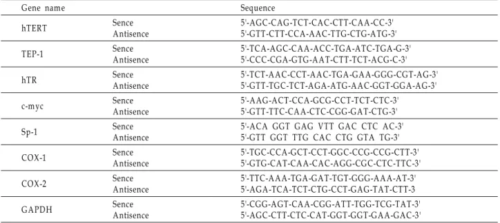
Table 1. Sequence of primers used for RT-PCR
Gene name Sequence
hTERT Sence
Antisence
5'-AGC-CAG-TCT-CA C-CTT-CAA -CC-3' 5'-GTT-CTT-CCA-AAC-TTG-CTG-A TG-3'
TEP-1 Sence
Antisence
5'-TCA-A GC-CAA-ACC-TGA -ATC-TGA -G-3' 5'-CCC-CGA-GTG-AA T-CTT-TCT-ACG-C-3'
hTR Sence
Antisence
5'-TCT-A AC-CCT-AA C-TGA -GA A-GGG-CGT-A G-3' 5'-GTT-TGC-TCT-A GA-ATG-AA C-GGT-GGA-A G-3'
c-m yc Sence
Antisence
5'-AAG-ACT-CCA-GCG-CCT-TCT-CTC-3' 5'-GTT-TTC-CA A-CTC-CGG-GA T-CTG-3'
Sp-1 Sence
Antisence
5'-ACA GGT GAG VTT GA C CTC A C-3' 5'-GTT GGT TTG CA C CTG GTA TG-3'
COX-1 Sence
Antisence
5'-TGC-CCA-GCT-CCT-GGC-CCG-CCG-CTT-3' 5'-GTG-CAT-CA A-CAC-A GG-CGC-CTC-TTC-3'
COX-2 Sence
Antisence
5'-TTC-A AA-TGA -GA T-TGT-GGG-AA A-A T-3' 5'-AGA -TCA-TCT-CTG-CCT-GA G-TAT-CTT-3
GAPDH Sence
Antisence
5'-CGG-A GT-CA A-CGG-ATT-TGG-TCG-TAT-3' 5'-AGC-CTT-CTC-CAT-GGT-GGT-GAA-GAC-3'
기영동으로 분리하였다. 분리된 단백질을 nitrocellulose membrane (Schleicher and Schuell, Keene, NH, USA)으로 전이시킨 후 특정 단백질에 대한 항체와 그에 대한 이차 항체, 반응을 실시한 후enhanced chemiluminoesence (ECL)용액
을 적용시킨 다음 에 감광
(Amersham Life Science) X-ray film 시켜 특정 단백질의 발현 양을 분석하였다.
활성 측정 Telomerase
정상 및 봉독이 처리된 배지에서 배양된 세포들을 모아 200 µl lysis reagent에1 x 10⁶개의 세포를 섞어서30분간 얼음 위에 서lysis를 실시하였다. TRAP반응을 위하여2 mg의 단백질이 함유된 2 µ 의 세포 추출액을l 25 µ 의l reaction mixture에 혼합 후 증류수를 첨가하여 최종, volume이50 µ 가 되도록 하였다l . PCR은primer elongation (25oC에서30분간), telomerase in- activation (94oC에서 분간5 ), product amplification (94oC에서 30 , 50초 oC에서30초 및72oC에서90초를30 cycles)의 순서로 진행이 되었으며 이를 이용하여, hybridization과ELISA re-
반응을 시켰다
action .
Prostaglandin E2의 측정
봉독 처리에 따른 PGE2생성량의 변화를 조사하기 위하여 준비된 세포를96-well plate에well 당 160 µ 의 배지에l 104 정도로 분주하여 시간 동안 배양한 후 봉독을 농
cell/well 24 ,
도별로 배지에 희석하여 처리하였다 이때 마지막 배지의 양. 을 모두 180 µ 로 통일시켰다l . 48시간 후 배지에2.5%의dode-
가 함유된 를
cyltrimethylammonium bromide buffer 20 µ 첨l 가하여 총 배지의 양이 200 µ 되게 한 후l lysis가 잘 일어나도 록pipetting을 수회 실시하였다 상온에 약. 10분간incubation 한 후trypan blue를 이용하여 암세포의 수를 계수하였다 약. 50 µ 의l lysate를 취하여kit에 준한protocol에 따라EIA를 실 시한 후 450 nm의 파장에서 얻어진 값을 기준으로PGE2의 양을 추정하였다.
결과 및 고찰
폐암세포의 증식에 미치는 봉독의 영향 A549
봉독에 의한A549 폐암세포에서PGE2생성 및 telomerase 활성 변화 연관 실험의 수행을 위한 조건 설정을 위하여, 48시 간 동안 다양한 농도의 봉독을 처리한 후MTT assay 및 형태 적 변화 관찰을 실시한 결과는Fig. 1에 나타낸 바와 같다 결과. 에서 알 수 있듯이 봉독 처리 농도의 증가에 따라A549세포의 형태변형 증가와 증식이 억제되어4 ug/ml 및5 ug/ml 처리 군에서는 대조군에 비하여 각각 약70% 및90% 이상의 증식억 제 효과를 보였으며 이는 다른 종류의 암세포에 나타난 선행, 연구의 결과들과 유사하였다[5,13,15,20-22].또한 이러한 봉독 의A549 세포 증식억제 효과가 세포주기 교란 및apoptosis
(A)
(B)
Fig. 1. Growth inhibition and morphological changes of A 549 human lung cancer cells after treatment with bee venom.
Cells were plated at 1x10⁵ cells per 60-mm plate, and incubated for 24 hr. The cells were treated with various concentrations of bee venom for 48 hr. (A ) The growth inhibition was m easured by the metabolic-dye-based M TT assay. Results are expressed as the m eans±S.E. of three independent experiments. (B) Exponentially grow- ing A549 cells were incubated with various concen- trations of bee venom for 48 hr. Cell m orphology was visualized by light microscopy. M agnification, X200.
유발과 연관성이 있는지를 조사한 결과Table 2에 나타낸 바와 같이 본 실험의 조건에서 봉독의 농도 증가에 따른 세포주기 특이적arrest유발은 관찰할 수 없었으나sub-G1기에 속하는 세포의 빈도가 다소 증가되어apoptosis 유발에 동반되었음을 알 수 있었다.
세포에서 의 생성에 미치는 봉독의 영향
A549 PGE2
는 세포의 분열이나 증식에 영향을 줌으 Prostaglandin (PG)
로서 각종 인체 질병의 유발과 진행에 중요한 역할을 함이 최근 밝혀진 바 있으며[6,19], PG의 합성에서 크게 가지2 COX 이 관여하고 있는데 대부분의 조직에서 일정한 수준
isoform ,
으로 발현되는 COX-1의 경우 인체의 항상성 유지와 연관된 기능수행에 관여하고 있다 그러나. COX-2는 다양한 성장인 자, cytokines,종양 촉진인자들 등의 자극에 의해 발현이 유도 되어 세포 증식을 촉진하고apoptosis를 억제하며 세포의 유 동성 및 부착성을 강화시킴으로서 각종 퇴행성 질환의 발병과
Table 2. Effect of bee venom on the progression of the cell cycle in A 549 human lung cancer cells
Dose (mg/ml)
% of cells
Sub G1 G1 S G2/M
0 1 2 3 4 5
0.91 1.74 1.87 2.24 2.83 4.97
57.05 55.91 55.17 57.66 55.93 55.72
23.03 22.86 22.05 16.32 19.51 17.54
19.43 19.85 21.58 24.25 22.20 22.31 The cells were treated with bee venom for 48 hr, collected, fixed, and stained with PI for flow cytometry analysis. Data are presented as the m ean values obtained from three in- dependent experiments.
진행에 중요한 역할을 한다[6,19].선행 역학적 조사와 여러 종류의 암 조직에서 COX-2가 높은 발현을 유지하는 것이 에 대한 저항성 획득과 염증반응과 연관된 세포의 apoptosis
암화에 밀접한 관련이 있을 것으로 보고되어 진 바 있으며[18], 의 과발현에 의해 암조직에서의 혈관신생 및 전이능이 COX-2
높아지고, COX-2의 선택적 억제제에 의한angiogenesis와 종 양형성 억제 등의 결과에서COX-2의 선택적 조절에 의한 암 예방 및 항암전략이 대두되고 있다.
따라서 봉독 처리에 의한A549세포의 증식억제가COX-2 의 발현 저하 및PG의 생성 변화와의 연관성이 있는지의 여부 를 조사하기 위하여COX-1 및-2의 발현 변화를RT-PCR 및
방법으로 조사하였다 및 의 결과에
Western blotting . Fig. 2A B
서 알 수 있듯이COX-1의mRNA 및 단백질 발현에는 봉독의 처리가 큰 영향을 미치니 못하였으나, COX-2의 경우 전사 및 번역 수준 모두에서 봉독 처리 농도의 증가에 따라 점차적인 발현의 감소를 보여주었다 이러한 봉독에 의한. COX-2의 선 택적 발현 저하가PG 중, PGE2의 생성 저하와 연관성이 있는
지를 조사하기 위하여 PGE2의 생성에 미치는 봉독의 영향을 조사한 결과는Fig. 2C에 나타낸 바와 같다. Fig. 2C에 나타낸 바와 같이 A549 세포에서 봉독 처리의 농도가 증가할수록 PGE2의 생성이 매우 감소되었으며, PGE2의 생성 감소 경향성 은COX-2의 발현 저하와 유사한 경향성을 보여주었다 따라. 서 봉독 처리에 의한A549세포의 증식 억제에는PGE2의 생성 저해와 연관성이 있으며 이는COX-2의 선택적 발현 억제에 의한 것임을 알 수 있었다.
조절인자들의 발현에 미치는 봉독의 영향 Telomere
는 진핵세포의 염색체 말단 부위에 존재하는
Ttelomere re-
peat sequences [(TTAGGG)n]로 이루어져 있고 이러한 반복, 구조의 형성 및 유지에 필수적으로 관여하는 효소가 telomer-
이다 대부분의 정상 체세포는 의 활성이 없
ase [16]. telomerase
기 때문에 세포가 분열할수록telomere의 길이는 짧아지게 되 지만 암세포의, 90% 이상에서는telomerase의catalytic sub-
단백질을 하는
unit coding human telomerase reverse tran-
가 과발현으로 높은 의 활성을 나
scriptase (hTERT) telomerase
타내고 있다 따라서. telomerase 활성 변화는 노화 혈관신생, 및 면역계질환 등에서 뿐 만 아니라 암의 발생과 진행과도 밀접한 연관성이 있으며 암의 진단과 진행의 정도를 나타내, 는 지표로 사용되고 있다[11,16].노화 조절의 측면에서 telo-
의 소실은 염색체의 안정성 소실을 야기하는 것으로
mere [14].
특히 암과 연관된 부분에서telomerase의 활성은hTERT 유전 자의 발현 조절에 의한 것이고, hTERT 유전자의 promoter 부위에는 다른 유전자의promoter보다 훨씬 더 많은 전사조 절인자의 결합부위를 보유하고 있어activator또는repressor 로 작용할 수 있다[4,16]. 따라서 암세포에서 높은 활성을 지니 는 telomerase의 활성을 선택적으로 억제함으로서 암세포의 성장과 분열을 억제하고자하는 시도는 새로운 항암제 개발을 위한 표적이 되고 있다.
(A) (B) (C)
Fig. 2. Inhibition of COX-2 expression and PGE2production by bee venom treatment in A549 human lung cancer cells. (A) After 48 hr incubation with bee venom, total RNA s were isolated and reverse-transcribed. The resulting cDNAs were subjected to PCR with COX-1 and COX-2 prim ers, and the reaction products were subjected to electrophoresis in a 1% agarose gel and visualized by EtBr staining. GAPDH was used as an internal control. (B) The cells were grown under the same conditions as (A ) and lysed and then cellular proteins were separated by SDS-polyacrylamide gels and transferred onto nitrocellulose mem branes. The m embranes were probed with anti-COX-1 and anti-COX-2 antibodies. Proteins were visualized using an ECL detection system. A ctin was used as an internal control. (C) The cells were treated with the indicated concentrations of bee venom for 48 hr and collected. The PGE2 accumulation in the medium was determined by an EIA kit as described in materials and methods. Results are expressed as the means±S.E. of three independent experiments.
(A)
(B)
Fig. 3. Effects of bee venom treatment on the levels of telomere regulatory genes in A549 human lung cancer cells. (A) After 48 hr incubation with bee venom, total RNAs were isolated and reverse-transcribed. The resulting cDNAs were subjected to PCR with the indicated primers, and the reaction products were subjected to electrophoresis in a 1% agarose gel and visualized by EtBr staining.
GA PDH was used as an internal control. (B) The cells were lysed and then cellular proteins were separated by SDS-polyacrylamide gels and transferred onto nitro- cellulose membranes. The membranes were probed with anti-hTERT antibody. Proteins were visualized using an ECL detection system. Actin was used as an internal control. (C) After 48 hr incubation with bee venom, telo- merase activity of cells was measured using a TRAP-ELISA kit as indicated in the protocol. Results are expressed as the means±S.E. of three independent experiments.
봉독 처리에 의한A549 세포의 증식억제가telomere 조절 인자들의 발현 변화와 어떤 연관성을 지니는지를 조사하기 위하여telomere길이에 조절에 가장 중요한 역할을 하는 가3 지 유전자[hTERT, human telomerase RNA (hTR)및telomer- 와 특히 의 발현 조절에 ase-associated protein (TEP)-1] hTERT
관여하는 두 가지 전사조절인자(c-myc 및Sp-1)의 발현에 미 치는 봉독의 영향을 조사하였다 이를 위하여 정상 및 봉독이. 처리된 배지에서48시간 동안 배양된A549 세포들을 대상으 로RT-PCR을 실시하였다. Fig. 3 의 결과에서 알 수 있듯이A 봉독의 처리 농도 증가에 따라 조사된 유전자 중 hTERT, hTR 및c-myc의mRNA 발현이 매우 감소되는 경향성을 보여 주었
으나, TEP-1 및 Sp-1의 발현에는 큰 변화가 없었다 이러한.
조절인자들의 발현 변화는 길이 조절에 직
telomere telomere
접적으로 중요한 조절 역할을 하는 telomerase 효소의 활성 변화와 연관성이 있을 것으로 추정되므로 이를 확인하기 위하 여 TRAP-ELISA를 통하여 봉독이 처리된A549 세포의telo- 효소 활성의 변화 여부를 조사하였다 의 결과
merase . Fig. 3B
에서 알 수 있듯이 봉독 처리된 배지에서 자란A549 세포의 활성은 봉독의 처리 농도가 증가될수록 매우 감소 telomerase
되는 경향성을 보여주어 봉독에 의한A549세포의 증식 억제 가 telomerase 활성 저하와 연관성을 지니며 이는, hTERT,
및 과 같은 조절 인자들의 발현 변화와
hTR c-myc telomere 연관되어 있음을 알 수 있었다.
요 약
본 연구에서는 봉독의 처리에 따른A549폐암세포의 증식 억제에서 PGE2생성 및telomerase 활성의 변화 관련성을 조 사하였다. A549세포의 증식은 봉독 처리에 의하여 유의적으 로 감소되었으며 이는, apoptosis 유발과 연관성이 있음을 알 수 있었다 봉독 처리 농도의 증가에 따라. COX-2의 발현이 전사 및 번역 수준에서 모두 감소되었으며 이에 따른 PGE2의 생성이 현저하게 감소되었으나, COX-1의 발현에는 큰 변화가 없었다 또한 봉독 처리에 따라. telomere 조절인자들 중,
및 의 발현이 억제되었으며 의
hTERT, hTR c-myc , telomerase 활성도 매우 감소되었다 본 연구의 결과는. PGE2생성과 telo-
활성 저하가 봉독의 항암 작용 표적인자로서 작용될 merase
수 있음을 보여준다.
감사의 글
이 연구는 지식경제부 부산광역시 지원 지역혁신센터사․ 업 동의대학교 블루바이오 소재 개발 및 실용화 지원 센터
지원에 의하여 이루어진 결과입니다
(RIC08-06-07) .
References
1. Ahn, C. B., C. W . Im, C. H. Kim , H. M . Youn, K. J. Jang, C. H. Song, and Y. H. Choi. 2004. Apoptotic cell death by melittin through induction of Bax and activation of caspase proteases in human lung carcinoma cells. J. Kor. Acupuncture Moxibustion Soc. 21, 41-55.
2. Ahn, C. B., C. W . Im, H. M . Youn, S. J. Park, and Y. H.
Choi. 2003. M elittin-induced apoptosis is associated with in- hibition of COX-2 and hTERT expression in hum an lung carcinoma A 549 cells. J. Kor. Acupuncture Moxibustion Soc.
20, 93-106.
3. Assem, E. S. and G. A tkinson. 1973. Histam ine release by M CDP (401), a peptide from the venom of the honey bee.
Bri. Pharmacol. 48, 337-338.
4. Cerni, C. 2000. Telomeres, telomerase, and myc. An update, Mutat. Res. 462, 31-47.
5. Choi, Y. H. 2005. Anti-proliferative effects of bee venom through induction of Bax and Cdk inhibitor p21W AF1/
CIP1 in human lung carcinoma cells. Kor. J. Oriental Physiol.
Pathol. 19, 167-173.
6. Giercksky, K. E. 2001. COX-2 inhibition and prevention of cancer. Best Pract. Res. Clin. Gastroenterol. 15, 821-833.
7. Habermann, E. 1971. Chemistry, pharmacology and toxicol- ogy of bee, wasp and hornet venoms. In venomous animals and their venom s. pp. 3-61, Academic Press.
8. Hwang, D. Y., H. H. Kim, C. J. Kim, and E. H. Kim. 2003.
Bee venom induces apoptosis and inhibits COX-2 in human osteosarcom a cell line M G-63. J. Kor. Acupuncture Moxibustion Soc. 20, 63-74.
9. Jang, M . H., M . C. Shin, S. Lim , S. M . Han, H. J. Park, I.
Shin, J. S. Lee, K. A. Kim, E. H. Kim, and C. J. Kim. 2003.
Bee venom induces apoptosis and inhibits expression of cy- clooxygenase-2 mRNA in human lung cancer cell line NCI-H1299. J. Pharmacol. 91, 95-104.
10. Kwon, K. R., H. K. Hoh, and C. H. Kim. 1994. The study of the introduction of bee venom acupuncture, biochemistry and pharm achology have been obtained the following results. J. Kor. Acupuncture Moxibustion Soc. 11, 159-171.
11. Kyo, S. and M . Inoue. 2002. Complex regulatory m echa- nisms of telomerase activity in normal and cancer cells:
How can we apply them for cancer therapy. Oncogene 21, 688-697.
12. M artikainen, P., K. Nym an, and T. J. Nevalainen. 1993.
Toxic effects of human pancreatic and snake and bee venom phospholipases A 2 on M CF-7 cells in culture. Toxicon 31, 835-843.
13. M oon, D. O., S. Y. Park, M . S. Heo, K. C. Kim, C. Park, W . S. Ko, Y. H. Choi, and G. Y. Kim. 2006. Key regulators in bee venom-induced apoptosis are Bcl-2 and caspase-3 in human leukemic U 937 cells through downregulation of
ERK and A kt. Int. Immunopharmacol. 6, 1796-1807.
14. Narayan, S., A. S. Jaiswal, A. S. M ultani, and S. Pathak.
2001. DNA damage-induced cell cycle checkpoints involve both p53-dependent and -independent pathways: role of te- lom ere repeat binding factor 2. Br. J. Cancer 85, 898-901.
15. Orsolić, N., L. Sver, S. Verstovsek, S. Terzić, and I. Basić.
2003. Inhibition of mammary carcinom a cell proliferation in vitroand tumor growth in vivo by bee venom. Toxicon 41, 861-870.
16. Poole, J. C., L. G. Andrews, and T. O. Tollefsbol. 2001.
Activity, function, and gene regulation of the catalytic sub- unit of telomerase (hTERT). Gene 269, 1-12.
17. Spoerri, P. E. 1973. Apamin from bee venom. Neurobiology 3, 207-214.
18. Surh, Y. J., K. S. Chun, H. H. Cha, S. S. Han, Y. S. Keum, K. K. Park, and S. S. Lee. 2001. M olecular mechanisms un- derlying chemopreventive activities of anti-inflamm atory phytochem icals: down-regulation of COX-2 and iNOS through suppression of NF- B activation.κ Mutat. Res.
480-481, 243-268.
19. Thun, M. J., S. J. Henley, and C. Patrono. 2002. Nonsteroidal anti-inflammatory drugs as anticancer agents: mechanistic, pharmacologic, and clinical issues. J. Natl. Cancer Inst. 94, 252-266.
20. Tu, W . C., C. C. W u, H. L. Hsieh, C. Y. Chen, and S. L.
Hsu. 2008. Honeybee venom induces calcium -dependent but caspase-independent apoptotic cell death in human m elanom a A2058 cells. Toxicon 52, 318-329.
21. W oo, H. J., H. J. Kim, S. H. Hong, S. H. Hong, B. T. Choi, Y. T. Lee, D. I. Park, and Y. H. Choi. 2007. Induction of apoptosis by bee venom in A549 human lung epithelial can- cer cells through m odulation of Bcl-2 and IAP family and activation of caspases. J. Life Sci. 17, 1596-1600.
22. Yeo, S. W ., J. C. Seo, Y. H. Choi, and K. J. Jang. 2003.
Induction of the growth inhibition and apoptosis by bee venom in hum an breast carcinom a M CF-7 cells. J. Kor.
Acupuncture Moxibustion Soc. 20, 45-62, 2003.