179
Correspondence to:Byung Soo Kim M.D., Ph.D.
Division of Hematology-Oncology, Department of Internal Medicine, Anam Hospital, Korea University Medical Center 126-1, Anam-dong 5-ga, Seongbuk-gu, Seoul 136-705, Korea Tel: +82-2-920-5031, Fax: +82-2-920-5031
E-mail: [email protected]
This study was supported by a grant (#SC2220) from Stem Cell Research Center of the 21st Century Frontier Research Program funded by Ministry of Science and Technology, Republic of Korea.
접수:2006년 5월 8일, 수정:2006년 6월 1일 승인:2006년 7월 14일
교신저자:김병수, 서울시 성북구 안암동 5가 126-1 136-705, 고려대학교 안암병원 내과 Tel: 02-920-5031, Fax: 02-920-5031 E-mail: [email protected]
Manipulation of Human Telomerase Activity in Cancer and Stem Cells: Application of siRNA-induced Inhibition of
Human Telomerase RNA (hTR)
Seok Jin Kim, M.D., Ph.D.1,2, Joon-Seok Song, Ph.D.1,2, Chang Hee Song1,2, Ji Hyun Yoo1,2, Young Do Yoo, Ph.D.3, Jun Suk Kim, M.D., Ph.D.1,2
and Byung Soo Kim, M.D., Ph.D.1,2
1Department of Internal Medicine, Korea University Medical Center, 2Institute of Korea University Stem Cell Research,
3Department of Medicine, Korea University Graduate School, Seoul, Korea
Background: We have determined the effects of human telomerase RNA inhibiton using siRNA in tumor cells and human embryonic and mesenchymal stem cells.
Methods: We selected the sequences against the predicted loop; these sequneces were comprised of nucleotides from 76 to 94 residues and from 143 to 163 residues as the target sequences, and we cloned these sequences into pU6sh75 and pU6sh143 cells. Three different kinds of cell lines were used: HeLa, SNUhES3, and human mesenchymal stem cells. The degree of inhibition of telomerase activity was as- sessed by TRAP assay and RT-PCR.
Results: The telomerase activity of the HeLa and SNUhES3 cells were 135.3±14.5 and 109.0±18.2;
these cells showed higher activity than human mesenchymal stem cells and Wi38 cells (46.3±5.0 and 26.0±12.0), which were control cells. When each of the types of cells was treated with siRNA-hTR, the transfection efficiency of pU6sh75 for the HeLa, SNUhES3, and human mesenchymal stem cells was 91.0±8.4%, 83.3±16.0% and 81.9±12.3%, respectively. In the case of pU6sh143, its transfection effici- ency was similar to pU6sh75; the HeLa, SNUhES3 and human mesenchymal stem cells tranfection effici- ency was 90.1±9.0%, 79.9±18.2% and 79.4±15.1%, respectively. After two days of transfection, the level of telomerase activity in the pU6sh75 transfected cells decreased to 64.3±10.1% and 56.0±11.0%
in the HeLa and SNUhES3 cells, respectively. When the cells were transfected with pU6sh143, the telomerase activity also decreased in the HeLa and SNUhES3 cells (71.3±9.1% and 61.6±8.3%, respec- tively). However, the difference of telomerase activity was not significant in the human mesenchymal stem cells: 43.0±7.2% with pU6sh75 and 46.0±9.0% with pU6sh143.
Conclusion: Telomerase RNA inhibiton with siRNA may be a feasible way to inhibit the telomerase activity of human tumor and embryonic stem cells. (Korean J Hematol 2006;41:179-185.)
Key Words: Telomerase, Tumor, Stem cell, siRNA
INTRODUCTION
Telomerase is a ribonucleoprotein involving the RNA component (hTR) and the telomerase catalytic subunit (hTERT).1-4) It is known that the activity of telomerase is higher in both tumor and stem cells than other somatic cells. That is why tumor and stem cells can self-renew and proliferate continuously. Among the components of telomerase, the hTR acts as a template for telomere synthesis.5) This hTR expression and telomerase activation may occur early in the pro- cess of human tumor development. Thus, the in- hibition of telomerase activity has been expected to play a role in the management of tumor as an anti-tumor mechanism. Increased activity of telo- merase is also reported in stem cells, thus high telomerase activity is known to be essential for the preservation of stem cells’ characteristics.6) The basic characteristics of stem cells are self- renewal and differentiation to multi-lineage cells.
For the clinical application of stem cells into cell- based therapy or regenerative medicine, the ma- nipulation of this peculiar characteristics of stem cells is required. Because the manipulation of telomerase activity may produce significant changes in biological characteristics of stem cells, several approaches targeting telomerase activity of stem cells have been tried to modulate the proliferation and differentiation of stem cells using various kinds of agents such as inhibitors of retroviral reverse transcriptase, peptide nucleic acid, cisplatin, hammerhead ribozyme, hTR anti- sense RNA, and hTR gene deletion.1-4) Recently, it has been demonstrated that synthesized anti- sense oligonucleotide against open part of hTR (2-5A-anti-hTR) showed cytotoxic effects on tu- mor cells.7) This effect of 2-5A-anti-hTR was sup- posed to be developed by an active induction of caspase-dependent apoptosis, which is indepen- dent of telomere length.8) RNA interference (RNAi) is a sequence-specific posttranscriptional gene silencing (PTGS) process by the siRNA
(short interfering RNA) in animals and plants.2) The dsRNA consisting of a sense and antisense strand of an endogenous mRNA, is rapidly pro- cessed by the RNase III type Dicer enzymes and assembled into RNA-induced silencing complex (RISC). The RISC results in the sequence-speci- fic degradation of homologous target sequence.
Chemically synthesized siRNA has been routinely used for gene knock-down, but the high cost, temporary and low efficiency gene silencing of synthetic siRNA, hinder the use of this strategy.
There have been some studies for the application of RNAi to inhibit telomerase activity in tumor cells.1,2) However, the application of RNAi to mo- dulate telomerase activity in stem cells has never been reported. In this study, we have tried to inhibit the human telomerase RNA (hTR) using small hairpin siRNAs, and determined the effects of telomerase inhibition on the biology of tumor and stem cells.
MATERIALS AND METHODS
1. Production of telomerase antisense by siRNA To knock-down a target gene, the target sites in the mRNA should not have mononucleotide repeats of more than 3 bases. Its GC contents should be 30~70%, and it should not be located at exon-intron boundaries or within the first 100 bases of the coding sequence that may have regulatory protein binding sites.9) To determine an optimal part fulfilling these criteria, the telo- merase RNA (hTR) structure was analyzed using synthetic siRNA design softwares of internet sites of invivogen, oligoengine, genescript, wister, amb- ion. The target part of the telomerase RNA (hTR) structure between residues 143 and 163 of the telomerase template sequence was selected. To maximize homologous binding, we also find the most ‘open’ part of the RNA molecule, thus we selected the hTR was between residues 94 and 76 of the telomerase template sequence. Because the previous analysis of telomerase RNA (hTR) struc- ture by Kondo et al has shown that the most ‘open’
Fig. 1. Sequences and predicted secondary structure of hairpin RNAs derived from the U6 promoter-driven expres- sion constructs. Mutant versions of each siRNA with 3 points mutation at the sequences of the target were also shown.
part of the hTR was between residues 94 and 76 of the telomerase template sequence.10) Therefore, we used the sequence against the predicted loop comprising nucleotides from 76 to 94 residues and from 143 to 163 residues in this study. Each pairs of oligonucleotides were synthesized, heated at 90oC for 10 minutes, and annealed at 55oC for 4 hours in 10 mM Tris-HCl (pH 8.0) and 1 mM EDTA. The resulting fragments included restri- ction endonuclease sites of EcoRI and XbaI at the 5' end and 3' end, respectively, and cloned into pU6shX (VCA-shPlasmid small hairpin RNA ex- pression vector, VectorCoreA, Korea) and named as pU6sh75 and pU6sh143 (Fig. 1). The mutant versions with 3 point mutations at the sequences, pU6sh75m and pU6sh143m were constructed the same way as above.
2. Transfection of telomerase antisense
We have used three different kinds of cells in this study: HeLa cell line (a human cervical ade- nocarcinoma cell line), human mesenchymal stem cells (MSCs) from human bone marrow aspirates, and SNUhES3 cell line (a human embryonic stem cells (ESCs)). A human normal fibroblast cell
line, Wi38 cell line was used as normal control.
The constructed plasmid vectors were transfected using FuGENETM6 (Roche, Germany) according to the manufacturer’s protocol. Briefly, 105 cells seeded in a 35-mm tissue culture dish were ex- posed to transfection mixture containing 2μg of siRNA expressing plasmids and 0.5μg of pSV-β- galactosidase control plasmid vector (Promega, USA) for 5 hr at 37oC. Then, 3mL of growth me- dia was added to the cells, followed by incubation for an additional 16hr.
3. Measurement of transfection efficiency Transfection efficiency was examined by galac- tosidase assay, thus, these vectors containing the galactosidase gene were tested after 24 hours of transfection. The percentage of transfected cells was calculated under an optic microscope (100X) and determined by the ratio between the number of galactosidease expressing cells (blue cells), and the number of total cells at the observed field.
Five distinct fields were recorded for each sam- ple. All experiments were performed in triplicate.
4. Measurement of telomerase activity
The cells were harvested 48 hr after the trans- fection to measure telomerase activity. The telo- merase activity was examined by a telomere re- peat amplification protocol (TRAP) assay with 2,000 cells using a TeloTAGGG Telomerase PCR ELISA Kit in accordance with the manufacturer’s instructions (Roche, Germany). After Primers TS (5’ AATCCGTCGAGCAGAGTT) and CX (5’ CC- CTTACCCTTACCCTTACCCTAA) were designed, thirty cycles of PCR were performed at 25oC for 30 min, at 94oC for 5 min, at 94oC for 30s, at 50oC for 30s, at 72oC for 90s, and a final extension at 72oC for 10 min. The PCR products were an- alyzed and defined positive when A>0.2 on the reading of the microplate reader. Telomerase ac- tivity of studied cells was relatively expressed on the basis of human epithelial U293 cell line’s telomerase activity as a value of 100.
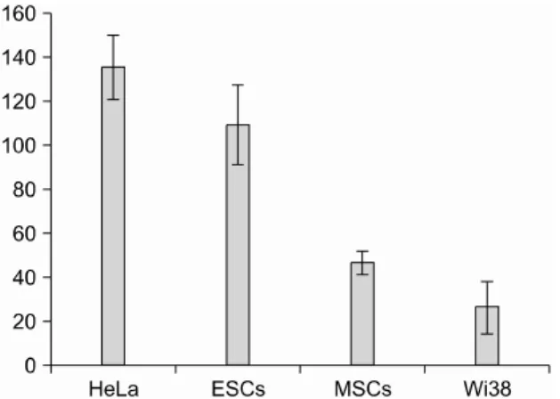
Fig. 2. Telomerase activity in experimental cell lines. The telomerase activity was assayed by a telomere repeat amplification protocol (TRAP) assay using 2,000 cells. The telomerase activity of the U293 cell line was considered as 100, with the telomerase activity of each cell line normali- zed to this. The means from at least three independent experiments are shown.
Fig. 3. Inhibition of Telomerase activity by siRNA. The ef- fects of siRNAs are expressed as the percentage of the telomerase activity compared with mutant versions (hTR75 m, hTR143m). The means from at least three independent experiments are shown.
5. Evaluation
Telomerase activity of each cells were com- pared between the pretreatment and post treat- ment of plasmid, respectively. And transfection degree and telomerase suppression effect of pU6sh75 and pU6sh143 were evaluated in each cell lines. All experiments were repeated nine times and the data were presented as mean±SEM.
Statistical significance was determined using a one way ANOVA test. Results were considered significant when the P value was less than 0.05.
RESULTS
Telomerase activity of each cell line was docu- mented before each cell line were treated with siRNA-hTR as shown in Fig. 2. HeLa and SNUh- ES3 showed significantly higher telomerase acti- vity than the Wi38, a control cell line. Thus, the telomerase activity of HeLa and SNUhES3 were 135.3±14.5 and 109.0±18.2, respectively. How- ever, human mesenchymal stem cells and Wi38 showed lower telomerase activity: 46.3±5.0 and 26.0±12.0, respectively (Fig. 2). When each cells were treated with siRNA-hTR, the transfection
efficiency of pU6sh75 for HeLa, SNUhES3, and human mesenchymal stem cells was 91.0±8.4%, 83.3±16.0% and 81.9±12.3%, respectively. In the case of pU6sh143, its transfection efficiency was similar to pU6sh75; HeLa, SNUhES3, and hu- man mesenchymal stem cells showed 90.1±9.0%, 79.9±18.2% and 79.4±15.1%, respectively. After two days of transfection, the TRAP assays were done with 2,000 cells. Compared to the telomer- ase activity of the mutant version transfected cells, the level of telomerase activity in the pU6sh75 transfected cells decreased to 64.3±
10.1% and 56.0±11.0% in HeLa and SNUhES3.
When the cells were transfected with pU6sh143, the telomerase activity also decreased in HeLa and SNUhES3: 71.3±9.1%, 61.6±8.3%, respecti- vely. However, the difference of telomerase acti- vity was not significant in the human mesenchy- mal stem cells compared to the baseline telomer- ase activity: 43.0±7.2% with pU6sh75, 46.0±9.0%
with pU6sh143 (Fig. 3). There was no significant difference of telomerase inhibition between pU6sh75 and pU6sh143 even though those of pU6sh143 transfected cells seemed to be higher than pU6sh75 transfected cells. To estimate the direct effect of siRNA, the transcriptional activity of target gene, hTR was measured by RT-PCR.
In the SNUhES3 transfected with pU6sh75 and
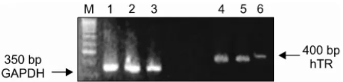
Fig. 4. Inhibition of mRNA expression of hTR. In the siRNA- hTR143 transfected ES, mRNA expression of hTR was decreased compared to control ES and siRNA-hTR143m transfected ES.
M, marker for molecular weight; lane 1 and 4, control ES;
lane 2 and 5, siRNA hTR143m transfected ES; lane 3 and 6, siRNA hTR143 transfected ES.
pU6sh143, mRNA expression of hTR was decre- ased compared to that of untransfected SNUhES3 and SNUhES3 transfected with pU6sh75m and pU6sh143m (Fig. 4).
DISCUSSION
Telomerase activity is usually considered to be low in somatic cells, however it is higher in cells replicating continuously especially, tumor cells.
Recently, the elevated telomerase activity of human stem cells have been highlighted because the role of telomerase have been suggested in the proliferation and differentiation of stem cells.3,4) Considering the role of telomerase for tumori- genesis and its exclusive expression in most tu- mor cells, telomerase can be a promising candi- date for a targeted therapy against tumor. For the clinical application of human stem cells as a tool of regenerative medicine, the manipulation of telomerase activity may be a feasible way to modulate the function of human stem cells. Telo- merase mainly consists of hTR and hTERT, and the expression of hTERT is closely associated with telomerase activity.11) Therefore, the inhibi- tion of hTR might be a possible target for the strategy to manipulate telomerase activity. Up to now, there have been several attempts to inhibit telomerase activity, for example, inhibitors of retroviral reverse transcriptase, peptide nucleic acid, hammerhead ribozyme, and hTR antisense RNA, and hTR gene deletion.12-14)
RNA interference (RNAi) is a sequence-specific post-transcriptional gene silencing mechanism, causing degradation of mRNAs homologous in sequence to the dsRNA and inhibiting specific gene expression effectively which is triggered by double-stranded RNA (dsRNA). RNAi technology has been a useful tools for investigating the fun- ction of genes and proteins, application of gene therapy against tumor, virus protection, drug target validation, or discovery screening.1,2) Gene therapy using a siRNA sequence could be an attractive approach for the treatment of viral infections, cancers, and genetic disorders. One of the major advantages of this approach is that it can inhibit the expression of the disease-associa- ted gene in a sequence specific-manner. Ever since the discovery that short siRNA could in- duce specific gene silencing, there have been numerous studies demonstrating the use of siRNA to modulate gene expression.12-14) There are two kinds of siRNAs, synthetic siRNA (in vitro pre- paration of siRNA) and expressed siRNA (in vivo expression of siRNA). For drug development, synthetic siRNA have been more commonly used due to the ease of dose control and more con- venience in clinical trials. However, synthetic siRNA has several disadvantages; it is unstable under the influence of nuclease, or toxin, and is not effective for the infusion into cells. Because such an expressed siRNA has various advantages over synthetic siRNA, it can be easily delivered by a viral vector and has a low toxicity. There have been several approaches that use expressed siRNA by siRNA expressing plasmid, knock- down of p53 or GαS, one of the ligand-dependent, G-protein-coupled signaling by siRNA expressing adenovirus.12-14)
In this study, we have tried to inhibit the human telomerase RNA (hTR) using a siRNA.
The selection of the target sequences was perfor- med using synthetic siRNA design softwares of internet sites and previous studies.15) The sele- ction criteria described by the Tuschl lab15) was used in this study: preferred GC contents <50%,
>50~100 nucleotide downstream of the start cordon, etc. The most probable part of hTR for siRNA knock-down was between residues 143 and 163, 20 nucleotides 3’ of the telomerase template RNA. BLAST searches of available databases for this region revealed that this region is homol- ogous only to the human hTR sequence in hu- man genome. Therefore, the sequence of 143~
163 region of hTR was selected for the design of siRNA expressing vector. In this study, the human U6 promoter which is a strong, ubiqui- tously active, and lacks essential promoter ele- ments within transcribed region, was used. In this study, the telomerase activity was higher in the human embryonic stem cell line, SNUhES3 than human mesencyemal stem cells extracted from human bone marrow aspirates. Because its baseline telomerase activity was low, the differ- ence between before and after siRNA treatment targeting hTRwas not significant in human mes- enchymal stem cells compared to SNUhES3. It is still not clear the reason why the telomerase activity of human mesenchymal stem cells is relatively low compared to human embryonic stem cells. However, the fact that mesenchymal stem cells are the group of heterogeneous cells including fibroblast, etc might be related with this difference from human embryonic stem cells.
Thus, the strategy using siRNA targeting telo- merase activity can be more useful in the mani- pulation of human embryonic stem cells than human mesenchymalo stem cells. Further study should be warranted to find the underlying mechanism of this difference in the future.
In conclusion, this study shows that the siRNA could inhibit telomerase activity of tumor as well as human embryonic stem cells. Therefore, the strategy using siRNA targeting telomerase activi- ty might be a feasible way to modulate the activi- ty of human stem cells and suppress the growth of tumor cells.
요 약
배경: 종양 세포와 줄기세포에서 텔로머라제 활성 도의 변화가 종양세포와 줄기세포의 생물학적 특성 을 변화시킬 수 있다. 본 연구에서는 siRNA를 이용 하여 텔로머라제 활성도를 억제하고자 하였다.
방법: 대상이 된 줄기 세포는 인간 중간엽 세포와 인간 배아줄기세포주(SNUhES3)를 시용하였고, 종 양세포주는 Hela 세포주를 이용하였다. 인간 텔로머 라제 억제를 위해 염기 서열 76에서 94까지의 뉴클 레오타이드와 143에서 163까지를 표적으로 하여 두 가지 anti-sense를 제조하였다(pU6sh75, pU6sh143).
결과: HeLa 세포주와 인간 배아줄기세포주에서 의 텔로머라제 활성도는 인간 중간엽 세포에 비해 의미있게 높았다(135.3±14.5, 109.0±18.2 vs. 46.3±
5.0). Transfection 효율은 pU6sh75의 경우, HeLa 세 포에서는 90.1±9.0%, 인간 배아줄기세포주에서는 79.9±18.2%, 그리고 인간 중간엽 세포에서는 79.4±
15.1%였다. siRNA 처리 후 텔로머라제 억제 정도는 pU6sh75 transfected 세포에서는 HeLa 세포주, 64.3±
10.1, 인간 배아줄기세포주, 56.0±11.0, 그리고 인간 중간엽 세포, 43.0±7.2%였다. 그리고 pU6sh143 tran- sfected 세포에서는 HeLa 세포주, 71.3±9.1, 인간 배 아줄기 세포주, 61.6±8.3, 그리고 이간 중간엽 세포, 46.0±9.0%였다.
결론: siRNA를 이용한 인간 텔로머라제 활성도의 억제는 종양세포주와 줄기세포주에서 효과적인 텔 로머라제 억제 방법으로 이용될 수 있겠다.
REFERENCES
1) Nakamura M, Masutomi K, Kyo S, et al. Efficient inhibition of human telomerase reverse transcriptase expression by RNA interference sensitizes cancer cells to ionizing radiation and chemotherapy. Hum Gene Ther 2005;16:859-68.
2) Blasco MA, Hahn WC. Evolving views of telomerase and cancer. Trends Cell Biol 2003;13:289-94.
3) Burns JS, Abdallah BM, Guldberg P, Rygaard J, Schroder HD, Kassem M. Tumorigenic heterogen- eity in cancer stem cells evolved from long-term cultures of telomerase-immortalized human mesen- chymal stem cells. Cancer Res 2005;65:3126-35.
4) Armstrong L, Saretzki G, Peters H, et al. Overex- pression of telomerase confers growth advantage,
stress resistance, and enhanced differentiation of ESCs toward the hematopoietic lineage. Stem Cells 2005;23:516-29.
5) Feng J, Funk WD, Wang SS, et al. The RNA com- ponent of human telomerase. Science 1995;269:
1236-41.
6) Liu L, DiGirolamo CM, Navarro PA, Blasco MA, Keefe DL. Telomerase deficiency impairs differen- tiation of mesenchymal stem cells. Exp Cell Res 2004;294:1-8.
7) Kondo Y, Koga S, Komata T, Kondo S. Treatment of prostate cancer in vitro and in vivo with 2-5A- anti-telomerase RNA component. Oncogene 2000;
19:2205-11.
8) Komata T, Kondo Y, Koga S, Ko SC, Chung LW, Kondo S. Combination therapy of malignant glioma cells with 2-5A-antisense telomerase RNA and re- combinant adenovirus p53. Gene Ther 2000;7:2071- 9.
9) Elbashir SM, Harborth J, Weber K, Tuschl T. An- alysis of gene function in somatic mammalian cells using small interfering RNAs. Methods 2002;26:
199-213.
10) Kondo S, Kondo Y, Li G, Silverman RH, Cowell JK.
Targeted therapy of human malignant glioma in a mouse model by 2-5A antisense directed against telomerase RNA. Oncogene 1998;16:3323-30.
11) Ito H, Kyo S, Kanaya T, Takakura M, Inoue M, Namiki M. Expression of human telomerase sub- units and correlation with telomerase activity in urothelial cancer. Clin Cancer Res 1998;4:1603-8.
12) Kosciolek BA, Kalantidis K, Tabler M, Rowley PT.
Inhibition of telomerase activity in human cancer cells by RNA interference. Mol Cancer Ther 2003;
2:209-16.
13) Shen C, Buck AK, Liu X, Winkler M, Reske SN.
Gene silencing by adenovirus-delivered siRNA.
FEBS Lett 2003;539:111-4.
14) Arts GJ, Langemeijer E, Tissingh R, et al. Adenoviral vectors expressing siRNAs for discovery and valida- tion of gene function. Genome Res 2003;13:2325- 32.
15) Elbashir SM, Harborth J, Weber K, Tuschl T. Analy- sis of gene function in somatic mammalian cells using small interfering RNAs. Methods 2002;26:
199-213.