Tryptophan Metabolite 3-Hydroxyanthranilic Acid Augments TRAIL-Induced Apoptosis in Activated T Cells
Su-Kil Seo*
Department of Microbiology and Immunology, College of Medicine, Inje University, Busan 614-735, Korea
Received January 19, 2011 /Accepted February 21, 2011Generation of tryptophan-derived metabolites by indoleamine 2,3-dioxygenase (IDO) is a potent im- munoregulatory mechanism in T cell responses. However, the mechanism remains unclear. We showed that 3-hydroxyanthranilic acid (3-HAA), the most potent metabolite, selectively induced apop- tosis in activated T cells, but not in resting T cells. This was not associated with cell cycle arrest. We found that TRAIL expression was selectively induced in activated T cells by treatment of 3-HAA.
Blockade of the TRAIL: DR4/DR5 pathway significantly inhibited 3-HAA-mediated T cell death. Our data suggest that TRAIL-induced apoptosis is involved in the mechanism of 3-HAA-mediated T cell death.
Key words : Indoleamine 2,3-dioxygenase, 3-hydroxyanthranilic acid, T cell, TNF-related apoptosis- inducing ligand
*Corresponding author
*Tel:+82-51-890-6434, Fax:+82-51-891-6004
*E-mail : [email protected]
서 론
Indoleamine 2,3-dioxygenase (IDO)는 트립토판(tryptophan) 분해효소로서 IFN-γ 및 염증사이토카인에 의해 특정 세포 및 조직에 발현하여 면역억제와 면역관용의 유도에 중요한 역할을 한다. IDO의 초기연구는 필수아미노산인 트립토판을 고갈시켜 세포내 기생균과 암세포의 증식을 억제하는 기능이 in vitro에서 보고되었다[21,22]. 1998년도에 Munn과 Mellor는 임신 된 마우스에 IDO 억제제를 투여하였을 때 모체의 T-세포 매개 면역반응에 의해 fetal rejection이 일어나는 실험결과를 통해 IDO의 면역반응 억제 기능을 최초로 보고하였다[15]. 그 후 암세포에 대한 T 세포의 면역관용발생[13,24], 자가면역질 환에서 자가반응 T 세포의 활성 억제 및 사멸유도[1,17], 동종 이식에서 이식편에 반응하는 T 세포의 활성 억제 및 사멸유도 [5,18,20], 알레르기반응 억제[6] 등 IDO의 강력한 면역억제 기 능이 보고되었다.
IDO의 발현은 수지상세포와 대식세포를 포함한 섬유아세 포, 외피세포, 내피세포 및 암세포 등의 다양한 세포에서 IFN- γ 에 의해 유도된다[12]. 생물학적 중요성에도 불구하고 여전 히 IDO의 T 세포 억제에 관한 분자생물학적 기전은 명확하지 가 않다. 크게 두 가지의 기전이 제기되고 있는데 하나는 특정 세포에 발현된 IDO가 T 세포의 증식과 활성에 필수적인 트립 토판을 고갈시켜 T 세포의 활성을 억제한다는 것이다. 최근에 IDO 발현 수지상세포에 의해 유도된 anergy T 세포에서 GCN2-kinase-dependent intergrated stress response (ISR)이
유도된다는 결과[14]를 통해 tryptophan 고갈이 활성화 T 세 포 내에 스트레스 반응을 유도하여 T 세포를 억제할 것으로 예상된다. 또 다른 기전은 IDO에 의해 생성된 트립토판 대사 체가 직접적으로 활성화된 T 세포의 고사를 매개하는 것이다 [12]. 트립토판 대사체인 L-kynurenine (L-kyn), 3-hy- droxykynurenic acid (3-HK), 3-hydroxyanthranilic acid (3-HAA), picolinic acid (PA)는 in vitro에서 T 세포의 증식을 억제하고 세포고사를 촉진시킨다[2,3,23]. 기존 연구들을 통해 가장 강력한 효능을 지닌 3-HAA가 비활성 T 세포의 기능적 손상을 미치지 않으면서 활성화 T 세포만을 선택적으로 사멸 시키는 선택적 T 세포 억제효능이 알려졌다[11]. 그러나 3-HAA의 강력한 면역조절 효능에도 불구하고 세포 내 작용기 전에 대한 연구보고는 아직 미비한 상태이다.
Tumor necrosis factor (TNF)-related apoptosis-inducing ligand (TRAIL)은 TNF superfamily의 한 구성원[26]으로서 종 양세포[25]와 바이러스 감염 세포[4]의 사멸을 유도한다. 또한 T 세포를 포함한 다양한 면역세포의 사멸에 중요한 기능을 갖는다[8,9]. 본 연구에서는 트립토판 대사체 3-HAA의 선택적 T 세포 억제 효과가 TRAIL-유도 세포사멸과 관련되어 있는지 를 조사하였다.
재료 및 방법
항체 및 reagents
다음의 항체는 BD Bioscience (San Jose, CA)에서 구매하였
다. PE-와 PE-Cy5-anti-human CD3 (UCHT1), PE-와
FITC-anti-human CD25 (BC96), PE-anti-human CD45RO
(UCHL1), PE-anti-human TRAIL (RIK-2), FITC-와 PE-와
PE-Cy5-mouse IgG, purified mouse IgG, purified anti- human TRAIL (RIK-2), PE-Annexin V, BrdU Flow kit, 7-AAD. PE-anti-human DR4 (DJR1)와 PE-anti-human DR5 (DJR2-2)는 e-Bioscience (San Diego, CA)에서 구매하였다.
3-HAA는 Sigma-Aldrich (St. Louis, MO)에서 구매하였다.
SEA는 Toxin Technology (Sarasota, FL)에서 구매하였다.
Carboxyfluorescein succinimidyl ester (CFSE)는 Invitrogen (Carlsbad, CA)에서 구매하였다.
세포분리 및 3-HAA 처리
인간말초단핵세포는 Ficoll-Histopaque (Sigma, St. Louis, MO)의 농도구배를 이용하여 정상인의 말초혈액으로부터 분 리하였다. 세포(1×10
7)를 세척 후 PBS완충액에 현탁시키고 5 μ M CFSE를 첨가하여 실온에서 8분간 반응시켰다. 차가운 5%
Fetal bovine serum (FBS)/PBS완충액으로 반응을 종결시킨 다음 Hank's buffered salt solution (HBSS)으로 세척하였다.
세포를 10% FBS (Invitrogen, Grand Island, NY), 100 U/ml penicillin (Cambrex, Baltimore, MD), 100 μg/ml streptomycin (Cambrex, Baltimore, MD)이 첨가된 RPMI 1640 세포배양액 에 현탁시키고 0.2 ml의 세포액을 96-well plate에 분주하였다.
0.15 μg/ml의 SEA를 분주된 세포액에 추가하여 세포를 자극 하였다. 배양 48 시간 후 100 μM의 3-HAA 혹은 대조군용액을 배양세포에 첨가하고 표시된 각 시간에 세포를 얻은 후 이후 진행될 실험에 사용하였다.
Flow cytometric analysis
배양 중인 세포를 채집하여 FACS용 tube (BD Falcon, Franklin Lakes, NJ)에 옮기고 FACS buffer (2% FCS/PBS + 0.01% NaN3)으로 세척하였다. Fc 수용체의 비특이적 결합을 방지하기 위해 purified human IgG를 첨가하고 4℃에서 10분 간 반응시켰다. 이후 PE-혹은PE-Cy5-anti-human CD3를 첨가 하고 4℃에서 25분간 반응시켰다. 세포를 FACS buffer로 세척 후 FACSCalibur flow cytometry (BD Bioscience)로 측정하고 CellQuest pro software를 사용하여 CD3+세포에서 CFSE의 감도를 분석하였다. 활성 T 세포의 분석은 세포에 PE-Cy5- anti-human CD3와 PE-anti-human CD25 혹은 -CD45RO로 이중 염색 후 flow cytometric analysis하였다. 세포사멸 분석 은 위와 같은 방법으로 채집된 세포에 PE-Cy5-anti-human CD3와 PE-Annexin V로 염색하고, CD3+CFSE
high와 CD3+
CFSE
low세포에서 Annexin V+ 세포의 %값을 측정하였다.
TRAIL/ DR4/ DR5 발현조사는 채집된 세포에 PE-Cy5-anti- human CD3와 PE-anti-human TRAIL, -DR4, 혹은 -DR5로 염 색하고, CD3+CFSE
high와 CD3+CFSE
low세포에서 TRAIL/
DR4/DR5+세포의 % 값을 측정하였다. 세포 주기 분석은 BrdU flow kit의 매뉴얼에 따라 실시하였다.
TRAIL 차단효과 분석
CFSE로 표시된 인간 말초단핵세포를 SEA로 48시간 동안 위와 동일한 방법으로 자극하였다. 3-HAA (100 μM)로 처리하 기 1 시간 전 purified anti-human TRAIL (2.5 μg/ml) 혹은 control IgG를 각각 배양 세포에 처리하였다. 3-HAA 처리 24 시간 후 세포를 채집하고 PE-anti-human CD3로 염색하였다.
세척 후 7-AAD를 첨가하고 flow cytometric analysis 하였다.
결 과
3-HAA의 활성 T 세포 사멸유도
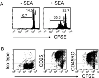
트립토판 대사체들이 세포고사를 촉진시켜 활성 T 세포를 우선적으로 억제함이 보고되었다[14-17]. 본 연구자는 최초 인 간 말초혈액 T 세포에서 3-HAA의 선택적 억제효과를 확인하 기 위해 CFSE로 표시된 인간 말초혈액단핵세포(PBMCs)를 SEA로 자극하였다. SEA는 세균의 초항원으로서 특정 TCR Vβ region을 갖는 T 세포를 활성화 시켜 증식을 강하게 유도 한다[23]. Fig. 1A에서 보듯 시 SEA에 반응하여 증식을 진행하 고 있는 T 세포는 CFSE의 intensity가 감소하게 되어 항원에 반응하지 않는 high intensity의 T 세포와 구별 할 수 있었다.
그리고 증식 중인 CFSE
lowT 세포(activated)들은 활성화 표면 단백질 CD25와 CD45RO가 모두 높게 발현되어 있어 CFSE
high비활성 T 세포(resting)와 확연히 구별되었다(Fig. 1B). 본 연구
A
B
Fig. 1.
In vitro
cell stimulation system. (A) Human PBMCs were labeled with CFSE and stimulated with SEA for 72 hr.The cells were then stained with PE-anti-CD3. Data represent the cell proliferation of gated 7-AAD-CD3+T cells, as determined by flow cytometry. The numbers indicate the percentages of gated T cells among total live cells. (B) Cell were stained with PE-Cy5-anti-CD3 and PE-CD25 or-CD45RO and analyzed by flow cytometry.
The data represent the expression levels of gated CD3+ T cells.
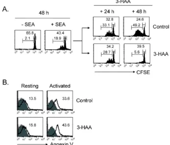
자는 구축된 in vitro 시스템을 이용하여 3-HAA의 선택적 억 제효과를 평가하였다[11]. SEA 자극 48 시간 후, 배양조건에 활성 T 세포(CD3
+CFSE
low, 19.2±3.2%)와 비활성 T 세포 (CD3
+CFSE
high, 40.2±5.4%)가 공존함을 확인하고, 3-HAA와 control vehicle를 배양세포에 처리하였다. Control vehicle처 리군에서는 T 세포 증식이 시간에 따라 계속 강화되지만(+24 h, 32.5±5.0%; +48 h, 43.7±6.8%) 3-HAA 처리군에서는 증식 억제 현상(+24 h, 26.3±3.9%; +48 h, 5.3±2.7% )이 뚜렷이 관찰 되었다(Fig. 2A). 3-HAA에 의한 증식억제 현상이 세포사멸 촉진에 의한 것인지를 조사하였다. 활성 T 세포는 3-HAA 처 리 시 control에 비해 세포사멸에 대한 감수성이 9.8±1.2% 증 가한 반면 비활성 T 세포의 사멸증감은 관찰되지 않았다(Fig.
2B). 이 결과를 통해 3-HAA는 선택적으로 활성 T 세포의 사멸 을 촉진시키는 효능이 있음을 알 수 있다.
세포주기에 미치는 효과
3-HAA의 활성 T 세포 사멸촉진 효과가 세포주기 억제와 관련된 것인지를 조사하였다. 분리된 PBMC를 SEA로 48시간 동안 자극한 후 3-HAA를 처리하고 24 시간 후에 BrdU + 7-AAD 방법으로 세포 주기를 측정하였다. 3-HAA 처리군의 활성 T 세포는 control 처리군과 유사한 세포 주기 형태를 지 니고 있었다(Fig. 3). 이를 통해 3-HAA에 의한 활성 T 세포
Fig. 2. 3-HAA selectively induces cell death in activated T cells.
(A) CFSE-labeled PBMCs were pre-stimulated with SEA for 48 hr prior to treatment with 100 μM 3-HAA for the indicated incubation periods. The cells were then stained and analyzed as in Fig.1A. (B) Cells were stained with PE-Cy5-anti-CD3 and PE-annexin V and analyzed by flow cytometry. The data represent the Annexin V expression levels of gated CFSElow or CFSEhighCD3+ T cells. Similar results were obtained with PBT cells from three individuals.
Fig. 3. 3-HAA-mediated cell death is not due to cell cycle arrest.
PBMCs were pre-stimulated with SEA for 48 hr prior to treatment with 100 μM 3-HAA for 24 hr. The cells were stained for incorporated BrdU and 7-AAD as described in Materials and Methods. The data represent the cell cycle ratio of gated FSChiCD3+ T cells (activated) or FSCloCD3+ T cells (resting), as determined by flow cytometry.
사멸효과는 세포주기 억제와 관련이 없음을 알 수 있다.
TRAIL 및 수용체 발현 증가 유도
TRAIL의 발현 증가는 활성T 세포의 사멸효과에 중요한 현 상학적 특징이다[21,22]. 본 연구자는 3-HAA의 활성 T 세포 사멸 유도 효과가 TRAIL-유도 세포사멸과 관련되어 있는지를 조사하였다. 3-HAA 처리 시 활성 T 세포에서 TRAIL의 발현 이 24.9±2.7% 증가하였다(Fig. 4A). 또한 TRAIL의 수용체인 DR4와 DR5의 발현증가도 3-HAA가 처리 된 활성 T 세포에서
Fig. 4. 3-HAA selectively induces TRAIL/DR4/DR5 expression on activated T cells. PBMCs were pre-stimulated with SEA for 48 hr prior to treatment with 100 μM 3-HAA for 24 hr. The cells were stained for TRAIL, DR4, or DR5.
The data represent the expression levels of gated CD45RO+CD3+ T cells (activated) or CD45RO-CD3+ T cells (resting), as determined by flow cytometry.
A
B
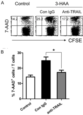
Fig. 5. Effect of TRAIL blocked on the 3-HAA-mediated activated T cell death. Pre-stimulated PBMCs were treated with anti-TRAIL mAb before 1 hr to treatment with 100 μM 3-HAA. After 24 hr in culture, cells were stained for 7-AAD. The data represent the 7-AAD positive cells gated total CD3+cells, as determined by flow cytometry. (B) Quantification of the percentage of 7-AAD positive T cells. *
p
<0.05, compared with the control treatment. The data are means±S.E. Similar results were obtained with PBT cells from three individuals.26.4±3.1% 높게 나타났다(Fig. 4B). 그러나 비활성 T 세포에서 의 발현증가는 관찰되지 않았다. 이 결과를 통해 3-HAA에 의 한 활성 T 세포의 선택적 사멸효과는 TRAIL과 관련되어 있을 것으로 추정할 수 있다.
TRAIL-유도 세포사멸 효과
3-HAA-매개 활성 T 세포 사멸현상에서 TRAIL 경로의 유 의성을 알아보았다. SEA로 자극된 배양조건에 3-HAA 처리 전 TRAIL 중화항체(anti-mouse TRAIL mAb)를 처리하여 TRAIL 수용체로의 사멸 신호전달을 차단하였다. . TRAIL 중 화항체 처리조건에서는 3-HAA에 의한 활성 T 세포의 사멸현 상이 유의성 있게(p=0.0328) 낮아졌다(Fig. 5). 이 결과를 통해 3-HAA에 의한 활성 T 세포 사멸유도 기전에 TRAIL이 부분적 으로 관련되어 있음을 알 수 있다.
고 찰
트립토판 고갈에 의한 세포 내 스트레스 반응 유도와 함께
트립토판 대사체 생성은 IDO-매개 T 세포 반응 억제의 주요 기전으로 알려져 있다[12]. 본 연구에서는 트립토판 대사체 3-HAA의 선택적 T 세포 억제 효능과 그 작용기전을 조사하였 다. 기전연구에 앞서 본 연구자는 3-HAA의 선택적 T 세포 억제 현상을 명확히 확인하기 위한 in vitro 실험 조건을 확립 하였다. 인간 말초혈액세포를 CFSE로 표지한 후 세균초항원 (SEA)으로 자극하고, 72 시간 후 배양세포를 flow cytometry 로 분석하여 활성 T 세포(CFSE
lowCD25
+CD45RO
+)와 비활성 T 세포(CFSE
highCD25
-CD45RO
-)를 명확히 구별하였다(Fig. 1).
이어 본 연구자는 활성과 비활성 T 세포가 공존하는 in vitro 조건에 3-HAA을 처리하였을 때, 활성 T 세포는 사멸이 촉진 되지만 비활성 T 세포는 영향을 받지 않는 선택적 억제효과를 확인하였다(Fig. 2). 이와 같은 결과는 3-HAA가 T 세포-매개 염증질환의 치료약물개발의 좋은 표적이 될 수 있음을 보여준 다. 현재 임상에서 사용되는 항-대사억제 및 칼시뉴린 저해 면역억제약물들은 초기 활성화 단계의 T 세포까지 억제대상 이 되므로 전반적 면역약화가 초래되어 감염 및 암발생 등의 부작용이 빈번히 발생된다. 그러므로 질병과 관련된 특정항원 에 과반응을 보이는 T 세포만을 선택적으로 표적할 수 있다면 부작용의 문제점을 최소화 할 수 있다. 최근, 3-HAA의 합성 유도체인 3,4-DAA를 다발성경화증(multiple screlosis, MS) 마우스모델에 경구 투여하였을 때 자가항원-반응 Th1 세포가 Th2 세포로 분화가 되면서 치료효과가 나타남이 보고되면서 이에 대한 신약개발의 관심이 높아지고 있다[24,25].
3-HAA의 강력한 면역조절 효능에도 불구하고 세포 내 작 용기전에 대한 연구보고는 아직 미비한 상태이다. Fallarino 등은 Fas-비의존적 기전에 의한 cytochrom c 방출-caspase8 활성화를 3-HAA가 촉진시켜서 Th1 세포를 사멸로 유도한다 고 보고하였으며[16], PDK-1 인산화를 억제하여 TCR- triggered NF-kB 활성화 억제 또한 하나의 기전으로 보고되었 다[26]. 본 연구자는 3-HAA에 의한 세포 내 glutathione (GSH) 고갈이 선택적 T 세포 억제의 주요 기전임을 보고하였다[17].
선행 연구보고들을 종합해보면, T 세포의 type, 활성상태 및
염증 환경의 조건에 따라 3-HAA의 주요 작용 기전은 다양해
질 수 있음을 알 수 있다. 본 연구에서는 TRAIL-유도 세포사멸
이 3-HAA의 선택적 T 세포 억제 기전 중의 하나임을 확인하
였다. TRAIL-유도 세포사멸은 생체 내에서 세포독성 T 세포
(CTL)의 암세포의 사멸유도기전 중의 하나이며, 일부 항암제
의 효능 기전으로 알려져 있다. 본 연구자는 활성과 비활성
T 세포가 공존하는 in vitro 조건에 3-HAA를 처리하여 활성
T 세포만 TRAIL과 그의 수용체의 발현이 증가되고(Fig. 4),
이들 상호작용을 차단하였을 때 3-HAA-매개 활성 T 세포사멸
효과가 유의하게 낮아짐을 확인하였다(Fig. 5). 이를 통해
TRAIL-유도 세포사멸이 3-HAA의 새로운 작용기전이며, 추
후 면역질환동물모델에서 3-HAA의 생리적 효과와의 관련성
을 조사해야 할 것이다.
감사의 글
This research was supported by the 2006 Inje University research grant.
References
1. Alexander, A. M., M. Crawford, S. Bertera, W. A. Rudert, O. Takikawa, P. D. Robbins, and M. Trucco. 2002.
Indoleamine 2,3-dioxygenase expression in transplanted NOD Islets prolongs graft survival after adoptive transfer of diabetogenic splenocytes.
Diabetes
51, 356-365.2. Fallarino, F., U. Grohmann, C. Vacca, R. Bianchi, C. Orabona, A. Spreca, M. C. Fioretti, and P. Puccetti. 2002. T cell apopto- sis by tryptophan catabolism.
Cell Death Differ
. 9, 1069-1077.3. Frumento, G., R. Rotondo, M. Tonetti, G. Damonte, U.
Benatti, and G. B. Ferrara. 2002. Tryptophan-derived catabo- lites are responsible for inhibition of T and natural killer cell proliferation induced by indoleamine 2,3-dioxygenase.
J. Exp. Med
. 196, 459-468.4. Gura, T. 1997. How TRAIL kills cancer cells, but not normal cells.
Science
277, 768.5. Grohmann, U., F. Fallarino, R. Bianchi, C. Orabona, C. Vacca, M. C. Fioretti, and P. Puccetti. 2003. A defect in tryptophan catabolism impairs tolerance in nonobese diabetic mice.
J.
Exp. Med
. 198, 153-160.6. Hayashi, T., L. Beck, C. Rossetto, X. Gong, O. Takikawa, K. Takabayashi, D. A. Carson, and E. Raz. 2004. Inhibition of experimental asthma by indoleamine 2,3-dioxygenase.
J.
Clin. Invest
. 114, 270-279.7. Hayashi, T., J. H. Mo, X. Gong, C. Rossetto, A. Jang, L. Beck, G. I. Elliott, I. Kufareva, R. Abagyan, D. H. Broide, J. Lee, and E. Raz. 2007. 3-Hydroxyanthranilic acid inhibits PDK1 activation and suppresses experimental asthma by inducing T cell apoptosis.
Proc. Natl. Acad. Sci. USA
104, 18619-18624.8. Janssen, E. M., N. M. Droin, E. E. Lemmens, M. J. Pinkoski, S. J. Bensinger, B. D. Ehst, T. S. Griffith, D. S. Green, and S. P. Schoenberer. 2005. CD4+ T-cell help controls CD8+
T-cell memory via TRAIL-mediated activation-induced cell death.
Nature
434, 88-93.9. Jeremias, I., I. Herr, T. Boehler, and K. M. Debatin. 1998.
TRAIL/Apo-2-ligand-induced apoptosis in human T cells.
Eur. J. Immunol.
28, 143-152.10. Kotzin, B. L., D. Y. Leung, J. Kappler, and P. Marrack. 1993.
Superantigens and their potential role in human disease.
Adv. Immunol.
54, 99-166.11. Lee, S. M., Y. S. Lee, J. H. Choi, S. G. Park, I. W. Choi, Y. D. Joo, W. S. Lee, J. N. Lee, I. H. Choi, and S. K. Seo.
2010. Tryptophan metabolite 3-hydroxyanthranilic acid se- lectively induces activated T cell death via intracellular GSH depletion.
Immunol. Lett.
132, 53-60.12. Mellor, A. L. and D. H. Munn. 2004 IDO expression by den- dritic cells: tolerance and tryptophan catabolism.
Nat. Rev.
Immunol
. 4, 762-774.13. Muller, A. J., J. B. DuHadaway, P. S. Donover, E.
Sutanto-Ward, and G. C. Prendergast. 2005. Inhibition of in- doleamine 2,3-dioxygenase, an immunoregulatory target of
the cancer suppression gene Bin1, potentiates cancer chemotherapy.
Nat. Med
. 11, 312-319.14. Munn, D. H., M. D. Sharma, B. Baban, H. P. Harding, Y.
Zhang, D. Ron, and A. L. Mellor. 2005. GCN2 kinase in T cells mediates proliferative arrest and anergy induction in response to indoleamine 2,3-dioxygenase.
Immunity
22, 633-642.15. Munn, D. H., M. Zhou, J. T. Attwood, I. Bondarev, S. J.
Conway, B. Marshall, C. Brown, and A. L. Mellor. 1998.
Prevention of allogeneic fetal rejection by tryptophan catabolism.
Science
281, 1191-1193.16. Platten, M., P. P. Ho, S. Youssef, P. Fontoura, H. Garren, E. M. Hur, R. Gupta, L. Y. Lee, B. A. Kidd, W. H. Robinson, R. A. Sobel, M. L. Selley, and L. Steinman. 2005. Treatment of autoimmune neuroinflammation with a synthetic trypto- phan metabolite.
Science
310, 850-855.17. Sakurai, K., J. P. Zou, N. I. Torres, J. R. Tschetter, H. S.
Kim, and G. M. Shearer. 2002. Study of the effect of indole- amine 2,3-dioxygenase on murine mixed lymphocyte re- actions and skin allograft rejection.
Transplant. Proc
. 34, 3271-3273.18. Sakurai, K., J. Zhou, J. Tschetter, J. Ward, and G. Shearer.
2002. Effect of indoleamine 2,3-dioxygenase on induction of experimental autoimmune encephalomyelitis.
J.
Neuroimmunol
. 129, 186-196.19. Schwarcz, R. 2004. The kynurenine pathway of tryptophan degradation as a drug target.
Curr. Opin. Pharmacol
. 4, 12-17.20. Seo, S. K., J. H. Choi, Y. H. Kim, W. J. Kang, H. Y. Park, J. H. Suh, B. K. Choi, D. S. Vinay, and B. S. Kwon. 2004.
4-1BB-mediated immunotherapy of rheumatoid arthritis.
Nat. Med
. 10, 1088-1094.21. Shimizu, T., S. Nomiyama, F. Hirata, and O. Hayaishi. 1978.
Indoleamine 2,3-dioxygenase: purification and some properties.
J. Biol. Chem
. 253, 4700-4706.22. Taylor, M. W. and G. Feng. 1991. Relationship between IFN- γ, indoleamine 2,3-dioxygenase, and tryptophan catabolism.
FASEB. J.
5, 2516-2522.23. Terness, P., T. M. Bauer, L. Rose, C. Dufter, A. Watzlik, H.
Simon, and G. Opelz. 2002. Inhibition of allogeneic T cell proliferation by indoleamine 2,3-dioxygenase-expressing dendritic cells: mediation of suppression by tryptophan metabolites.
J. Exp. Med
. 196, 447-457.24. Uyttenhove, C., L. Pilotte, I. Theate, V. Stroobant, D. Colau, N. Parmentier, T. Boon, and B. J. Van den Eynde. 2003.
Evidence for a tumoral immune resistance mechanism based on tryptophan degradation by indoleamine 2,3-dioxygenase.
Nat. Med
. 9, 1269-1274.25. Vidalain, P. O., O. Azocar, C. Rabourdin-Combe, and C.
Servet-Delprat. 2001. Measle virus-infected dendritic cells develop immunosuppressive and cytotoxic activities.
Immunobiology
. 204, 629-638.26. Wiley, S. R., K. Schooley, P. J. Smolak, W. S. Din, C. P.
Huang, J. K. Nicholl, G. R. Sutherland, T. D. Smith, C.
Rauch, and C. A. Smith. 1995. Identification and character- ization of a new member of the TNF family that induces apoptosis.