1
원유 및 젖샘조직 내 osteopontin의 동정
강재윤1·김희철·김동식1·지영흔·신태균*
제주대학교 생명자원과학대학 수의학과, 1(주) 제주우유 (게재승인: 2007년 1월 16일)
Identification of osteopontin in milk and in the mammary glands of cows
Jaeyoun Kang
1, Heechul Kim, Dong-Sik Kim
1, Youngheun Jee, Taekyun Shin*
Department of Veterinary Medicine, Cheju National University, Jeju 690-756, Korea
1
Jeju milk Co., Ltd, Jeju 690-191, Korea
(Accepted: January 16, 2007)
Abstract : The importance of milk for the growth and health of a newborn offspring is well known. Milk contains immunoglobulin G (Ig G), Ig A, lactoperoxidase, lactoferin, cytokines, and growth factors.
Osteopontin, one of the multifunctional proteins, is secreted by macrophages, T cell, and epithelial cells.
Although osteopontin production in bovine milk have been documented, the precise expression levels in bovine milk have not been clarified. The aim of this study was to observe the expression of osteopontin, in bovine milk during the lactation period or bovine mammary glands. Western blot analysis detected that osteopontin was expressed in bovine milk whey and mammary glands. The expression level of osteopontin in colostrum whey was higher than those in early milk and mature milk whey. Immunohistochemistry showed that osteopontin was detected in the glandular epithelium and epithelial cells of intralobular duct of mammary glands. These findings suggest that osteopontin transiently shows high expression in colostrum and plays a potential role in the immunological development of breast-fed calves.
Key words : colostrum, mammary glands, milk, osteopontin
서 론
우유는균형된영양소를공급하는완전식품으로알려 져있을뿐아니라
,
다양한면역활성물질도포함하고있다
[9].
주요한 우유단백질 중에는 면역글로불린G(IgG), IgA, lactoperoxidase,
락토페린등이알려져있 다[4].
또한우유와 초유에는interleukin(IL)-1
β, IL-6, IL-10, tumor necrosis factor
α, granulocyte-macrophage colony-stimulating factors, epidermal growth factor, trans- forming growth factor
α, vascular endothelial growth factor
등다양한싸이토카인과성장인자들이 함유되어있다
[9].
이와같은물질들은세포의활성화에관여할뿐만아니라생체를보호하는면역방어역할을하는중요한
인자들이다
[5].
우유 내에는이상에서 설명한유효한 성분외에도
cDNA microarray
등과같은첨단기술에의거하여새로운물질이꾸준히확인되고있다
[7].
즉,
사람모유에서싸이토카인과성장인자유전자를
cDNA microarray
로분석한결과
240
여개의유전자가확인되었는데그중에는이전에주목받지못하던
osteopontin
이확인되기도하였다
[7].
Osteopontin
은무기물화된소의뼈에서처음으로발견되었고
[2],
그후사람의뼈[1],
생쥐의신장[14]
등에 서확인되었으며우유에서도발견되었다[12].
그러나osteopontin
을많이함유하고있는소의원유및젖샘조직내에서
osteopontin
의분포와변화에관한연구는많*Corresponding author: Taekyun Shin
Department of Veterinary Medicine, Cheju National University, Jeju 690-756, Korea [Tel: +82-64-754-3363, Fax: +82-64-756-3354, E-mail: shint@cheju.ac.kr]
지않다
[7, 8, 12].
이연구에서는우유내유용물질의확인차원에서비 유시기별로원유내
osteopontin
의발현을조사하고아 울러젖샘조직내osteopontin
의출현여부를western blot
과면역조직화학방법으로확인하고자하였다
.
재료 및 방법
실험재료
원유내
osteopontin
의양적분석을위해인근목장에서
3-5
년생홀스타인젖소15
마리에서원유를무균처리 된병에채취하였다.
젖소의비유시기는출산후1
일, 15
일에서20
일사이, 1
개월에서2
개월사이, 3
개월에서5
개월사이, 6
개월에서8
개월사이이며,
각시기당3
마 리의 젖소에서 원유를 채취하였다.
채취한 원유는western blot
분석을위해−70
oC
냉동고에보관하였다.
젖샘조직은인근도축장에서도축된
5
년생홀스타인 젖소의젖샘조직을채취하여,
분리된일부조직은통상 적인방법에따라포르말린에고정하였고,
일부조직은western blot
분석을위해−70
oC
냉동고에보관하였다.
Western blot 분석
원유내단백질분석을위해
,
원유를12,000 rpm
으로20
분간원심분리후지방층을제거하여탈지유를분리하였다
.
분리된 탈지유를Bradford assay
법(Bio-Rad, USA)
에따라단백질을정량하였다.
또한
,
분리된젖샘조직은leupeptin(0.5 µg/ml), PMSF(1
mM), aprotinin(5 µg/ml)
등의단백질분해억제제가포함된40 mM Tris-HCl, pH 7.4, 120 mM NaCl, 0.1% Nonidiet P-40(polyoxyethylene [9] p-t-octyl phenol)
의완충액에서완전히균질화하였다
.
균질화된조직을14,000 rpm
으로
20
분간원심분리하여상층액을회수후단백질을 정량하였다.
정량된원유와젖샘조직을
10% sodium dodecyl sulfate-
polyacrylamide
겔에서전기영동하고,
겔상의단백질밴드를다시
nitrocellulose transfer membranes(Schleicher and Schuell, USA)
로100V
에서2
시간동안전사시켰다.
단 백질이 흡착된membrane
은ponceau solution(Sigma Chemical, USA)
으로염색한 후immunoblot
을실시하였다
.
Osteopontin
을확인하기위해monoclonal mouse anti- osteopontin IgG(Santa Cruz Biotechnology, USA)(1 : 1000)
을
1
차항체로이용하였다. 2
차항체로는horseradish peroxidase-conjugated goat anti-mouse IgG(Vector, USA) (1 : 2,000)
로실온에서60
분간 반응시켰다.
또한osteo- pontin
면역반응을 확인한membrane
을mouse anti-
β-
actin(Sigma Chemical, USA)
으로재면역반응시켜원유샘플에서세포성분의유무를확인하였다
.
면역반응이끝난
membrane
은WEST-one Detection solution(iNtRON Biotech, Korea)
로1
분간반응시켜, X-ray
필름에노출시키고
,
그결과를GS-700 Imaging Densitometer(Bio-Rad, USA)
로 측정하였다.
그리고western blot
의 결과는Student-Newman-Keuls’
방법에의한다중비교법을이용하여유의성을검정하였다
.
조직표본 준비와 조직 검사
포르말린에고정한젖샘조직은
10%
중성포르말린으 로고정하고에탄올과크실렌으로탈수와투명화과정 을거쳐파라핀에포매한후5
µm
두께로조직절편을만들어
H-E
염색을실시하였다. Western blot
과면역조직화학에이용한젖샘조직은조직학적소견상병변이 없는것을이용하였다
.
면역조직화학 염색
젖샘조직에서
osteopontin
의분포및발현양상을관찰하기위해면역조직화학염색을실시하였다
.
면역조직화 학염색은파라핀포매조직을이용하여통상적인avidin- biotin peroxidase
방법을이용하였다.
먼저젖샘조직에서파라핀을제거하고
,
내재성peroxidase
를제거하기위해
0.3% H
2O
2를포함한메탄올에20
분간반응시켰으며,
각조직을비특이적반응을방지하기위해
10% normal
horse serum
으로1
시간반응시켰다. 1
차항체로mono- clonal mouse anti-osteopontin IgG(Santa Cruz Biotechnology,
USA)(1 : 400)
를 실온에서1
시간 이상 반응시킨 후biotinylated anti-mouse IgG(Vector, USA)
로45
분간반응 시켰다.
이어서avidin-biotin peroxidase complex Elite kit(Vector, USA)
로실온에서45
분간반응시켰다.
각단 계가끝나고PBS(pH 7.4)
로5
분간3
회충분히세척했으 며,
면역반응이 끝난조직절편은3,3-diaminobenzidine tetrahydrochloride substrate kit(Vector, USA)
를활용하여발색시켰다
.
그리고헤마톡실린용액으로대조염색을한 후,
에탄올과크실렌으로탈수와투명과정을거쳐봉입하여광학현미경으로관찰했다
.
결 과
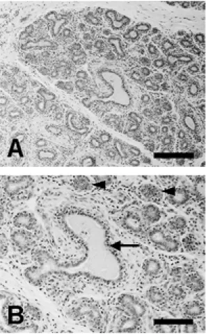
젖샘의 조직학적 구조
홀스타인젖소의젖샘을조직학적으로관찰한결과 많은 수의 샘소엽이결합조직사이에관찰되었다
(Fig.
1A).
샘소엽부위를고배율로관찰한결과샘포들이관찰되었으며일부샘포내에는분비물이관찰되었다
(Fig.
1B).
샘포사이에는분비도관들도관찰되었다.
원유 및 젖샘조직 내 osteopontin 검출
홀스타인 젖소의원유및 건유기의젖샘조직에서
osteopontin
을확인하기위하여western blotting
을실시하였다
. Osteopontin
은원유와젖샘조직에서면역반응을보였으며
,
분자량은약60 kDa(50 kDa
와93 kDa
사이)
정도로 나타났다
(Fig. 2B).
아울러 젖샘조직에서도osteopontin
이관찰되었다(Fig. 2B, mammary gland).
세포성분의유무를확인하기위하여
osteopontin
항체를반응시킨
membrane
을β-actin
항체로반응시킨결과원유내에서는β
-actin
의면역반응이관찰되지않았으며젖샘조직에서만나타났다
(Fig. 2B).
비유시기별 osteopontin의 양적 비교
우선
membrane
을ponceau
염색하여모든비유기원유의 단백질이 동량인 것을 확인하였다
(Fig. 3A).
Osteopontin
은모든비유기의원유에서관찰되었으며그분자량은약
60 kDa (50 kDa
와93 kDa
사이에서관찰)
이었다
(Fig. 3B). Osteopontin
의 면역반응을GS-700 Imaging Densitometer
로측정한결과출산후1
일의원유
(
초유)(Fig. 3B, colostrum)(density osteopontin ratio/mm
2value, 43.830
±4.605)
에서아주강한반응을나타내었으 며,
출산 후15
일에서20
일사이의 원유(Fig. 3B, early milk)(5.020
±1.881)
에서osteopontin
은초유보다 의미있 는감소를보였다(*,
p< 0.001).
그후다른시기의원 유,
즉출산후1
개월에서2
개월사이의원유(Fig. 3C, mature milk, a)(3.637
±2.137),
출산후3
개월에서5
개월 사이의원유(Fig. 3C, mature milk, b)(3.337
±0.995),
그리고출산 후
6
개월에서8
개월사이의원유(Fig. 3C, mature milk, c)(1.880
±0.806)
에서osteopontin
은초유보 다유의성있게 감소된 것으로 나타났다(*,
p< 0.001).
Fig. 1. Histological findings of a Holstein cow mammary glands (A and B). There are gland lobules with alveoli (B, arrowheads) and large irregular intralobular duct (B, arrow).
Hematoxylin and eosin staining. Scale bars = 200 µm (A);
100 µm (B).
Fig. 2. Western blot analysis for osteopontin (OPN) in the bovine milk and mammary glands. (A) Protein component of milk and mammary glands in 10% SDS-PAGE. Ponceau solution staining. Left lane indicates molecular weight standard.
(B) A representative photograph of Western blot analysis of osteopontin and
β-actin.
Fig. 3. Western blot analysis for osteopontin (OPN) in the bovine milk during the lactation period. (A) Ponceau staining shows that loading protein is similar in all lanes in 10 % SDS-PAGE. Left lane indicates molecular weight standard. (B) A representative photograph of Western blot analysis of osteopontin. (C) A semiquantitative analysis of osteopontin in the milk during the lactation period. Colostrum (within the first 24 h postpartum), early milk (from 15 to 20 days postpartum), and mature milk (more than 1 month after delivery) were collected from health Holstein cow. Mature milk samples were further classified into three groups, mature milk (a) (approximately 1-2 months postpartum), (b) (approximately 3-5 months postpartum) and (c) (approximately 6-8 months postpartum). Data were obtained from three different experiments and are shown as mean
±SE.
Fig. 4. Immunohistochemical localization of osteopontin in the bovine mammary glands and milk cells. No specific reaction
product is seen in sections incubated with non-immune sera (A). Osteopontin was detected in the glandular epithelium
(B, arrowheads) and in epithelial cells of intralobular duct (C, arrowheads). Osteopontin was immunostained in the milk
(C, arrow). Osteopontin was positive for some cells in bovine milk (C, inset). A-C: Counterstained with hematoxylin. Scale
bars: in A-C, 40 µm; in inset, 10 µm.
젖샘조직과 원유에서 osteopontin의 면역조직화학 적 염색
젖샘조직에서
osteopontin
의면역조직화학염색반응은주로샘포내상피세포에서관찰되었으며일부샘포 내분비물에서도양성반응을보였다
(Fig. 4B).
젖샘포관 상피세포및함유물에서osteopontin
의면역반응을관찰할수있었으나소엽속결합조직에서
osteopontin
염색 반응은미약하게관찰되었다(Fig. 4C).
또한원유를슬 라이드에도말하여면역조직화학염색한결과일부세포에서
osteopontin
의면역반응이관찰되었다(Fig. 4C,
inset).
고 찰
Osteopontin
은처음뼈에서발견된이래여러조직,
특히여러부위의상피세포에서발견되고있다
.
이실험에서는원유내에서
osteopontin
이있는지를조사하였던바원유에서
osteopontin
이있음을확인하였다.
유즙내
osteopontin
의확인은소[12]
와사람[7]
에서 알려져왔으나,
이논문에서는비유시기별로osteopontin
의차이를처음으로확인하였다
.
즉비유시기별로조사한결과초유에서가장많았고시간이경과함에따라다 소감소하였으나비유기간중지속적으로
osteopontin
이 관찰되었다.
초유에서
osteopontin
이많은이유는잘알려져있지않으나
osteopontin
의발현을촉진할수있는인자중하나인비타민
D [14]
와관련성이있을것으로추정하고있다
.
초유에서비타민D
가성숙우유보다더많이존재하는것으로알려져있으며
[6]
이들비타민D
가젖샘조직내
osteopontin
의생산을촉진할수있을것으로생각된다
.
Osteopontin
의주된관찰부위는결합조직섬유또는세포내로알려져있다
[14]. Osteopontin
의발현세포를조사한결과젖샘조직의샘포상피세포에서관찰되었으며 아울러원유로부터분리된세포를염색한결과이들세
포에서도
osteopontin
양성세포가확인되었다.
즉우유내
osteopontin
은상피세포에서분비되었거나또는원유내에포함된세포성분
(
큰포식세포또는상피세포)
등에서분비된것으로생각된다
.
Osteopontin
은다양한기능을가지는단백질로알려져있다
.
면역세포인큰포식세포,
상피세포등은외부의자극을받을경우
osteopontin
을생산분비하며세포매개성면역반응을유도하고
,
염증세포이동에도관여하기 도한다[3].
아울러osteopontin
은세포독성을가지는nitric oxide
를억제함으로써항염증작용에도관여하는것으로알려지고있다
[11].
이와같은기능을유추하여볼때우유중포함된
osteopontin
은장내에서이와같은면역반응에관여할수있을것으로판단된다
.
Osteopontin
의 기능을 확인하기 위해osteopontin
knockout
생쥐에로타바이러스를감염시킨결과wildtype
생쥐에비해장염이심하였다고한다
[10].
즉우유내osteopontin
은섭취후곧소장및대장점막과접촉하는것을고려할때이들은장내병원체의점막접촉을제 어하는기능을할수있을것으로생각된다
.
이실험에 서비유시기별로차이가있을뿐만아니라초유에서osteopontin
이많은점은면역기능이불완전한신생동물의장기능활성에도움을줄것으로생각된다
.
즉우유는다양한영양분이외에도
osteopontin
과같은면역조절물질이포함되어있는점은우유의영양성을더욱높 여주는것으로판단된다
.
젖샘조직에서
osteopontin
의역할은아직명확히규명된바가많지않지만
osteopontin knockout
생쥐의경우젖샘발육의장애가있는것으로보아
osteopontin
은젖 샘조직분화에필수적인물질일것으로알려지고있다[8].
아울러osteopontin
의세포접착기능은유방암조직의전이에도깊이관여하는것으로알려지고있다
[13].
즉젖샘에서생성된
osteopontin
은우유내에포함되어다양한생리활성기능을갖지만조직내에서비정상적 으로분화된젖샘세포에작용할경우전이를촉진할수 있을것으로생각된다
.
이상의결과를종합하여볼때젖소의원유에서는전 비유시기에걸쳐
osteopontin
이포함되어있으며,
특히초유에
osteopontin
이더많이존재하는점은신생동물의장내면역력획득에
osteopontin
이중요한인자로작용할것으로생각되었다
.
결 론
우유내다양한생리활성을가지는
osteopontin
의유무를소의원유와젖샘조직에서
western blot
과면역조 직화학방법으로조사하였다.
그결과osteopontin
은원유와젖샘조직에서확인되었다
.
비유시기별원유에서osteopontin
의발현을비교한결과초유에서osteopontin
의발현이다른시기의원유보다높게나타났다
.
면역조직화학염색결과젖샘조직에서
osteopontin
은주로샘포 세포와젖샘포관상피세포에서양성반응을보였다.
또 한,
원유중포함된세포성분(
큰포식세포또는상피세포
)
에서도osteopontin
이강하게발현된점으로보아이들세포들이
osteopontin
의주된분비세포로생각되었다.
아울러
osteopontin
의기능을고려할때원유내에존재하는
osteopontin
은장점막보호및감염체로부터생체를보호하는역할을할것으로생각되었다