371
Phylogenetic Status of an Undiscovered Zygomycete Species, Syncephalastrum monosporum, in Korea
Tham Thi Duong, Thi Thuong Thuong Nguyen, Sun Jeong Jeon and Hyang Burm Lee*
Division of Food Technology, Biotechnology and Agrochemistry, College of Agriculture and Life Sciences, Chonnam National University, Gwangju 61186, Korea
ABSTRACT : During a survey of undiscovered taxa in Korea, two zygomycete fungal isolates, EML-BT5-1 and EML-BT5-2, were isolated from the seed of a pumpkin (Cucurbita pepo) fruit in Korea. Based on their morphological characteristics and a sequence analysis of four genes, ITS1-5.8S-ITS2, 18S, 28S rDNA, and EF-1α, the isolates were confirmed to be Syncephalastrum monosporum in the family Syncephalastraceae. To our knowledge, the zygomycete fungal species S. monosporum has not been previously described in Korea.
KEYWORDS : Mucorales, Multigenes, Pumpkin seed, Syncephalastrum species, Undiscovered taxa
The genus Syncephalastrum with the type species Synce- phalastrum racemosum was described by Schröter (1886) [1]. This genus currently includes two species, S. monos- porum and S. racemosum, according to Index Fungorum (www.indexfungorum.org). The species belonging to this genus in the family Syncephalastraceae are characterized by the production of cylindrical merosporangium on the surface of fertile vesicles [2]. Syncephalastrum spp. are fre- quently isolated from soil, dung, plant materials, and org- anic substrates [2, 3]. Species of Syncephalastrum have been reported to have important biotechnological applic- ations as producers of endoglucanase, xylanase, and asp- artic proteinase (syncephapepsin) [4-6]. In addition, S.
racemosum has shown great potential to produce chitosan used as film support for lipase immobilization [7]. How- ever, some of these species have been implicated in muc- ormycosis in humans and animals [8, 9].
In Korea, only one species of S. racemosum has been
recorded [10]. During studies of the diversity of fungi in the order Mucorales isolated from a pumpkin (Cucurbita pepo) seed sample in Korea, a zygomycete species, Synce- phalastrum sp. was isolated.
In recent years, several multi-loci analyses of the nuc- lear 18S ribosomal RNA small subunit (SSU), nuclear large subunit 28S ribosomal RNA (LSU), and translation elongation factor-1α (EF-1α) have been conducted to eva- luate the phylogeny of mucoralean species [11-15]. How- ever, only a few phylogenetic analyses of the genus Syn- cephalastrum have been performed.
The objectives of the present study were to analyze the phylogenetic status of S. monosporum, based on a multi- loci sequence analysis and to describe the morphological characteristics of this species in detail.
C. pepo seeds were collected from pumpkin fruit collec- ted on the campus of Chonnam National University, Gwangju, Korea. The samples were transported to the laboratory in plastic bags. Hyphal tips were transferred to potato dextrose agar (PDA) plates, using a sterile nee- dle under a stereomicroscope. The plates were incubated at 25°C for 3~5 days in the dark until colonies could be distinguished. Pure isolates were maintained in PDA slant tubes and stored in 20% glycerol at -80°C in the Environ- mental Microbiology Laboratory Fungal Herbarium, Chon- nam National University, Gwangju, Korea, under deposi- tion number (EML-BT5-1). The strain was also deposited in glycerol at -80°C in the Culture Collection of National Institute of Biological Resources (NIBR), Incheon, Korea (KOSPF0000133913).
*Corresponding author E-mail: [email protected] Received October 29, 2016 Revised November 27, 2016 Accepted December 15, 2016
This is an Open Access article distributed under the terms of the Creative Commons Attribution Non-Commercial License (http://
creativecommons.org/licenses/by-nc/3.0/) which permits unrestricted non-commercial use, distribution, and reproduction in any medium, provided the original work is properly cited.
Kor. J. Mycol. 2016 June, 44(2): 371-376 http://dx.doi.org/10.4489/KJM.2016.44.2.371 pISSN 0253-651X • eISSN 2383-5249
© The Korean Society of Mycology
Genomic DNA was directly extracted from mycelia, using the HiGene Genomic DNA prep kit for fungi (Bio- pact Corp., Daejeon, Korea). The internal transcribed spacer (ITS), small subunit of 18S rDNA and large sub- unit of 28S rDNA, and translation elongation factor-1α (EF-1α) sequences were amplified with the primer pairs ITS1, ITS4 [16]; NS1, NS4 [14] LROR, LR5F [17]; and MEF-11, MEF-41 [14], respectively.
The sequences were aligned using ClustalX [18] and edited manually [19]. Phylogenetic analyses were perfor- med in MEGA 6 [20] with the default settings. Maximum likelihood (ML) phylogenetic trees were constructed from
individual datasets of the ITS and 28S rDNA sequences and combined datasets of the 18S and 28S rDNA and EF-1α gene sequences. The nearest-neighbor-interchange was selected as the ML heuristic method, and the initial ML tree was set automatically. Umbelopsis isabellina and U. ramanniana sequences were used as outgroups. The ITS, 18S, 28S, and EF-1α sequences of strains EML-BT5- 1 and EML-BT5-2 were deposited in the GenBank data- base with accession numbers KY047152, KY047155, KY 047158, and KY047154 for EML-BT5-1; and KY047143, KY047156, KY047157, and KY047153 for EML-BT5-2, respectively. A BLASTn search showed that the ITS se-
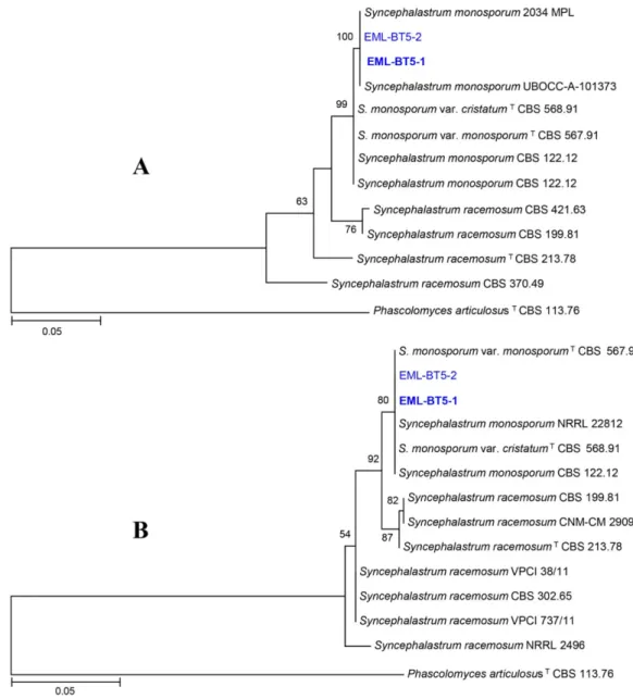
Fig. 1. Phylogenetic tree of EML-BT5-1 and EML-BT5-2 based on a maximum likelihood analysis of internal transcribed spacer (A) and 28S (B) rDNA sequences. Sequence of Phascolomyces articulosus was used as an outgroup. Bootstrap values with more than 50% support in 1,000 replications are shown above branches. The bar indicates the number of substitutions per nucleotide position.
quence of strains EML-BT5-1 and EML-BT5-2 were most closely related to representative S. monosporum (GenBank accession no. KP233744 and KF225035) with nucleotide identities of 99.5% (428/430 bp) and 98.7% (462/468 bp), respectively. Based on the 28S rDNA sequence analysis, strains EML-BT5-1 and EML-BT5-2 showed 99.8% (584/
585 bp) identity value with S. monosporum sequence (Gen- Bank accession no. AF157215). The 18S rDNA sequences of EML-BT5-1 and EML-BT5-2 showed 99.9% (1000/1001 bp) and 99.2% (934/942 bp) identity values with sequen- ces of S. monosporum var. pluriproliferum (GenBank acce- ssion no. AF157161) and S. monosporum var. pluriprolife- rum (GenBank accession no. JX644490), respectively. The EF-1α gene sequence of strains EML-BT5-1 and EML- BT5-2 showed 99.9% (696/697 bp) and 99.8% (564/565 bp) identity values with S. monosporum var. pluriproliferum (GenBank accession no. AF157294) and S. monosporum var. pluriproliferum (GenBank accession no. JX644588), respectively. The ITS, 18S, 28S, and EF-1α phylogenetic analyses indicated that isolates EML-BT5-1 and EML-BT 5-2 were nearly identical to S. monosporum in the family Syncephalastraceae (Figs. 1, 2).
To examine morphological characteristics and growth rate, EML-BT5-1 was cultured in PDA (39 g in 1 L deio- nized water; Difco, Detroit, MI, USA), synthetic mucor
agar (SMA; 40 g dextrose, 2 g asparagine, 0.5 g KH2PO4, 0.25 g MgSO4·7H2O, 0.5 g thiamine chloride, and 15 g agar, in 1 L of deionized water), and malt extract agar (MEA; 33.6 g in 1 L deionized water; Difco), and incub- ated at 10°C, 15°C, 20°C, 25°C, 30°C, 35°C, and 40°C in the dark for 7 days. Samples were observed under a light microscope (DFC290; Leica Microsystems, Wetzlar, Ger- many) and by field emission scanning electron micro- scopy (SEM; Hitachi S4700; Hitachi, Tokyo, Japan). For SEM, the isolate was fixed in 2.5% paraformaldehyde- glutaraldehyde in 0.05 M phosphate buffer (pH 7.2) for 2 hr, washed with cacodylate buffer (Junsei Chemical, To- kyo, Japan), and then fixed in 1% osmium tetroxide (Elec- tron Microscopy Sciences, Hatfield, PA, USA) for 1 hr.
After this, the blocks were dehydrated with graded etha- nol and dried in a fume hood. Finally, samples were sput- ter-coated with gold and observed by SEM at the Korea Basic Science Institute, Gwangju, Korea.
Syncephalastrum monosporum R.Y. Zheng, G.Q. Chen
& F.M. Hu, Mycosystema 1: 39 (1988) (Fig. 3).
Description: On SMA, the colonies grew rapidly and attained a diameter of 85~86 mm after 4 days at 30°C.
The color of colonies was cotton-white. The reverse side of the colony was also white. Sporophores were hyaline, rarely brownish, with septae, 8.5~16.0 μm in width, and
Fig. 2. Phylogenetic status of EML-BT5-1 and EML-BT5-2 in the Syncephalastraceae clade based on a maximum likelihood analysis of a combined dataset of 18S and 28S rDNA and translation elongation factor 1-alpha sequences. Umbelopsis isabellina and U. ramanniana sequences were used as outgroups. Bootstrap values with more than 50% support in 1,000 replications are shown above branches. The bar indicates the number of substitutions per nucleotide position.
variable in length. Vesicles were globose, subglobose, ovoid, or subpyriform, sometimes rounded or papillate at the apices, measuring 15.0~34.5 μm in diameter. Sporan- giola developed on vesicles and covered their entire sur- faces, were rod-shaped, ovoid to subglobose, rounded at the apices, uniform in width throughout or becoming broader at the top, occasionally narrower in the middle portion, and measuring 12.5~32.0 × 4.5~7.5 μm. The spo-
rangial wall was subpersistent, slowly dissolving or broken to release spores. Single sporangiospores were ovoid to subglobose, measuring 4.0~6.5 μm in diameter. Zygospo- res were not observed, and rhizoids were not well devel- oped. The isolates grew at different rates according to the temperature and medium. The average diameters of EML- BT5-1 on SMA, PDA, and MEA were 21.5 mm, 17 mm, and 15 mm at 24 hours, respectively. The optimal temp- Fig. 3. Morphology of Syncephalastrum monosporum EML-BT5-1. A, D, colony in synthetic mucor agar; B, E, colony in potato dextrose agar; C, F, colony in malt extract agar; G, growth of mycelia on pumpkin seed surfaces; H, M, development of sporan- giola on vesicles and young sporangiola (white arrow); I, J, N, O, vesicles bearing mature sporangiola; K, P, sporophores with a septa (purple arrow), old vesicles, and scars remaining after detachment of sporangiola (yellow arrow); L, sporangiospores; A~C, obverse view; D~F, reverse view; H~M, light microscopy; M~P, scanning electron microscopy (scale bars: H~K = 50 μm, L~N, O = 10 μm, P = 20 μm).
erature range for growth was 25~30°C. Slow growth was observed at 40°C, and no growth was observed at 5°C.
SMA was the best medium for mycelial growth, followed by PDA; the growth of colonies was the slowest on MEA (Fig. 4). The morphology and physiology of the isolate was generally similar to those previously described for the species by Zheng et al. [21].
Recently, Hoffman et al. [11] have performed a phylo- genetic evaluation of some species belonging to the genus Syncephalastrum and several species in the class Zygomy- cetes, using 18S and 28S rDNA, EF-1α, and actin gene sequences. This study showed that there was only a single genus, Syncephalastrum, with two species in the family Syncephalastraceae. In the ITS and 28S trees, our strains, EML-BT5-1 and EML-BT5-2, were not distinct from S.
monosporum. Furthermore, based on the phylogenetic ana- lysis using a combined dataset of 18S and 28S rDNA and EF-1α sequences (Fig. 2), EML-BT5-1 and EML-BT5-2 were located within a clade that included S. monosporum and S. racemosum in the family Syncephalastraceae.
Based on the morphological, physiological and molec- ular analyses, the fungus was identified as S. monosporum.
Herein, S. monosporum is described as a new record of zygomycete fungi belonging to undiscovered taxa in Korea.
Acknowledgements
This work was supported by the Graduate Program for the Undiscovered Taxa of Korea, and by the Project on Survey and Discovery of Indigenous Species of Korea funded by NIBR of the Ministry of Environment (MOE),
and in part by a fund from National Institute of Animal Science under Rural Development Administration, Rep- ublic of Korea.
REFERENCES
1. Schröter J. Kryptogamen-Flora von Schlesien: Pilze. Breslau: J.
U. Kern; 1908.
2. Benjamin RK. The merosporangiferous Mucorales. Aliso 1959;
4:321-433.
3. Benny GL. The methods used by Dr. R. K. Benjamin, and other mycologists, to isolate Zygomycetes. Aliso 2008;26:37- 61.
4. Ho HC, Shiau PF, Wu SL. Single-column purification of syn- cephapepsin: an aspartic proteinase from Syncephalastrum rac- emosum. Protein Expr Purif 1998;12:399-403.
5. Sapre MP, Jha H, Patil MB. Purification and characterization of a thermostabile-cellulase free xylanase from Syncephalastrum racemosum Cohn. J Gen Appl Microbiol 2005;51:327-34.
6. Wonganu B, Pootanakit K, Boonyapakron K, Champreda V, Tanapongpipat S, Eurwilaichitr L. Cloning, expression and characterization of a thermotolerant endoglucanase from Syn- cephalastrum racemosum (BCC18080) in Pichia pastoris. Pro- tein Expr Purif 2008;58:78-86.
7. Amorim RV, Melo ES, Carneiro-da-Cunha MG, Ledingham WM, Campos-Takaki GM. Chitosan from Syncephalastrum racemosum used as a film support for lipase immobilization.
Bioresour Technol 2003;89:35-9.
8. Pavlović MD, Bulajić N. Great toenail onychomycosis caused by Syncephalastrum racemosum. Dermatol Online J 2006;12:7.
9. Schlebusch S, Looke DF. Intraabdominal Zygomycosis caused by Syncephalastrum racemosum infection successfully treated with partial surgical debridement and high-dose Amphotericin B lipid complex. J Clin Microbiol 2005;43:5825-7.
10. Yang S, Lee J, Kwak J, Kim K, Seo M, Lee YW. Fungi associa- ted with the traditional starter cultures used for rice wine in Korea. J Kor Soc Appl Biol Chem 2011;54:933-43.
Fig. 4. Effect of temperature and culture medium on mycelial growth of Syncephalastrum monosporum EML-BT5-1. Mycelia were grown on synthetic mucor agar (SMA), potato dextrose agar (PDA), and malt extract agar (MEA) at different temperatures.
11. Hoffmann K, Pawłowska J, Walther G, Wrzosek M, de Hoog GS, Benny GL, Kirk PM, Voigt K. The family structure of the Mucorales: a synoptic revision based on comprehensive multi- gene-genealogies. Persoonia 2013;30:57-76.
12. Li GJ, Hyde KD, Zhao RL, Hongsanan S, Abdel-Aziz FA, Ab- del-Wahab MA, Alvarado P, Alves-Silva G, Ammirati JF, Ari- yawansa HA, et al. Fungal diversity notes 253-366: taxonomic and phylogenetic contributions to fungal taxa. Fungal Divers 2016;78:1-237.
13. Nguyen TT, Lee SH, Bae S, Jeon SJ, Mun HY, Lee HB. Charac- terization of two new records of zygomycete species belong- ing to undiscovered taxa in Korea. Mycobiology 2016;44:29- 37.
14. O'Donnell K, Lutzoni FM, Ward TJ, Benny GL. Evolutionary relationships among mucoralean fungi (Zygomycota): evidence for family polyphyly on a large scale. Mycologia 2001;93:286- 97.
15. Walther G, Pawłowska J, Alastruey-Izquierdo A, Wrzosek M, Rodriguez-Tudela JL, Dolatabadi S, Chakrabarti A, de Hoog GS. DNA barcoding in Mucorales: an inventory of biodiversity.
Persoonia 2013;30:11-47.
16. White TJ, Bruns TD, Lee SB, Taylor JW. Amplification and direct sequencing of fungal ribosomal RNA genes for phylog- enetics. In: Innis MA, Gelfand DH, Sninsky JJ, editors. PCR protocols: a guide to methods and applications. San Diego:
Academic Press; 1990. p. 315-22.
17. Vilgalys R, Hester M. Rapid genetic identification and mapp- ing of enzymatically amplified ribosomal DNA from several Cryptococcus species. J Bacteriol 1990;172:4238-46.
18. Thompson JD, Gibson TJ, Plewniak F, Jeanmougin F, Higgins DG. The CLUSTAL_X windows interface: flexible strategies for multiple sequence alignment aided by quality analysis tools.
Nucleic Acids Res 1997;25:4876-82.
19. Hall TA. BioEdit: a user-friendly biological sequence alignm- ent editor and analysis program for Windows 95/98/NT. Nuc- leic Acids Symp Ser 1999;41:95-8.
20. Tamura K, Stecher G, Peterson D, Filipski A, Kumar S. MEGA 6: Molecular Evolutionary Genetics Analysis version 6.0. Mol Biol Evol 2013;30:2725-9.
21. Zheng RY, Chen GQ, Hu FM. Monosporus varieties of Synce- phalastrum. Mycosystema 1988;1:35-52.