─ 75 ─ eISSN 2287-1683
pISSN 1738-8767
Journal of Trauma and Injury Vol. 28, No. 2, June, 2015
� Case Report �
� Address for Correspondence : Sang-Bong Chung, M.D.
Department of Neurosurgery, National Medical Center, Euljiro 6-Ga, Jung-Gu, Seoul 100-799, Korea
Tel : 82-2-2260-7183, Fax : 82-2-2276-2386, E-mail : [email protected] Submitted : February 4, 2015 Revised : March 19, 2015 Accepted : May 15, 2015
진단이 늦어진 기관삽관 후 발생한 턱관절 탈구
국립중앙의료원 신경외과 정상봉, 전형배, 김태관
- Abstract -
Unrecognized Bilateral Dislocation of Temporomandibular Joint during Orotracheal Intubation
Sang-Bong Chung, M.D., Hyoungbae Jeon, M.D., Taikwan Kim, M.D.
Department of Neurosurgery, National Medical Center, Seoul, Korea
Mandible dislocation during endotracheal intubation is an unusual occurrence but easy to be overlooked for its unfa- miliarity. We recently had a case of iatrogenic bilateral dislocation of a temporomandibular joint during orotracheal intubation and emphasize the importance of an early awareness of this possible complication. [ J Trauma Inj 2015; 28:
75-78 ]
Key Words: Temporomandibular joint, Orotracheal intubation
I. Introduction
Temporomandibular joint (TMJ) dislocation during orotracheal intubation is a rare occurrence.(1-6) TMJ dislocation that occurs during operations is often unnoticed until patients become aware of pain and swelling.(1) We recently had a case of iatrogenic bilateral dislocation of a TMJ during orotracheal intubation. This is a case report of delayed diagnosis of TMJ dislocation during intubation in an uncon- scious patient who stayed comatose for several days.
II. Case
A 65-year-old female patient was transferred to emergency room with a history of sudden collapse just before. The patient was comatose at the initial presentation. Head computed tomography (CT) demonstrated intraventricular hemorrhage with hydrocephalus. She was given etomidate for anes- thetic induction and immediately intubated without difficulty by an attending doctor. After confirming that the orotracheal tube was well positioned, we
─ 76 ─
- Journal of Trauma and Injury Vol. 28, No. 2 -
attempted to insert extraventricular drainage catheter for decompression of increased intracranial pressure. The next day, the patient regained the consciousness progressively and could obey doctor’s commands. The on-call resident decided to extubate the orotracheal tube and did it without difficulty.
During the night, however, the patient’s con- sciousness was rapidly deteriorated; she had to be intubated again and ventilated. The patient was treated in intensive care unit (ICU) with complicated indwelling catheters for drainage of intraventricular
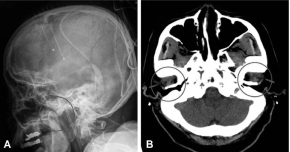
hematoma and mechanical ventilator support. On the 3th day in ICU, she was suspected of protruding her tongue much compared with those of other patients who were intubated. We reviewed all previ- ously scanned skull X-ray and head CT imaging.
Then, we noticed that we missed the detection of bilateral TMJ dislocation after orotracheal intuba- tion (Fig. 1).
As we were not familiar with the technique of reduction of dislocated jaw, a specialist was consult- ed. The bilateral TMJ dislocation was reduced with
Fig. 1. Skull X-ray lateral view (A) demonstrating anterior dislocation of the temporomandibular joint. Head CT axial view (B) depicting dislodgement of the condyles from glenoid fossa.
A B
Fig. 2. Skull X-ray lateral view (A) demonstrating normal position of the temporomandibular joint after reduction. Head CT axial view (B) depicting the reduced condyles into glenoid fossa.
A B
the traditional transoral approach without any form of anesthesia by emergency room clinician. He reset the dislocated jaw, then the awkwardly protruded mouth and tongue was reduced. Imaging confirmed the normal position of dislocated TMJ after manual reduction (Fig. 2).
The patient was treated well and discharged 1 month later without any problem in mouth opening.
III. Discussion
TMJ dislocation is the dislodgement of the condyle from its normal position in the squamo-temporal portion of the cranial base.(7,8) Anterior and anteromedial dislocations are the most common ones observed. Subluxation refers to a condition in which the TMJ is transiently displaced without complete loss of articulating function, and is usually self- reduced by the patient. It could occur spontaneously or by trauma, endotracheal intubation, dental pro- cedures, endoscopy, transesophageal echo probe placement, yawning, laughing, vomiting and seizure.(3) Such TMJ instability including TMJ sub- luxation and hypermobility has a prevalence of up to 25~50% in the general population, and is most com- mon in middle-aged females.(9) These patients may have no signs and symptoms or may present with clicking, pain, or chronic dislocations. Frequent dis- location may also be seen in patients with connec- tive tissue disease, such as Ehlers-Danlos syndrome or muscular dystonias.(7,10,11)
The diagnosis is usually obvious by the character- istic wide open mouth with the inability to close it.
Spontaneous movement of the mandible, if present at all, is usually restricted.(5) Acute TMJ dislocation is associated with pain and severe tenderness over the TMJ areas. Awake patients will often complain of pain in the area of the TMJ, which quickly sug- gest the diagnosis.
However, in patients with a decreased level of consciousness due to brain pathology or sedation like our patient, one should be highly aware of this possible complication in order to detect it early;
otherwise, the dislocation might be overlooked. On palpation, the condyles of the mandible could be felt below the zygomatic arch. Palpation of the condyle
below the zygomatic arch could reveal its absence from the glenoid fossa. Radiologic findings of the TMJ will confirm the diagnosis.
It is uncertain the exact time at which the dislo- cation occurred in our patient, but the on-call resi- dent seemed to be encountered difficulty during the orotracheal intubation after he was notified that the patient’s neurologic status was rapidly deteriorated.
A rigorous forward thrust of the jaw and maximal opening of the mouth may cause dislocation of a meniscus in TMJ.(7,12) The doctor should have determined that the mandible is restored after it has been widely opened. However, its diagnosis was overlooked not only by the doctor but also by our team who were all unfamiliar with this pathology.
Unfortunately for our patient, the TMJ dislocation was unrecognized for several days. Delay of diagno- sis for over a month has been reported in patients with head trauma or sedation after cardiac surgery.(3,13)
The dislocation can often be reduced by manual technique. Operator in front of the patient places his thumbs near the posterior teeth with his fingers grasping the lower edge of the mandible. Downward pressure on the posterior teeth and upward move- ment of the chin, along with posterior displacement of the entire mandible should be applied allowing an immediate simple manual reduction. Dislocations should be reduced early, since spasm of the external pteygoid and other muscles may ankylose the jaw.(5) If left untreated for longer than 14 days, fibrosis and even fractures become increasingly apparent, which may require surgical treatment.(13)
Considering this unusual presentation, we would like to emphasize the importance of an early aware- ness of the potential for TMJ dislocation during rou- tine intubation maneuvers. Furthermore, clinicians who deal with airway management should be given some information in resetting the dislocated TMJ.
REFERENCES
01) Han I, Kim TK, Yoo JH, Park JH, Chung EY. Dislocation of the temporomandibular joint following general anesthesia.
Korean J Anesthesiol 2014; 67: S113-4.
02) Oofuvong M. Bilateral temporomandibular joint dislocations during induction of anesthesia and orotracheal intubation. J
─ 77 ─
Sang-Bong Chung, et al.: Unrecognized Bilateral Dislocation of Temporomandibular Joint during Orotracheal Intubation
Med Asso Thailand 2005; 88: 695-7.
03) Quessard A, Barriere P, Levy F, Steib A, Dupeyron P.
Delayed diagnosis of a postanaesthesia temporomandibular joint dislocation. Annales francaises d’anesthesie et de reani- mation 2008; 27: 846-9.
04) Rattan V, Arora S. Prolonged temporomandibular joint dislo- cation in an unconscious patient after airway manipulation.
Anesthesia and analgesia 2006; 102: 1294.
05) Sosis M, Lazar S. Jaw dislocation during general anaesthesia.
Can J Anaesthesiol 1987; 34: 407-8.
06) Sriganesh K, Farooq S, Byrappa V. Temporomandibular joint dislocation during tracheal intubation in a patient with Sjogren syndrome. J Neurosurg Anesthesiol 2015; 27: 82-3.
07) Luyk NH, Larsen PE. The diagnosis and treatment of the dis- located mandible. Am J Emerg Med 1989; 7: 329-35.
08) Mangi Q, Ridgway PF, Ibrahim Z, Evoy D. Dislocation of the mandible. Surg Endosc 2004; 18: 554-6.
09) Awsare AN, Prakash N. Temporomandibular dislocation:
should every doctor be trained in resetting the jaw? The British J Oral & Maxillofac Surg 2006; 44: 339.
10) Akinbami BO. Evaluation of the mechanism and principles of management of temporomandibular joint dislocation.
Systematic review of literature and a proposed new classifica- tion of temporomandibular joint dislocation. Head Face Med 2011; 7: 10.
11) Prabhakar V, Singla S. Bilateral anterosuperior dislocation of intact mandibular condyles in the temporal fossa. International J Oral Maxillofac Surg 2011; 40: 640-3.
12) Kurita K, Mukaida Y, Ogi N, Toyama M. Closed reduction of chronic bilateral temporomandibular joint dislocation. A case report. Int J Oral Maxillofac Surg 1996; 25: 422-3.
13) Pillai S, Konia MR. Unrecognized bilateral temporomandibu- lar joint dislocation after general anesthesia with a delay in diagnosis and management: a case report. J Med Case Rep 2013; 7: 243.
─ 78 ─
- Journal of Trauma and Injury Vol. 28, No. 2 -