─ 192 ─ elSSN 2287-1683
plSSN 1738-8767
Journal of Trauma and Injury Vol. 27, No. 4, December, 2014
� Case Report �
� Address for Correspondence : Sang Cjeol Lee, M.D.
Department of Thoracic and Cardiovascular Surgery, Kyungpook National University Hospital, Kyungpook National University School of Medicine, 130 Dongdeok-ro, Jung-gu, Daegu, Republic of Korea
Tel : 82-53-200-6527, Fax : 82-53-426-4765, E-mail : [email protected]
Submitted : June 11, 2014 Revised : August 31, 2014 Accepted : September 22, 2014
흉부 둔상으로 골절된 늑골로 인해 발생한 좌심실 천공
경북대학교병원 흉부외과, 경북대학교 의학전문대학원 흉부외과학교실 오탁혁, 이상철, 이덕헌, 조준용
- Abstract -
Penetrating Injury to the Left Ventricle from a Fractured Rib Following Blunt Chest Trauma
Tak-hyuk Oh, M.D., Sang Cjeol Lee, M.D., Deok Heon Lee, M.D., Joon Yong Cho, M.D.
Department of Thoracic and Cardiovascular Surgery, Kyungpook National University Hospital, Kyungpook National University School of Medicine, Daegu, Republic of Korea
The perforation of a cardiac chamber by a fractured rib after blunt trauma is a rare event. Here, we report the case of patient who was referred for multiple rib fractures after a fall from a height. The patient was found to have a penetrat- ing cardiac injury which was detected on a computed tomography chest scan. Computed tomography is a useful screen- ing tool for victims of blunt chest trauma. Once cardiac perforation has been confirmed or is highly suspected, it is important to preserve the patient’s vital signs until reaching the operating room by minimally manuplating the chest wall and permitting hypotension, which also prevents exsanguinating hemorrhage. For the same reasons, early cardiac tamponade may also improve the patient’s survival. [ J Trauma Inj 2014; 27: 192-5 ]
Key Words: Trauma, Heart injury, Rib fracture
I. Intrduction
Penetrating cardiac injury has been a big chal- lenge in trauma surgery. Although cardiac surgery has evolved considerably, the mortality rate of traumatic cardiac perforation remains high. The need for rapid decision-making and prompt thora-
cotomy to save the lives of cardiac trauma victims cannot be overemphasized. Herein, we report a rare case of left ventricular perforation by a fractured rib, which was successfully treated by expeditious management.
─ 193 ─
Tak-hyuk Oh, et al.: Penetrating Injury to the Left Ventricle from a Fractured Rib Following Blunt Chest Trauma
II. Case
A 45-year-old man fell from a height of 2 m while working on a construction crew. He complained of severe left chest pain and shortness of breath after the impact, but there was no external bleeding or loss of consciousness.
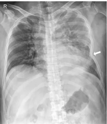
An hour after the trauma, the patient was brought to our emergency department. Upon arrival, he was still alert without any neurologic deficit, and his hemodynamic status was stable. A physical exami- nation revealed a focal paradoxical movement in the left chest wall. Initial laboratory findings showed a hemoglobin level of 12.6 g/dL and a platelet count of 287,000 /μL. Chest radiography (CXR) showed mul- tiple rib fractures on the left side, with depression of the sixth rib but no definite findings of intratho- racic organ damage (Fig. 1). Initially the patient was managed conservatively including bed rest and analgesics. Fifty minutes later, after the pain had been controlled, computed tomography (CT) of the chest was performed, revealing a large left hemoth- orax and hemopericardium (Fig. 2). Shortly after the patient returned from CT room, a vital signs check showed the following measurements: a blood pres- sure, 53/26 mmHg; heart rate, 99 beats/min; and respiration rate, 20 breaths/min. Volume replace- ment was started immediately, and the patient was sent to the operating room (OR) as soon as possible, without undergoing any additional evaluations or
invasive bedside procedures. In approximately fif- teen minutes, the patient was transported from the emergency room to the OR.
After anesthesia induction, signs of cardiac tam- ponade developed and a rapid left anterolateral tho- racotomy was performed. The pericardium was found to be torn longitudinally, anterior to the phrenic nerve, and the opening was partially
Fig. 1. Initial chest radiography shows left multiple rib frac- tures with a depressed sixth rib (arrow).
Fig. 2. Axial (A) and coronal (B) views from the chest CT show the presence of a hemothorax and hemopericardium. The anterior end of the fractured sixth rib is medially displaced and located in the pericardium.
A B
occluded by blood clots. As the pericardium was opened and the blood clots were extracted, massive quantities of blood were pumped out through a 1-cm long perforation in the left ventricle posterolateral wall. While compressing the bleeding site with fin- gers, primary closure was attempted with pledgeted sutures. After the bleeding had been controlled, the repair site was buttressed with a fibrin-collagen fleece and fibrin glue. Concomitant lung laceration in the left lower lobe was repaired with absorbable sutures and the fragmented ribs were fixated with metal plates and screws. Transesophageal echocar- diography was performed before wound closure to search for any additional intracardiac injuries or abnormalities, but the findings were negative.
The patient was admitted to the general ward after recovering from anesthesia and discharged on hospital day 18 without any sequelae.
III. Discussion
Penetrating cardiac trauma is one of the most lethal injuries. Cardiac penetration is usually asso- ciated with a stab or gunshot wound. Although rib fracture is commonly accompanied by blunt or non- penetrating chest trauma, penetration of the heart by a fractured rib is rare. There have been few spo- radic case reports of cardiac laceration or perfora- tion by a fractured rib following blunt trauma.(1,2)
To save the lives of patients with cardiac perfora- tions, particularly those caused by blunt trauma, two important points must be considered: how to detect the cardiac injury as early as possible, and how to move the patients to the OR as quickly as possible. In our hospital, we perform routine chest CT on blunt thoracic trauma victims, provided that the patient’s hemodynamic status permits it.
Although CT scanning entails the burden of moving and monitoring severely injured multiple trauma patients, plain chest radiography alone cannot detect every pathological finding and may even miss a critical injury such as an aortic lesion.(3) In con- trast to plain radiography and ultrasonography, CT scanning with contrast media can provide informa- tion concerning the patient’s entire anatomical structure, the type and degree of the damaged
organs, and the presence of vascular injury, through objective medical images that facilitate the planning of treatment and setting of priorities. Recently, focused assessment with sonography for trauma (FAST) has been considered to be a useful screening tool for detecting free intraabdominal and pericar- dial fluid and it has played an increasing role in the initial assessment of severely injured patients.(4) Regrettably in our case, however, we did not per- form FAST because the initial clinical feature and CXR finding of the patient appeared to be benign.
Once cardiac perforation has been confirmed or is strongly suspected, it may be best to move the patient immediately to the OR for thoracotomy without performing any further evaluation or pro- cedures. Time delay at a critical moment has a great adverse impact on survival. An invasive procedure such as chest tube insertion, which is usually indi- cated for evacuation of a hemothorax, may be dan- gerous in a patient with cardiac perforation because rough manipulation of the chest can cause exsan- guinating hemorrhage, and emergent surgery can- not be avoided in any case. Even a simple position change can cause abrupt massive hemorrhage.(2) In our case, given the concern about the possibility of exsanguinating hemorrhage, we did not perform any further evaluations or bedside procedures in the emergency department although we believe that the CT scanning destabilized the patient. For the same reason, although our patient suffered a profound state of shock, he was deliberately kept in a low blood pressure condition until thoracotomy, with a systolic pressure of approximately 90 mmHg.
The effect of cardiac tamponade on survival is controversial; however, Degiannis et al.(5) claimed that it offers a significant survival advantage. It also appears that, in our case, early tamponade after the initial hemorrhage may have helped to preserve the patient’s vital signs until he reached the OR. Sometimes, emergency department thoraco- tomy (EDT) is indicated for patients with penetrat- ing chest injuries who lack vital signs, or who are in extremis at presentation.(4) Unfortunately, however, our hospital is not equipped with the necessary facilities and experienced personnel for EDT.
Asensio et al.(6) reported a higher survival rate in
─ 194 ─
- Journal of Trauma and Injury Vol. 27, No. 4 -
─ 195 ─
Tak-hyuk Oh, et al.: Penetrating Injury to the Left Ventricle from a Fractured Rib Following Blunt Chest Trauma
their OR thoracotomy group than in the patients undergoing EDT (70% versus 16%). Accordingly, we consider that, for now, the best solution is to take the patient to the OR for thoracotomy as soon as possible, without delay. Additionally, we recommend that echocardiography should be conducted in the OR, after controlling major bleeding, to determine whether there are additional cardiac injuries.
Conclusively, cardiac perforation by a fractured rib is a rare event, and early detection and prompt surgery are essential for saving lives. Performing CT screening for blunt thoracic trauma patients, avoid- ing invasive chest wall manipulation, and permitting hypotension may also improve survival rates.
REFERENCES
01) Kaul P, Somsekhar G, Macauley G. Secondary left ventricular
injury with haemopericardium caused by a rib fracture after blunt chest trauma. J Cardiothorac Surg 2006; 1: 8.
02) Roth T, Kipfer B, Takala J, Schmid RA. Delayed heart perfo- ration after blunt trauma. Eur J Cardiothorac Surg 2002; 21:
121-3.
03) Exadaktylos AK, Sclabas G, Schmid SW, Schaller B, Zimmermann H. Do we really need routine computed tomo- graphic scanning in the primary evaluation of blunt chest trau- ma in patients with “normal” chest radiograph? J Trauma 2001; 51: 1173-6.
04) Patel NY, Riherd JM. Focused assessment with sonography for trauma: methods, accuracy, and indications. Surg Clin North Am 2011; 91: 195-207.
05) Degiannis E, Loogna P, Doll D, Bonanno F, Bowley DM, Smith MD. Penetrating cardiac injuries: recent experience in South Africa. World J Surg 2006; 30: 1258-64.
06) Asensio JA, Murray J, Demetriades D, Berne J, Cornwell E, Velmahos G, et al. Penetrating cardiac injuries: a prospective study of variables predicting outcomes. J Am Coll Surg 1998;
186: 24-34.