688
Copyright © 2020 The Korean Society of Fisheries and Aquatic Science pISSN:0374-8111, eISSN:2287-8815
Vibrio vulnificus
는해양에서식하며전세계적으로해수,
뻘 이나어패류에서검출되는그람음성간균이다(Tamplin et al., 1982; DePaola et al., 1994; Nascimento et al., 2001; Baffone et al., 2006; Lee et al., 2019; Park et al., 2019).
전세계적으로 수온이9°C
에서31°C
사이의해안가또는해수와담수가만나 는하구에서대개발견되고, 18°C
이상일때잘증식하며10°C
이하에서는개체수를측정할수없는수준으로감소한다.
염분 은15-25 psu
에서잘증식하고30 psu
이상에는개체가감소하 는것으로보고되어있다(Kaspar and Tamplin, 1993; Motes et al., 1998; Strom and Paranjpye, 2000).
비브리오패혈증은이 균에의한감염병으로균에오염된어패류등해산물을 날로 또는덜조리된상태로섭취하거나피부의상처부위가해수에 노출되면감염되어위장염과원발성패혈증(primary septice-
mia)
또는창상감염(wound infection)
이일어나며,
원발성패 혈증의경우치명률이50%
이상에이른다(Hlady and Klontz, 1996; Shapiro et al., 1998; Oliver, 2005a). V. vulnificus
는생 물형(biotype)
에따라분류할때3
가지유형이있으며1
형은가 장보편적이고전세계해수에서발견되며, 50%
이상의치명 률을나타내는 원발성패혈증을포함하여 이균에의한질환 의대부분을차지한다. 2
형은유럽의장어양식장해수에서발 견되는형으로주로장어에심각한병원성이있으나드물게사 람에게상처감염을유발한다. 3
형의경우이스라엘어류양어 장의양식수와관련있으며절단이요구되는심각한연조직감 염을일으키지만치명률은8%
미만으로알려져있다(Horse- man and Surani, 2011).
유전형(genotype)
은임상분리균주와 환경분리균주에서확인되는유전자서열에근거하여잠재적가덕도 연안 해수에서 Vibrio vulnificus의 분포 및 분리균주의 병원성 유 전자 특성
오희경 ·정희진
1·김영목
1*
부경대학교 4차산업융합바이오닉스공학과, 1부경대학교 식품공학과
Distribution and Molecular Characteristics of Vibrio vulnificus Isolated from Seawater Along the Gadeok Island Coast
Hee-Kyung Oh, Hee-Jin Jeong 1 and Young-Mog Kim 1 *
Department of Industry 4.0 Convergence Bionics Engineering, Pukyong National University, Busan 48513, Korea
1Department of Food Science and Technology, Pukyong National University, Busan 48513, Korea
Vibrio vulnificus is a Gram-negative marine bacterium known to cause septicemia. This study was conducted to investigate the distribution of V. vulnificus along the coast of Gadeok Island in Korea and to determine the mo- lecular characteristics of isolated strains sampled between March and November 2019 from seawater. The strains were mostly detected between July and September, when the average water temperature and average salinity were 22.2-26.2°C and 14.2-29.9 psu, respectively. V. vulnificus was not detected in seawater below 15°C. In September, the highest population of V. vulnificus was observed at 2,100 MPN (most probable number)/100 mL, attributable to decreased salinity from heavy rains. In addition, the detection rate of V. vulnificus was higher at the sampling station near the Nakdong River. Virulence-related genes were also identified among the isolates, such as vvhA (97.1%), viuB (44.1%), and vcgC (57.4%). In particular, viuB and vcgC were only observed in V. vulnificus isolated from June to September, when the detection rate was high and water temperature was above 20°C, suggesting the role of seasonal characteristics.
Keywords: Vibrio vulnificus , Virulence gene, vvhA, viuB, vcgC
*Corresponding author: Tel: +82. 51. 629. 5832 Fax: +82. 51. 629. 5824 E-mail address: [email protected]
This is an Open Access article distributed under the terms of the Creative Commons Attribution Non-Commercial Licens (http://creativecommons.org/licenses/by-nc/3.0/) which permits unrestricted non-commercial use, distribution, and reproduction in any medium, provided the original work is properly cited.
Received 22 May 2020; Revised 26 June 2020; Accepted 19 August 2020 저자 직위: 오희경(대학원생), 정희진(대학원생), 김영목(교수) https://doi.org/10.5657/KFAS.2020.0688
Korean J Fish Aquat Sci 53(5), 688-693, October 2020
인악성
V. vulnificus
에대한환경에서의 분포를파악하는데이용된다
. 16S rRNA
서열에근거하여임상유래유전자를가진
B
형과환경유래의A
형이있고,
독성관련유전자(virulence- correlated gene)
기준으로임상유래의C
형과환경유래의E
형 이있다(Rosche et al., 2005; Jones and Oliver, 2009; Han and Ge, 2010).
우리나라에서비브리오패혈증은2000
년에감염병 으로지정되어매년100
명미만의환자가신고되고있고,
대개6-7
월초에첫환자발생이보고되었으며,
주로오염된어패류섭취를통해발생하고있다
.
사망통계자료가있는2011
년부터
2018
년까지모두418
명이감염되어203
명이사망하였고,
지역별누적사망수는경기35
명,
경남26
명,
전남25
명,
부산23
명,
서울22
명,
인천18
명,
충남16
명등서해안과남해안지 역이동해안지역보다높았다(KCDC, 2018a).
이처럼매년V.
vulnificus
감염에의한환자와사망자가지속적으로발생하고있어
V. vulnificus
감염관리를위한지속적인모니터링과안전 관리가필요하다.
하지만, V. vulnificus
감염관리를위한연구 는미미하여국내환경중에서V. vulnificus
의검출현황및분 포를조사하고분리된균주들의병원성관련유전자특성을분 석하고자진행하였다. V. vulnificus
균주분리를위한모델해 역으로선정된가덕도는낙동강하구아래위치하고있으며섬 동부연안으로낙동강본류와서낙동강이넓은면적에걸쳐유 입되고있다.
이지역은굴종묘생산과물김및개량조개양식 이주를이루고,
가덕도동부전역에걸쳐갯바위낚시활동이 많이이루어지고있다.
낙동강은우리나라에서두번째로큰강 으로하구인근연안해수의염분농도폭이넓고, V. vulnificus
의생육에유리한조건이조성될것으로추측되나이지역의V.
vulnificus
출현등에대한연구는미비한실정이다.
이에가덕 도해역에서V. vulnificus
의검출현황및분포정도를조사하고 분리균의병원성관련유전자특성을살펴보았다.
재료 및 방법 시료 채취
실험에사용된해수시료는낙동강유입이있는가덕도동부
연안에설정된지점
9
개소와담수유입이상대적으로적은가 덕도서부연안1
개소지점에서매월채취하여사용하였다(Fig.
1).
멸균된채수병에250 mL
의해수를채취하여7-10°C
를유지 하면서실험실로운반후즉시실험에사용하였다.
해수의수온,
염분및pH
는YSI 1030 (YSI Inc, Yellow Springs, OH, USA)
를이용하여시료채취시현장에서측정하였다.
V. vulnificus의 분리 및 정량
해수시료 중의
V. vulnificus
정량은 미국Food and Drug Administration (FDA)
의Bacteriological Analytical Manual (USFDA, 2018a)
을 응용하여3 tube MPN (most probable number)
법으로실시하였다. 2%
의NaCl
을함유한2
배농도의alkaline peptone water (APW, pH 8.5) 3
개의시험관에별도 의전처리를하지않은해수를10 mL
씩접종하고, 1
배농도의APW
에해수1
및0.1 mL
를단계별로접종하였다.
필요에따 라phosphate buffered saline (USFDA, 2018b)
으로십진계열 희석하여100
배, 1,000
배희석액을만들어1 mL
씩접종하고35°C
에서18-24
시간정치배양하였다. APW
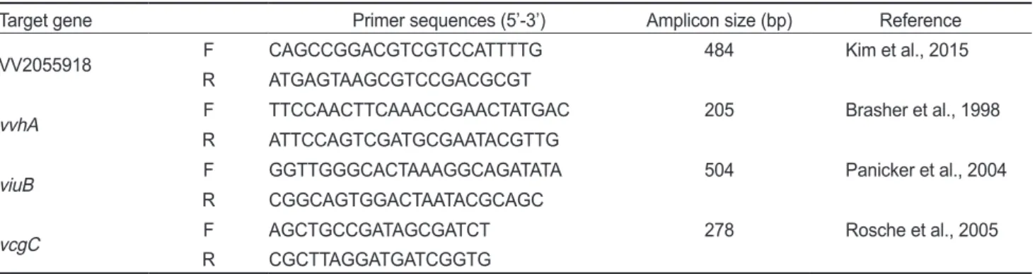
배양양성관에대Table 1. Primers used for PCR
Target gene Primer sequences (5’-3’) Amplicon size (bp) Reference
VV2055918 F CAGCCGGACGTCGTCCATTTTG 484 Kim et al., 2015
R ATGAGTAAGCGTCCGACGCGT
vvhA F TTCCAACTTCAAACCGAACTATGAC 205 Brasher et al., 1998
R ATTCCAGTCGATGCGAATACGTTG
viuB F GGTTGGGCACTAAAGGCAGATATA 504 Panicker et al., 2004
R CGGCAGTGGACTAATACGCAGC
vcgC F AGCTGCCGATAGCGATCT 278 Rosche et al., 2005
R CGCTTAGGATGATCGGTG
PCR, polymerase chain reaction.
Fig. 1. Sampling stations of sea water at Gadeok Island coast in Korea.
해
CHROMagar
TMVibrio plate (CHROMagar, Paris, France)
에획선하고37°C
에서24
시간배양후V. vulnificus
로추정되 는파란색집락을PCR (polymerase chain reaction)
분석한결 과양성에대하여최확수표(most probable number, MPN)
를 이용하여100 mL
당MPN
값으로나타내었다.
이후PCR
양성 균주는TCBS (thiosulfate citrate bile salts sucrose) agar plate (BD Difco, Franklin Lakes, NJ, USA)
에획선하고35°C 18-24
시간배양하여집락을확인하고, Tryptic soy broth (2% NaCl)
에접종배양하여추출(Genomic DNA extraction kit; Bioneer, Daejeon, Korea)
한DNA (deoxyribonucleic acid)
를 주형으 로PCR
반응을실시하여병원성관련유전자(vvhA, viuB
및vcgC)
특성을확인하였다.
PCR 조건
V. vulnificus
균수의정량을위해사용된primer
와PCR
조건 은Kim et al. (2015)
에따라수행하였다. V. vulnificus
의특이primer (species specific primer)
를사용하였고(Table 1), PCR
조건은94°C
에서5
분간열변성(denaturation)
후, 94°C
에서30
초, 60°C
에서30
초, 72°C
에서30
초간순환반응(annealing)
을25
회실시하고,
최종적으로72°C
에서10
분간신장반응(elon-
gation)
을수행하였다.
병원성관련인자특성을확인하기위한
PCR
조건으로vvhA
와viuB
는94°C
에서3
분간열변성후, 94°C
에서1
분, 65°C
에서1
분, 72°C
에서1
분간30
회순환반응 실시후, 72°C
에서5
분간신장반응을수행하였다(Panicker et al., 2004). vcgC
의경우94°C
에서5
분간열변성, 94°C
에서40
초, 56°C
에서40
초, 72°C
에서1
분간35
회순환반응실시후, 72°C
에서7
분간신장반응을수행하였다.
증폭된DNA
산물은1.5% agarose gel
에서전기영동하고green star staining solu- tion
으로염색하여UV Transilluminator (DAIHAN Scientific, Daejeon, Korea)
로확인하였다.
결과 및 고찰
가덕도 동부 연안 해수의 이화학적 특성
2019
년1
월부터12
월까지낙동강과가까운가덕도동부연안 의9
개지점과담수유입이상대적으로적은가덕도서부연안 의1
개지점등모두10
개지점에서월1
회채취하였다.
일반 수질 조사결과 월별평균 수온은
9.7-26.2°C
로8
월이 가장높았고, 1
월이가장낮았다.
월별평균염분은14.2-33.8 practical salinity units (psu)
로3
월이 가장높았고9
월이가 장낮았으며,
하계인7, 8
및9
월이23.7±3.68, 29.9±2.06
및14.2±0.46 psu
로다른계절보다낮았다(Fig. 2).
평균pH
는3
월이
8.33
으로가장높았고월별유의차는크지않았으나9
월이
7.99±0.10
으로가장낮았다(
자료미제시). 9
월은태풍으로인한
150 mm
이상의강우에의해다량의낙동강하천수가유입되어낮은염분과
pH
를초래한것으로추정된다.
용존산소(dissolved oxygen, DO)
는월별평균값이6.84-10.78 mg/L
범 위였다(
자료미제시).
조사지점별측정값은수온과pH
의경우 평균17.2-17.8°C
와8.13-8.19
로유의차가크지않았으나(
자료 미제시)
염분의경우가덕도동부9
개지점이평균28.1-30.5 psu
로가덕도서부10
번지점의32.4 psu
보다낮았고,
낙동강 본류와가까운6
번과7
번지점은28.1±6.87
과28.4±6.90
으 로다른조사지점보다더낮았다(Table 3).
V. vulnificus의 검출
2019
년가덕도연안10
개지점에서매월채취한총120
개해 수시료에서확인된V. vulnificus
의검출은Table 2
에나타내 었다.
월별로3
월과6-11
월에3.6-2,100 MPN/100 mL
범위 에서검출되었고, 1, 2, 4, 5
및12
월은검출되지않았다.
검출 율은6
월부터40%
로증가하여하계인7-9
월은검출율이80- 100%
이었는데이때수온및염분이평균22.2-26.2°C
및14.2- 29.9 psu
로다른조사월보다수온은높고,
염분은낮았다.
특 히9
월은모든조사지점에서검출되었고그수치도100-2,100 MPN/100 mL
으로큰폭으로증가하였는데,
이때평균염분은14.2±4.10 psu
로태풍에의한많은비로인해낙동강하천수가다량유입되어
V. vulnificus
의수치가크게증가한것으로판 단된다. 8
월은평균수온이26.2°C
로7
월과9
월보다높았고,
염 분도평균29.9 psu
로7
월및9
월의평균염분23.7 psu
및14.2
psu
보다높았으며검출율은7
월과9
월보다낮았다. Motes et
al. (1998)
은V. vulnificus
의개체수가수온26°C
까지는증가하 고이후30
까지는높은수준에서정지하며, 15-25 psu
의염분 에서높은수준으로검출된다고하였고, Parvathi et al. (2004)
은열대성하구에서V. vulnificus
의밀도가수온보다염분에의 해더조절된다고보고한바있다.
본연구결과를살펴보면일 정수준이상의수온에서는균의생육에염분이더큰영향을 미치는것으로평가된다.
검출율이10-20%
이었던3, 10
및11
월은평균수온이15.2-20.5°C
이었고,
그외검출한계미만으 로나타난조사월은평균수온이15°C
미만나타났다.
이는수 Fig. 2. Temperature and salinity of seawater collected at Gadeok Island coast in 2019. ○, temperature (°C); ●, salinity (psu).온
15°C
이하에서는해수와패류에서V. vulnificus
의검출한 계이하이었다는보고와유사하였다(Lee et al., 2019).
한편우 리나라에서V. vulnificus
의검출은주로6
월부터11
월에검출되고동계에는검출율은낮으나보고되는사례가있으며최근 에는연안해수에서
1
월과2
월에도검출되고있다(Park et al., 2019; KCDC, 2020).
조사지점별
V. vulnificus
의검출율은Table 3
에나타내었다.
가덕도동부연안9
개지점에서의검출율은25.0-50.0%
로평 균염분은28.1-30.5 psu
이었고,
시료의8.3%
가검출된가덕도 서부10
번지점의경우평균염분이32.4 psu
로나타나30-34 psu
에서개체수가감소한다는보고와유사하였다(Motes and DePaola, 1996; Motes et al., 1998).
특히낙동강유입지점과 가까운지점의검출율이더높았고,
평균염분은상대적으로낮 아염분이V. vulnificus
의생육에영향을미쳤음을알수있다. 분리균의 특성
2019
년1
월부터12
월까지매월채취한시료에서분리한V.
vulnificus
에대하여확인한병원성관련유전자특징은Table 4
에나타내었다. 3
개시험관법(3 tube MPN)
으로최소3
단계의십진계열 희석으로접종한
APW
배양결과양성관에대하여
CHROMagar
TMVibrio plate
에획선한후파란색을띠는집 락을APW
양성관당하나또는둘이상선택하였다. PCR
결 과양성인균주는TCBS agar plate
에서성상을확인하고이후vvhA, viuB
및vcgC
의병원성관련유전자에대한PCR
분석 을실시하였다.
그결과동일한지점에서분리된균주사이에집 락형태및병원성관련유전자특성결과가같은경우는동일균 주가중복분리된것으로간주하여제외하였고,
최종적으로채 취지점기준에서서로다른특성을보이는균주는총68
균주로 확인되었다.
V. vulnificus
는TCBS agar plate
에서전형적으로녹색집락 을형성하는sucrose
분해음성이지만일부는sucrose
분해하는 것으로보고되고있다(Oliver, 2005b). Chromagarvibrio me- dium
은β-galactosidase activity
를기반으로하여병원성vibrio
균을분리하므로sucrose
분해및비분해V. vulnificus
를모두 분리할수있다(Monget and Robichon, 2011).
분리한총68
균 주중TCBS agar plate
에서녹색집락을나타내는sucrose
비 분해균주는57
균주(83.8%)
이었고, 11
균주(16.2%)
는노란색 집락의sucrose
분해균주이었다.
이는V. vulnificus
의15%
는sucrose
양성을나타낸다는기존연구결과와국내에서분리한임상균주중
16.7%
가sucrose
분해양성이었다는보고와유사 Table 2. Monthly detection of Vibrio vulnificus from seawater col-lected at Gadeok Island coast
Month No. of positive (%) MPN/100mL No. of samples
Jan 0 (0.0) <3.0-<3.0 10
Feb 0 (0.0) <3.0-<3.0 10
Mar 1 (10.0) <3.0-3.6 10
Apr 0 (0.0) <3.0-<3.0 10
May 0 (0.0) <3.0-<3.0 10
Jun 4 (40.0) <3.0-210 10
Jul 9 (90.0) <3.0-23 10
Aug 8 (80.0) <3.0-21 10
Sep 10 (100.0) 30-2,100 10
Oct 2 (20.0) <3.0-3.6 10
Nov 1 (10.0) <3.0-3.6 10
Dec 0 (0.0) <3.0-<3.0 10
Total 35 (29.2) <3.0-2,100 120
Table 4. Characteristics of virulence genes in Vibrio vulnificus isolated from seawater at Gadeok Island coast by month in 2019
Virulence enes Jan Feb Mar Apr May Jun Jul Aug Sep Oct Nov Dec Total
vvhA 0
(0.0) 0
(0.0) 1
(100.0) 0
(0.0) 0
(0.0) 11 (100.0) 16
(100.0) 15 (93.8) 20
(95.2) 2 (100.0) 1
(100.0) 0
(0.0) 66 (97.1)
viuB 0
(0.0) 0
(0.0) 0
(0.0) 0
(0.0) 0
(0.0) 5
(45.5) 5
(31.3) 11 (68.8) 9
(42.9) 0
(0.0) 0
(0.0) 0
(0.0) 30 (44.1)
vcgC 0
(0.0) 0
(0.0) 0
(0.0) 0
(0.0) 0
(0.0) 8
(72.7) 7
(43.8) 13 (81.3) 11
(52.4) 0
(0.0) 0
(0.0) 0
(0.0) 39 (57.4)
No. of isolation 0 0 1 0 0 11 16 16 21 2 1 0 68
Table 3. Relationship of Vibrio vulnificus detection from seawater samples and salinity at Gadeok Island coast according to sampling station
Sample
station Salinity
(psu) No. of
positive (%) MPN/100mL No. of samples 1 28.8±5.30 5 (41.7) <3.0-2,100 12 2 28.9±1.73 3 (25.0) <3.0-1,200 12
3 29.3±1.73 3 (25.0) <3.0-640 12
4 30.5±5.68 3 (25.0) <3.0-430 12
5 29.5±5.96 4 (33.3) <3.0-750 12
6 28.1±6.87 6 (50.0) <3.0-210 12
7 28.4±6.90 4 (33.3) <3.0-230 12
8 29.6±4.26 3 (25.0) <3.0-200 12
9 28.9±5.85 3 (25.0) <3.0-390 12
10 32.4±3.03 1 (8.3) <3.0-30 12
한수치로확인되었다
(Oliver, 2005b; Kim et al., 2006).
병원성관련유전자는병원성요인으로작용할수있는인자 중
V. vulnificus
에특이적인vvhA, viuB
및vcgC
에대하여분 석하였다. vvhA
는V. vulnificus
의hemolysin/cytolysin
유전자 이다.
철흡수관련유전자는hut, vct, vib
등여러가지가있으 나viuB
는V. vulnificus
가생산하는ferric vulnibactin receptor
로서독력이강한균주는대체로많은vulnibactin
을생산하며 철이결핍된환경에서균이증식하는데가장큰영향을미친다 고보고되어있다(Shin, 2013; Chung et al., 2016). vcgC
는해 수분리균으로부터사람에게잠정적으로병원성균으로작용할 수있는임상균주를추정하기위하여분석하였다. PCR
분석결 과97.1%
인66
균주에서vvhA
가확인되었다. viuB
는30
균주(44.1%)
가양성이었고, vcgC
는39
균주(57.4%)
에서확인되었 다.
분리균주로부터vvhA
만확인된균주는27
균주(39.7%)
이 었고, vvhA, viuB
및vcgC
가모두검출된균주의경우도27
균 주(39.7%)
이었다.
그외는vvhA
와viuB, vvhA
와vcgC
등일 부유전자를가진것으로확인되었다. viuB
와vcgC
는6
월부 터9
월까지검출되어평균수온이20°C
이상으로상승하고V.
vulnificus
의검출율이높은달과기간이일치하였다. Han et al.
(2009)
은clinical genotype (vcgC)
이viuB
의존재와관련있 으며,
따뜻한달에더많이검출된다고보고한바있다. Çam et al. (2019)
은Galveston bay
의V. vulnificus
연간조사에서16S rRNA A
형에서16S rRNA B
형으로우세한형이바뀌는계절 적변화를확인하였고, vcgC
와16S rRNA B
형이서로강한상 관관계가있다고하였다.
감염병감시연보(KCDC, 2018b)
에 의하면우리나라에서발생한비브리오패혈증환자는주로6
월 부터시작하여11
월까지확인되고특히7, 8, 9
및10
월의발생 률이높은것으로확인된다.
우리연구결과와비교해보면수온 이20°C
이상인6
월부터V. vulnificus
가증가하는동시에병원 성관련유전자를가진균주의비율이높아패혈증환자가발생 하는것으로추정할수있다.
가덕도동부연안은낙동강유입으로
V. vulnificus
의생육에 좋은여건을제공하고수온및염분등환경조건이충족될경 우급증하는경향이확인되며,
검출율이증가하는계절에잠재 적인병원성균주의비율이높게확인되었다.
가덕도는낙동강 하구연안의위생학적상태와인근연안에영향요인으로작용 할수있는만큼지속적인모니터링이필요할것으로생각된다.
사 사
이논문은
2020
년해양수산부재원으로해양수산과학기술진흥원의지원을받아수행된연구임
(
수산환경및수산물중미 생물학적위해요소현장형진단시스템개발).
References
Baffone W, Tarsi R, Pane L, Campana R, Repetto B, Mariot-
tini GL and Pruzzo C. 2006. Detection of free-living and plankton-bound vibrios in coastal waters of the Adriatic Sea (Italy) and study of their pathogenicity-associated prop- erties. Environ Microbiol 8, 1299-1305. https://doi.org/
10.1111/j.1462-2920.2006.01011.x.
Brasher CW, DePaola A, Jones DD and Bej AK. 1998. De- tection of microbial pathogens in shellfish with multiplex PCR. Curr Microbiol 37, 101-107. https://doi.org/10.1007/
s002849900346.
Çam S, Brinkmeyer R and Schwarz JR. 2019. Quantitative PCR enumeration of vcgC and 16S rRNA type A and B genes as virulence indicators for environmental and clinical strains of Vibrio vulnificus in Galveston bay oysters. Can J Microbiol 65, 613-621. https://doi.org/10.1139/cjm-2018-0399.
Chung HY, Kim YT, Kim SY, Na EJ, Ku HJ, Lee KH, Heo ST, Ryu SR, Kim HB, Choi SH and Lee JH. 2016. Complete genome sequence of Vibrio vulnificus FORC_017 isolated from a patient with a hemorrhagic rash after consuming raw dotted gizzard shad. Gut Pathog 8, 1-6. https://doi.
org/10.1186/s13099-016-0104-6.
DePaola A, Capers GM and Alexander D. 1994. Densities of Vibrio vulnificus in the intestines of fish from the U.S. Gulf Coast. Appl Environ Micrbiol 60, 984-988. https://doi.
org/10.1128/aem.60.3.984-988.1994.
Han F and Ge B. 2010. Multiplex PCR assays for simultaneous detection and characterization of Vibrio vulnificus strains.
Lett Appl Microbiol 51, 234-240. https://doi.org/10.1111/
j.1472-765X.2010.02887.x.
Han F, Pu S, Hou A and Ge B. 2009. Characterization of clinical and environmental types of Vibrio vulnificus isolates from Louisiana oysters. Foodborne Pathog Dis 6, 1251-1258.
https://doi.org/10.1089/fpd.2009.0343.
Hlady WG and Klontz KC. 1996. The epidemiology of Vibrio infections in Florida, 1981-1993. J Infect Dis 173, 1176- 1183. https://doi.org/10.1093/infdis/173.5.1176.
Horseman MA and Surani S. 2011. A comprehensive review of Vibrio vulnificus: an important cause of severe sepsis and skin and soft-tissue infection. Int J Infect Dis 15, e157-e166.
https://doi.org/10.1016/j.ijid.2010.11.003.
Jones MK and Oliver JD. 2009. Vibrio vulnificus: disease and pathogenesis. Infect Immun 77, 1723-1733. https://doi.
org/10.1128/IAI.01046-08.
Kaspar CW and Tamplin ML. 1993. Effects of temperature and salinity on the survival of Vibrio vulnificus in seawater and shellfish. Appl Environ Micrbiol 59, 2425-2429. https://doi.
org/10.1128/AEM.59.8.2425-2429.1993.
KCDC (Korean Centers for Disease Control and Prevention).
2020. Infectious disease portal. Retrieved from http://www.
cdc.go.kr/npt/biz/npp/portal/nppVbStatsMain.do on Apr 3, 2020.
KCDC (Korean Centers for Disease Control and Prevention).
2018a. Reported cases of Vibrio vulnificus sepsis by year
and province. In: Infectious Disease Surveillance Yearbook, 2018. KCDC, Cheongju, Korea, 98.
KCDC (Korean Centers for Disease Control and Prevention).
2018b. Reported cases of Vibrio vulnificus sepsis by year and month. In: Infectious Disease Surveillance Yearbook, 2018. KCDC, Cheongju, Korea, 184.
Kim HJ, Ryu JO, Lee SY, Kim ES and Kim HY. 2015. Multi- plex PCR for detection of the Vibrio genus and five patho- genic Vibrio species with primer sets designed using com- parative genomics. BMC Microbiol 15, 1-11. https://doi.
org/10.1186/s12866-015-0577-3.
Kim SM, So HA, Song KM, Lee YY, Lim CW, Lee JH, So HS, Kim JK, Park RK and Park SD. 2006. Comparison of biological and genetic characteristics between sucrose-fer- menting and sucrose-nonfermenting Vibrio vulnificus iso- lates. J Bacteriol Virol 36, 221-228. https://doi.org/10.4167/
jbv.2006.36.4.221.
Lee DS, Shin HY, Park KS and Shin IS. 2019. Distribution of Vibrio parahaemolyticus and Vibrio vulnificus in seawater and shellfish at Gomso Bay. Korean J Fish Aquat Sci 52, 114-120. https://doi.org/10.5657/KFAS.2019.0114.
Monget D and Robichon D. 2011. Culture medium for detecting and identifying Vibrio bacteria. US7892783B2. U.S.A.
Motes ML, DePaola A, Cook DW, Veazey JE, Hunsucker JC, Garthright WE, Blodgett RJ and Chirtel SJ. 1998. Influence of water temperature and salinity on Vibrio vulnificus in Northern Gulf and Atlantic Coast oysters Crassostrea vir- ginica. Appl Environ Microbiol 64, 1459-1465. https://doi.
org/10.1128/aem.64.4.1459-1465.1998.
Motes ML and DePaola A. 1996. Offshore suspension relaying to reduce levels of Vibrio vulnificus in oysters Crassostrea virginica. Appl Environ Microbiol 62, 3875-3877. https://
doi.org/10.1128/aem.62.10.3875-3877.1996.
Nascimento SMM do, Vieira RHS dos F, Theophilo GND, Ro- drigues D dos P and Viera GHF. 2001. Vibrio vulnificus as a health hazard for shrimp consumers. Rev Inst Med Trop Sao Paulo 43, 263-266. https://doi.org/10.1590/S0036- 46652001000500005.
Oliver JD. 2005a. Wound infections caused by Vibrio vulnificus and other marine bacteria. Epidemiol Infect 133, 383-391.
https://doi.org/10.1017/S0950268805003894.
Oliver JD. 2005b. Vibrio vulnificus. In: Oceans and health:
Pathogens in the marine environment. Belkin S and Colwell RR. Springer, Boston, MA, U.S.A., 253-276. https://doi.
org/10.1007/0-387-23709-7_10.
Panicker G, Call DR, Krug MJ and Bej AK. 2004. Detection of pathogenic Vibrio spp. in shellfish by using multiplex PCR and DNA microarrays. Appl Environ Microbiol 70, 7436- 7444. https://doi.org/10.1128/AEM.70.12.7436-7444.2004.
Park K, Mok JS, Kwon JY, Ryu AR and Shim KB. 2019. Sea- sonal and spatial variation of pathogenic Vibrio species iso- lated from seawater and shellfish off the Gyeongnam Coast
of Korea in 2013- 2016. Korean J Aquat Sci 52, 27-34.
https://doi.org/10.5657/KFAS.2019.0027.
Parvathi A., Kumar H.S., Karunasagar I. 2004. Detection and enumeration of Vibrio vulnificus in oysters from two estu- aries along the southwest coast of India, using molecular methods. Appl Environ Microbiol 70, 6909-6913. https://
doi.org/10.1128/AEM.70.11.6909-6913.2004.
Rosche TM, Yano Y and Oliver JD. 2005. A rapid and simple PCR analysis indicates there are two subgroups of Vibrio vulnificus which correlate with clinical or environmen- tal isolation. Microbiol Immunol 49, 381-389. https://doi.
org/10.1111/j.1348-0421.2005.tb03731.x.
Shin SH. 2013. Multiple high-affinity iron-uptake systems of Vibrio vulnificus. J Bacteriol Virol 43, 168-176. https://doi.
org/10.4167/jbv.2013.43.3.168.
Shapiro RL, Altekruse S, Hutwagner L, Bishop R, Hammond R, Wilson S, Ray B, Thompson S, Tauxe RV and Grif- fin PM. 1998. The role of Gulf Coast oysters harvested in warmer months in Vibrio vulnificus infections in the United States, 1988-1996. J Infect Dis 178, 752-759. https://doi.
org/10.1086/515367.
Strom MS and Paranjpye RN. 2000. Epidemiology and patho- genesis of Vibrio vulnificus. Microbes Infect 2, 177-188.
https://doi.org/10.1016/S1286-4579(00)00270-7.
Tamplin M, Rodrick GE, Blake NJ and Cuba T. 1982. Isolation and characterization of Vibrio vulnificus from two florida es- tuaries. Appl Environ Microbiol 44, 1466-1470. https://doi.
org/10.1016/0198-0254(83)90264-9.
USFDA (U.S. Food and Drug Administration). 2018a. BAM chapter 9: Vibrio. Retrieved from https://www.fda.gov/food/
laboratory-methods-food/bam-chapter-9-vibrio on Nov 30, 2019.
USFDA (U.S. Food and Drug Administration). 2018b. BAM R59: Phosphate-buffered saline (PBS), pH 7.4. Retrieved from https://www.fda.gov/food/laboratory-methods-food/
bam-r59-phosphate-buffered-saline-pbs-ph-74 on Nov 30, 2019.