군 f 니
Famesol 의 HL-60 세포에 대한 세포득성과 활성산소 및
항산화효소 활성 변화
임 소 윤 • 박시원후 상명대학교 자연과학대학 화학과
(Received November 6. 2006; Revised December 15, 2006)
Reactive Oxygen Species and Antioxidant Enzyme Activities in Accordance with the Cytotoxicity of Farnesol Against HL-60 Cells
So Yoon Lim and Sie Won Park 쌓
Department of Chemistry, College of Natural Science, Sangmyung University, Seoul 110-743, Korea
Abstract —— Farnesol in fruits, vegetables, herbs and leaves acts as bioactive component related with prevention of cancer and psychological malaise. We investigated the cytotoxic effects of famesol on human leukemic cell, HL-60 cells, by MTT assay using 3-(4,5-dimethylthiazol-2-yl)-2,5-diphenyltetrazoliumbromide. Famesol (0.1—50 exhibited cytotoxicities against HL-60 cells in concentration and culture period dependent manner. In the cytotoxic condition induced by farnesol against HL-60 cells, the generation of reactive oxygen species such as 0 율 and H2O2 were found to be considerably increased.
The most prominent augmentations of 0 율 and HgOg were over five folds of controls. In an attempt to explore the response of HL-60 cells to the increased 0 율 and HgOg, superoxide dismutase (SOD), glutathione peroxidase (GPx) and catalase activ
ities of HL-60 cells treated with famesol were measured. SOD and GPx activities were found to be remarkably elevated by addition of famesol showing the best results of 273% and 167% of controls, respectively. All data suggest that farnesol may have played as an apoptosis inducer in HL-60 cells via production of reactive oxygen species (ROS) and HL-60 cells may have failed to overcome the damage of ROS on account of still deficient ROS scavengers including SOD and GPx, Keywords □ famesol, cytotoxicities, HL-60 cells. ROS, antioxidant enzymes
Terpene계 화합물은 isopreneCCs)의 중합유도체로 식물에서는 주로 탄소수가 적은 monoterpenes(Ciy)이나 sesquiterpenes (C15)으로 존재하며, 수목, 꽃, 야채, 과일 등의 향기와 풍미성분 으로 알려겨 있다. 이둘은 공기 중의 nonmethane 탄화수소 (NMHC)의 원료로서 대기 중의 다 밍 번 웅 맥 참여하며 , ozone 발생 및 토양의 질소와 탄소의 순환^ 등 지구 환경과 관련하여 중요한 역할을 담당하고 있으며, 산업적으로도 그 졸은 향기와 항균성으로 인해 향수, 화장품, 의약품 동_o로 널러 사용되어 왔 다 3> 더욱이 이들이 식물과 인간을 포함한 동물체내에서 산소득 성이나 지질과산화반응의 억제^'®' 등 개체방어에도 필수적으로 작용하고 있옴으로 보아, 생명체내에서 긴요한 생리 화학에 관
본 논문에 관한 문의는 저자에게로 (전화) 02-2287-5147 (팩스) 02-2287-0070 (E-mail) parksw^i smu.ac.kr
여하고 있옴이 시사되고 있다.
Faraesolfi- sesquiterpenes 계열의 isoprenyl alcohol로서 꽃과 파일 등의 정유 성분으로, 동물에서는 mevalonat^cholesterol 생 합성 경로의 유도체로 존재한다. Farnesol은 항균작용,은'가 후각신 경 작용^^ 그리고 함^^작용 등 다양한 작용이 보고 되고 었으며, 항암작용으로는 암세포 증식억제,®*페 암세포사멸 유도^^•의 그러 고 암세포 증식파 발육억제1크^ 있다. 이러한 다잉한 farnesol 의 항암작용의 작용기작으로는 nuclear hormone receptor인 femesoid X receptor(FXR)의 활성화/ 스^ peroxisome proliferator- activated receptors-a(PPARa) 및 -"/(PPARy) 활성 화페 그러고 thyroid hormone receptor(THR) (31 mRNA expression 유도 동 gene transcription 조절과 관련 었는 것으로 보고 되고 있다.오®그 이외에도 post translation modification의 한가지로 단백질에 fames이이나 geranylgeranyl을 부가하는 prenylation반응은 signal transduction, cytoskeletal regulation, cell proliferation
372
그러고 apoptosis에 매우 중요하며 이 과정의 조절 유견자는 ras oncogene으로 알려져 있다. 그런데 ras oncogene에 이성어 생기 면 protein prenylation이 장애률 받고 당연허 이러한 ras 단택질 특히 K-ras 단백질의 이상미 약 30%의 인간 암에서 밝혀진 것 으로 보아 farnesol은 암의 생러학과 적결되어 있옴을 알 수 있 다.미 그런가 하면 femesyl-0 -acetyl hydroquinon은 cholesterol 합성경로의 3-hydroxy-3-methyI glutaryl CoA(HMG-CoA) reductase 의 활성을 억제함으로써 대장암세포증식을 억제하는 것이 밝혀졌다.^^^
한편 암의 발생원인중 활성산소(reactive oxygen species, ROS)에 의한 oxidative stress가 주요 원인으로 알려져 왔으며, 암세포 사멸에도 활성산소가 밀접하게 관계되는 것이 보고 되어 왔다.19) 활성산소란 원자패도나 분자괘도에 한 개나 또는 그이 상의 unpaired electron울 가진 화합물로서 반응성이 매우 높은 것이 특정이다. ROS의 종류로는 hydrogen peroxide(H202), superoxide radical(Op, hydroxyl radical(OH) 그리고 singlet oxygen('02) 등이 있으며 이들은 강한 빈음성 때문에 갖가지 유 해 작용을 유발하여 oxidative stress롤 일으킨다. 이 ROS는 인 체에 이롭기도 하고 해롭기도 하는 양면성을 지닌 물질로 알려 져 있다. 인체 내의 해로운 작용으로는 oxidative stress를 일으 켜 세포내 signal cascade에 있어서 secondary messengers로 작 용하여 암세포를 유발하는 가하면, 위괘앙, Alzheimer, 관절염, 뇌졸중둥의 직접적인 원인이 되기도 하며, 2^ 이 로 운 작용 으로는 감염된 세균이나 neoplastic 세포를 neutrophils이나 phagocytes 같은 면역계 세포가 박멸할 때 ROS인 0 ^를 신속하 게 다량 발생시켜 사멸시키는 도구로 사용하는 경우가 있다.^^'으^^
한편 인체 내에서는 해로운 내인성 ROS가 자연스럽게 생성되는 데, 예률 들어 peroxisome에서 long chain fatty acids가 산화되 면서 H2O2가 부산물로 생성되며, lipooxygenase가 arachidonic acid를 전환하는 중에도 생성되고, 또 다른 주요 경로인 mitochondria의 respiratory chain에서 지속적으로 일어나는데 보 통 mitochondria의 electron transport chain을 지나는 견자중의 약 1^ 2% 정도가 홀러나와 분자산소(0 ,)와 쉽게 결합종}쉬 02를 형성한다고 한다.2산'2구^ 외인성 ROS는 자동차 배기가스와 같은 환 경오염물질, 담배연기, 이온 복사파, iron salts, 자외선, 자연식품 의 phenol성 화합물 등에서 각각 생성되므로 ROS는 우러 주번의 다밍권' source로부터 생성되는 것을 알 수 었다.^^^ 한편 홍머 있 는 것은 발암의 주원인인 ROS가 많은 함함제의 작용기작으로도 작용한다는 점이다. 예률 돌어 cisplatin, doxorubicin, vincristine, cytosine arabinoside, methotrex, bleomycin 등의 항암제는 암 세포의 apoptosis률 유발하는 활성산소를 생성시켜 암세포가 사 멸하게끔 유도하는 것으로 보고 되고 있다.^®
현재 사용되고 있는 항암•제는 그 부작용이 심하식 환자의 삶 의 질이 떨어지고 완처율도 평균 50% 정도에 불과하며 말기 암
환자의 완치율은 5% 이내로 떨어지는 것으로 알려져 있다. 따라 서 각국은 가능하면 무득성이거나 득성이 매우 약하면서도 항암 작용이 우수한 힘■제 개발에 심혈을 기울이고 있으나 아직까지 도 이상적인 항암■제개발은 이루어지지 않고 있다. 본 연구는 식 물의 정유성분이며 sesquiterpene계 화합물인 farnes이의 항암효 과를 인간 백혈병 암세포인 HL-60 세포률 대상으로 세포득성 효 파률 검색하고, 이 세포득성 효과의 작용기작을 구명하고자 farnes이에 의해 유도된 활성산소 생성량의 번화와 활성산소 소 거효소의 활성번화률 측정함으로써 famesol의 항암효과의 작용 기작을 활성산소와 연계하여 규명하고자 시행되었다.
실험 방법
시약 및 기기
RPMI 1640, fetal bovine serum(FBS), streptomycin mixture 는 GIBCO BRL(Grand Island, New York USA)에서 구입하여
용*하였다. trans, trans-farnesol, ferricytochrome c, xanthine, xanthine oxidase, superoxide dismutase, glutathione(reduced), glutahione reductase, phorbol 12-myistate 13-acetate(PMA), M^utylhydropcroxidc, nicotinamide adenine dinucleotide pho- sphate(reduced), glutathione peroxidase, glutathione reductase, lipopolysaccharide(LPS)(£. coli, 055:B5)는 Sigma Chemical Co.(St Louis, MO, U .S A )의 제품을 사용하였고 5-FU(CAS No.
51-21-8)은 Wako Pure Chemicals Industries Ltd.(Osaka, Japan)의 제품을 구입하여 사용하였다. 단백질 정량시약은 Bio- Rad kit(Hercules, CA, USA)률 사용하였고 그러고 기타 시약은 analytical grade 제품을 구입하여 사용하였다. 20 m/와 50 m/의 culture flask 와 24 well plate는 Falcon Co.(Dickenson Co.
France), CO2 incubator(New Brunswick Scientific Inc.), UV/
Visible spectrophotometer^ Varian Carry 3(Varian, PTY LTD, Australia), ELISA microplate reader(ELX800, BIO-TEK instruments)등을 사용하였다.
HL-60 세포 배양
한국 세포주 은행 (Korean Cell Line Bank, KCLB, Seoul, Korea)으로부터 분양받았으며, 배양액은 10% heat-inactivated fetal bovine serum(FBS)을 함유한 RPMI 1640 배지에 100 U/
m/ penicillin/100 |ig/m/ streptomycin을 첨 가하여 37^C, 5%
CO2, humidified incubatoi에서 배양하였으며 계대배양S: 약 70%
의 confluence가 되는 2—3일 마다 시행하였다. Farnesol은' 실험 직전에 dimethyl sulfoxide(DIVISO)에 용해시켜 사용하였으며 대 조군(control)과 시료군(sample)의 DMSO의 최종능도는 0.05%
이었다. 항산화효소 활성을 측정하기 위한 효소액 조제를 위한 HL-60 세포의 대량 배앙은 20 m/ 또는 50 m/의 culture flask돌
Vol. 50, No. 6;2006
374 임소윤 • ^•시원
사용하여 시행하였다.
MTT 법에 의한 세포독성 측점
Fames이에 의한 HL-60 세포득성은MTT 법®으로 측정하였 다. HL-60 세포는 24 well plate에 ixio® cells/m/로 접종하고, 다양한 농도의 farnesoKO.l, 0.5, 1, 5, 10, 50, 100 |ig/m/)을 처 리하여 12, 24, 48시간 동안 배양하였으며 , contr이에는 vehicle 로써 DMSO룔 동량 첨가하였다. 배양 후 새 배지로 바꿔주고 10 10/ 의 MTT(5mg/m/>틀 첨가하여 4시간 동안 배양하였다. 배 앙 후 800rpm에서 10 분간 원심분리 하여 상등 액은 버리고 200^1/의 DMSO를 가하여 생성된 formazan을 용해시킨 후, 570 nm에서 microplate reader틀 이용하여 몹광도를 죽정하였다.
계산법: % cytotoxicity= (A570 of control cells-A570 of treated cells)/A570 of control c e lls x 100%.
HL-60 세포의 O j 이온 정량
HL-60 세포에 femesd을 첨가하쉬 배양 후 생성된 O2 이온의 정량은 Markert피 법에 의한 ferricytochrome c의 환원반응을 활용하였다. 요약하면, HL-60 세포는 femes이을 농도별(0.1, 1, 10, 50 ng/m/)로 처러하식 단시 간 인 10, 30 그리고 60 분간 배 앙한 다옴 세포들을 800 rpm으로 10분간 원심 분러한다. 상등 액은 버리고, 세 포는 HBSS(Hank's balanced salt solution) buffer로 부유시켜 24 well plates에 ix 10** cells/mZ로 접종한 다 옴 PMA(20 nM)를 5 yd 첨가하고 9Q초 후에 cytochrome c 용액 의 최중농도가 150 nM이 되도록 가한다. 이 반응 액을 37°C에 서 1시간 동안 incubation한 다옴 1,000rpm에서 원심 분리하여 상등 액을 취하여 550nm에서 흡광도률 10초 간격으로 5분간 측정하여, 02 생성량을 O2(nmol/m])=47.7xA550nm 식으로부터 계산하였다. Fames이을 장시간(12 hr) 처리하는 경우에 생성되 는 0 ^ 생성량은 단시간의 경우와 동일한 방법으로 시행하였다.
HL-60 세포의 H^O;^ 정량
Famesol에 의해 HL-60 세포에서 생성되는 HjOjj는 Paglia and Valentine크®* 방법을 변형하여 측정하였다. HL-60 세포는 famesol 을 0.1, 1, 10, 50 ng/m/ 농도로 처러하고, 단시간인 10, 30 그리 고 60분간 배양한 다옴 세포들을 800 rpm으로 10분간 원심 분 리한 후 상등 액은 제거한다. 세포는 HBSS buffer로 부유시켜, 1 X 10® cells/m/로 세포수률 조정하고, cuvette에 1.0 m/의 세포 와 표준 시약(20mM GSH, 1.25mM NADPH, l.OU/mi GSH- Px and 2.0 U/m/ GSSG reductase 함유)을 2.0 ml 가하여 반응 을 개시한다. Spectrophotometer는 0.2—0.6 O.D.(340nm)가 되 도록 조절하고 흡광도는 full scale mode로 400초간 37 X 에서 monitoring 한다. 이와 같이 12시간 처러 설험의 경우에도 동일 한 방법으로 시행하였다. 생성된 H2O2양은 NADPH의 산화에
의한 340 nm에서의 롭광도 감소를 monitoring 하는 동안 얻어 진 직선의 기울기로부터 계산하식 nmol NADPH oxidized/min/
mg protein으로 표시하였다.
SOD, GPx. catalase 효소액 조제
HL-60 세포는 femesol을 0.1, 1, 10, 50|ig/m/의 농도로 처리 하며 12시간 배양한 다옴 800 rpm으로 10분간 원심 분리하여 세 포를 포집한다. 세포는 HBSS로 세 차례 세척하고 Im /의 등장 액 Tris • HC1 buffer에 부유시켜 freezing, thawing을 반복하여 lysate률 만든다. 이 lysate률 4°C에서 8,000 rpm으로 10분•간 원 심분러 하여 핵과 조직 잔류물을 제거하고 상등액을 취하여 superoxide dismutase(total SOD), glutathione peroxidase (GPx), catalase 활성을 각각 측정하였다.
SOD 활성 측정
SOD활성은 xanthine/xanthine oxidase에 의해 생성된 Oj에 의한 cytochrome c의 환원 반응을 SOD가 억제하는 정도률 측 정하는 것으로, McCord and Fridovich체와 Floeh 방법크늘 이 용하였다. 반응 액(0.02 M phosphate buffer, pH 7.4, 0.1 mM EDTA, 50 |iM xanthine, 10 |iM cytochrome c)에 조제된 효소 액을 첨가하여 25°C에서 15분간 방치한 다옴 xanthine oxidase (XOD>를 첨가하며 반응을 개시하였으며 550 nm에서 10초 단위 로 5분간 흡광도를 측정하였다. Xanthine oxidase 첨가량은 효 소 액을 함유하지 않은 반응 액의 흡광도 흡수가 분당 최소한 0.025가 되도록 조절하여 첨가하였다. SOD 활성의 표시는 cytochrome c의 환원속도룰 50% 억제하는 양을 lim it로 책정 하여 units/mg protein으로 나타내었다.
Glutathione Peroxidase(GPx) 활성측정
GPx의 활성측정은 Gimzler et a/.®의 방법을 적용하였다. 배 양된 세포로부터 조제된 25 Mi의 효소 시료를 900 \d Tris/EDTA (50mM Tris, pH 7.6, Im M EDTA, 4mM sodium azide)에 가 한 다옴 2 0 ^ glutathione(0.15 M), 20 \d glutathione reductase (208 units/m/), 2 0 ^ NADPH(8.4 mM) 그리고 20 p/ /-butyl- hydroperoxide(30 |iM)를 가하여 총량 1 m/로 하여 340 nm에서 흡광도감소를 1분간 측정하였다. 효소활성은 nmoles NADPH oxidized/min/mg protein으로 정의하였다.
Catalase 활성측정
Catalase 활성 측정은 Claiborne^®^의 방법에 준하였다. 반응액
■?r 1.95 m/ phosphate buffer, 1 ml H202(0.09 mol//) 그 51 고 효 소 액이 함유된 총량 3 mi으로 이루어져 있다. 240 nm에서의 흡 광도 번화를 10초단위로 측정하였으며, catalase 활성은 nmoles H2O2 consumed/min/mg protein로 표시하였다.
J. Pharm. Soc. Korea
통계처리
실험 결과는 3회 ~4회 실험횟수에 대한 평균과 평균오차 값을 mean±S.E.M.으로 표시하였다. 유의 성 검증은 Student Mest 률 시행하여 control 값에 대하식 *p<0.05와 **p<0.01로 표시 하였다.
실험결과 및 고찰
HL-60 세포에 대한 famesol의 세포득성
HL-60 세포에 farnes이을 0.1-100 의 농도로 처리하고 12, 24, 48시간 배양 후, 살아었는 세포 변화를 MTT 법으로 측 정하였다. Table I에 제시한바와 같이 각 농도조건에서의 세포는 femes이의 농도에 따라 대부분 능도와 배양기간 의존적인 감소 경향을 나타내었다. 12시간과 24시간에 나타난 세포득성은 저 농 도인 0.1~1.0 ng/m/에서는 0.7~6.3% 정도로 미약했으나, 농도 가 높아짐에 따라 61.7%까지 나타났다. 배앙시간이 48식간인 경 우에는 모든 농도에서 24시간보다 더 높은 세포득성을 나타내었 으며, 1.0|xg/m/에서 38.5%로 나타난 것으로 보아 famesol의 HL-60 암세포에 대한 세포득성은 현저한 것으로 관단되었다.
ICjo 값은 48시간 기준으로 7.81 서 positive controls. 사 용한 5^FU(IC5o=0.72 ^1평/111/)와 비교하여 세포득성효파가 낮으나, 정상임파구에 대한 득성 실험결과 거의 세포득성을 보이지 않았 다(결과 제시하지 앉옴). 이 결과로부터 HL-60 암세포에 대한 femes이의 세포득성 효과는 이미 보고된 monoterpenes과
sesquiterpenes 화합물의 세포득성 효 과 ® 와 도 일치하며 farnesol이 인체 내에서 상당히 안전하게 암세포제거 역할을 할 수 있는 가능성을 시사한 것으로 볼 수 있다.
Famesol에 의한 HL>60 세포의 0 오 생성
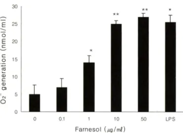
함"?]■제의 작용기작중에 항암제가 암 세포내에 활성산소률 유 발하며 apoptosis^®'®!" 일으켜 의해 암세포가 사멸하는 경우가 다양하게 보고 되고 있다. 이에 본 설험에서도 famesol에 의해 야기된 HL-60 세포의 세포득성의 작용기작을 활성산소와 연계 s H 규명해보고자 일차적으로 다옴 실험을 시행하였다. HL-60 세포는 famesoli: 농도별(0, 1.0, 5.0, 50.0 (xg/m/)로 처리하고■, 단 시간(10, 30, 60분) 및 12시간 동안 생성되는 02의 잉=을 측정하 였다. 02은 분자심:소(Og)로부터 생성되는 일련의 활성산소 전환 반응의 첫 번째 분자2 4 로서 활성산소 연구의 핵심 분자라 할 수 있다. Table II에는 단시간 처리하여 얻어진 결과를 제시 하였으며 Fig. 1에 12시간 장기간 처러하여 얻어진 결과를 제시 하였다.
Table II의 결과에 따르면 femesol은 1분 ~60분 처리 시 농도 증가에 따른 Oj 생성량은 control과 비교하쉬 미약한 수준에서의 증가밍상을 보이고 있으나, 12시간 처리 시 famesol의 능도에 따 라 현저한 0^ 생성 증가가 나타났다(Fig. 1). 특히 10.0|_ig/m/의 농도에서는 control 값의 5배 이상의 0 ^가 생성 되었으며, 처리 된 franensol의 농도가 증가할수록 생성량 또한 증가하는 것으로 측정되었다(Fig. 1). 참고로 본 실험에서는 02의 생성을 야기한
Table I - Cytotoxicities induced in HL-60 cells incubated with indicated farnesol concentrations
Incubation Farnesol (|ig/m/)
time (hr) 0 . 1 1 . 0 1 0 . 0 50.0 1 0 0 . 0 5-FU
1 2 1.5±1.6 8.5±3.1
Cytotoxicity (%)
14.9+8.7 21.5±7.6 29.5±5.8* 21.8±6.9
24 7.2±2.5 31.6±6.4 45.4±9.7 49.5±5.6* 61.7±9.3* 64.5±5.7*
48 11.5±3.6 38.5 ±7.3* 58.3 ±7.4 68.4±9.5 91.3+5.2** 88.6±9.7*
72 19.6±6.4 37.9±6.4* 77.5±9.7* 87.6±8.7* 97.6±4.2** 95.7±7.5**
Data shown are cytotoxicities±S.E.M. of four independent experiments. 5-FU (l^g/m/) was used as positive control. Cells remaining after farnesol exposure were determined using the MTT assay. % cytotoxicity—(A570 of control cells-A570 of treated cells)/A570 of control cellsX100%. *p<0.05 and **p<0.01 represent significant differences compared with control values.
Table II - Effect of farnesol on the generation of 0^ by PMA stimulated HL-60 cells
Incubation Farnesol (ng/m/)
time (min) 0 . 0 0 . 1 1 . 0 1 0 . 0 50.0 LPS
1 0 5.4+0.8 5.6±0.9
O2 (nmol/m/)
6.7±0.2** 6.3±0.7* 5.8 ±1.2 8.3 ±1.6
30 6 . 6 ±1.4 7.1+2.3 5.7±0.3* 4.6±1.8 6.2+1 . 6 11.9+0.4**
60 6.7±2.4 7-l±2.3 5.9+3.1 5.3 ±0.6** 6.7±1.2 14.8±L7*
Cells were incubated with indicated concentrations of farnesol for 10, 30 and 60 min. Harvested cells were suspended at a density of 1x10난 cells/m/ and then incubated for 15 min. LPS (1 ng/m/) was used as posoitive control. Ninety seconds after stimulation with 5 \d
of PMA solution (2 0 } iM ), 50 ^of cytochrome c solution (1.0 mM) was added. After 1 hr incubation, the supernatants thus obtained was used for measuring 62 by absorbancy at 5 5 0 nm. 0 율 produced was expressed as nmol/min/mg protein. Results are mean±S.E.M. of four independent experiments. *p<0.05 and **p<0.01, significantly different from non treated cells.
Vol. 50, No. 6, 2006
*★ ★
0 0.1 1 10 50 LPS
F a r n e s o l U g / m l)
0 0.1 1 10 50
F a r n e s o l U g /m 오)
Fig. 1 - Effect of farnesol on the generation of 0 으 in HL-60 cells.
After cells were incubated with indicated concentrations of farnesol for 1 2 hr. harvested cells were suspended at a density of 1 x 1 0^ cells/m/ and incubated for 1 hr at 37^C after 90 sec of PMA (1 i^M) stimulation. The background absorbancy of 0^ released from non stimulated cells were substracted from all data. Values are the mean±S.E.M. of four determinations. *p<0.05 and **p<0.01, significantly different from non treated culture cells.
다고 알려진 PMA률 가하여 02 생성량을 측정하였으나, 부수적 으로 PMA률 가하지 않은 대조 실험을 수차례 반복 시행한 걸 과 PMA 첨가률 하지 않아도 02 생성량은 크게 달라지지 않았 다.(결과 제시하지 않옴) Table I에서 보였던 farnesol에 의한 HL- 60 세포의 생존율 감소 현상은 세포내 Og 생성의 증가에 기인할 수도 었을 것으로 추정된다.
Farnesol에 의한 HL*60의 생성
02는 반감기가 1(T®초 정도로 매우 짧아서 곧 다옴 반응으로 진행하여 를 생성하게 되러라고 간주된다. 따라서 franesol 에 의해 0 ^ 다옴으로 생성될 것으로 관단되는 H2O2의 생성량 변 화률 측정하였다. Farnesol 처리 후 생성된 량은 생성량 측정과 동일한 조건하에서 실험하였는데 이는 생성된 0 ?1 단시 간에 H A 로 전환되러라 생각하였지만 축적되 지 앉았을 가능성 노 있으므로 분 단위의 단시간 검색을 하여보았고, 아울러 0 게 서와 같이 시간 단위의 장시간에서 비로소 HgOa가 축적될 수도 있기 때문에 두 가지 조건에서 모두 검색하였다. 우선 farnesol 을 단시간 처리 후 생성된 H2O2앙은 Table II에 제시되었듯이 control 값에 비하여 큰 번화는 없었다. 물론 fames이의 농도가 증가할수록 또는 반응시간에 따라 H A 의 생성량이 증가하는 양 상을 보였으나 그 정도는 10~20% 정도의 증가에 불과하였다.
12시간의 장기 처리 실험에서는 farnesol 농도 증가에 비례하여 H2O2의 생성량도 크게 증가하였으며 , 50 ng/m/ 고농도를 처리한 경우에는 control과 비교하여 5배의 큰 증기률 나타내었다(Fig.
Fig. 2 - Effect of farnesol on the generation of in HL-60 cells.
Cells were incubated with indicated concentrations of farnesol for 12 hrs. Harvested cells(lml) were suspended at a density of 1 x 1 0® cells/m/ and reagent ( 2 ml) containing 20 mM GSH, 1.25 mM NADPH, 1.0 U/m/ GPx and 2.0 U/
m/ GSSG reductase was added to the cell suspension.
produced was determined by the decrease of absorbancy at 340 nm and expressed as ^mol NADPH oxidized/min/mg protein. Values are the mean±S.E.M. of four determina
tions. *p<0.05 and **p<0.01, significantly different from non treated cells.
2). 이 Fig. 2의 결과는 02 생성량 실험인 Fig. 1의 결과와 매우 유사한 것으로 farnesol을 HL-60 세포에 가하면 활성산소인 Oj 와 H /)2가 모두 증가한다는 사실을 시사 하는 것이며, 아울러 H2O2 증가가 생각보다 시간단위의 장시간에서 나타나는 현상 에는 아마도 이돌 분자의 세포내 소기관이나 세포막의 통과과정 이나 또 다른 원인이 관계될 가능성도 제시된다. 여하튼 본 실 험 결과는 femesol의 처리로 인하여 야기되는 HL-60 세포의 세포득성의 원인이 활성산소가 apoptosis의 족발인자각^'찐^ 라는 기존의 걸과돌을 근간으로 02와 H^O;;에 의 했을 가능성이 큰 것으로 간주되었다. 이 사실은 doxorubicine을 위시한 다수의 항 암제의 작용 기작이 활성 산소 유발 및 그에 따른 apoptosis 촉 발에 의해 암세포가 사멸한다는 보고2 9 들과도 일치하는 결파 이다.
Farnesol에 의한 HL*60 세포내 SOD 효소활성 변화 Farnesol 처리 후 생성되는 유득한 활 성 산 소 H2O2)에 대 해 HL-60 세포의 대응 방법이 당면히 관심 사항어 된다. 첫째로 farnesol을 HL-60 세포에 처리하였을 때 0 ^의 양이 크게 증가하 였으므로, C>2을 전환시키는 효소인 superoxide dismitas(SOD) 활성을 측정하식 보았다. SOD는 활성산소 전환반응의 첫 번째 단계에서 02률 H2O2로 전환시키는 효소로서 포 동 물 의 경우 세 포질에는 CuZnSOD, 미토콘드리아에는 MnSOD가 존재하는 것 으로 알려져 있다. 본 실험에서는 fernesol의 세포득성이 재현성
/. Pharm. Soc. Korea
376 임소윤 • ^•시원
05050532211
{ILU/IOLUU
j U0!15^U
이 다 2 0
0.1 1 10 50
F a r n e s o l Ug/m£)
- Effect of famesol on the glutathione peroxidase (GPx) activities of HL-60 cells. The reaction mixture (1 m/) contained 0.1 mol// Tris-HCl buffer (pH 8.0), 0.4 mmol/7 EDTA, 1.0 mmol// NaN^, 1.0 mmol/1 ^-butylhydroperoxide, 1.0 mmol// glutathione (GSH), 0.15 mmol// NADPH, 1 unit of glutathione reductase and 1 0 0 |o/ enzyme extract, t-
butylhydroperoxide was added to start the reaction. GPx activity was determined by the rate of NADPH oxidation at 340 nm via a spectrophotometer. Enzyme activity was expressed as |요mole NADPH oxidized/mg protein/min.
*p<0.05 and **p<0.01, significantly different from non treated cells.
12
조
Q 1 0
C&
E 8
To
c
ZJ 6
>
> 4
0CD
Q 2
0
CO 0
0.1 1 1 0
F a r n e s o l (/ig /m 으)
50
Fig. 3 ■ Effect of farnesol on the SOD activities of HL-60 cells. The reaction mixture (1.1 ml) contained 0.02 M phosphate buffer, pH 7.4, 0.1 mM EDTA, 50 |iM xanthine, 10 |^M cytochrome c and enzyme solution. The reaction was initiated by adding xanthine oxidase and the rate of cytochrome c reduction was monitored at 550 nm spectro
photometrically. The enzyme activity was expressed as units/mg protein according to the definition of 1 unit as the amount of SOD necessary for inhibiting cytochrome c reduction by 50%. *p<0.05 and **p<0.01, significantly different from non treated cells.
Vol. 50, No. 6, 2006
Table III - Effect of farnesol on the generation of H2O2 in HL-60 cells
Incubation Farnesol
time (min) 0 . 0 0 . 1 1 . 0 1 0 . 0 50.0
1 0 0.6±0 . 1
H2O2 (nmol/m/)
0.7±0.5 0.6 ±0.4 0.7+0.5 0.8±0 . 2
30 0.7±0.8 0.8±0.3 0.7±0.4 0.5±0.3 0.6±0.7
60 0.5 ±0.6 0.6+0.7 0.8 ±0.3 0.9 ±0.2 0,8±0.5
Cells were incubated with indicated concentrations of farnesol for 10, 30 and 60 min. Harvested cells (1 ml) were suspended at a density of 1x10^ cells/m/ and 2 m/ of reagents containig 20 mM GSH, 1.25 mM NADPH, 1.0 U/m/ GPx and 2.0 U/m/ GSSG reductase was added to the cell suspension. H2O2 produced was determined
NADPH oxidized/min/mg protein,
by the decrease of absorbancy at 340 nm and expressed as jo.mol
있게 나타나는 조건으로 famesol을 농도별로 HL-60 세포에 처 리하여 24시간 배양한 다움 야기되는 total SOD 활성을 측정하 였다. Fames이에 의한 HL-60 세포의 세포득성이 증가하는 경향 과 유사하게 SOD 활성이 증가하였으며, 10 ng/m/ 농도에서는 control파 비교하여 약 273% 정도의 활성증가률 보이고 있다.
(Fig. 3). 따라서 famesol을 첨가할 때 증가한 유해 활성산소인
©2는 HL-60 암세포 자신에게도 해롭기 때문에 이 0 붉 제거하 기 위해 HL-60 세포의 SOD 활성이 증가 하는 것으로 판단된다.
한편 이 07> SOD에 의해 모두 또는 층분히 제거되지 않는다면 0 ^ 이미 보고 된 바와 같이 caspase 활성화감결2볼 통하여 HL- 60 세포의
는 득성을 활성 값의
apoptosis률 촉발시킬 것이며 그에 따라 HL-60 입고 사멸할 것으로, 02 생성량과 이률 제거하는 상관관계가 중요할 것으로 생각된다.
세포 SOD
Famesol에 의한 HL^60 세포내 GPx 효소활성 변화
이상의' 걸과로부터 farnesol이 HL-60 암세포 내에 활성산소인 02와 H2O2를 증가시켰으며 02를 소거하는 SOD 활성도 증가시 켰으므로, 다 옴 로 H2O2률 제거하는 효소인 GPx와 catalase의 활성을 각각 측정하였다. GPx와 catalase는 모두 HgOa률 소거하 는 효소로서 GPx의 반응에서는 견자 주게로 GSH(reduced glutathione)가 사용되고, catalase의 경우에는 H2O2 단득으로 반 응에 참여하며 특히 이 효소는 peroxisome에 존재하는 점이 특 징이라 할 수 있다.**^'**^* Famesol에 의해 증가된 SOD 효소는 당 연히 종산물인 를 control 보다 더 많이 생성할 것이고 이 H2O2는 보고 된 바신도'"*요화 같어 caspase 활성화에 의해 apoptosis 률 일으키므로 HL-60 세포에 하다 할 수 있으B로 이 암세 포는 어떻게든 이 H2O2률 제거하기 위해 GPx 또는 catalase 활
505050
XI 2
2
1 1
4
U!0jojd
6 e /
&
U!E/P0Z!P!XO
X
^ Q
<
Z
loErj
378 임소윤 • ^■시원
성을 증가시킬 가능성이 있다고 관단된다. 따라서 세포득성이 나 타나거나 또는 나타나지 않는 조건을 설정하여 GPx와 catalase 효소활성을 죽정하였다. 우선 catalase 활성은 control과 동일한 변화 양상을 보이며(결과 제시하지 않옴), 이는 아마도 catalase 는 peroxisome에 국한 되어 존재하는 효소이므로 farnesol에 의 해 생성된 H2O2는 catalase가 아닌 세포질의 GPx 에 의해서 주 로 견환되는 것으로 생각된다. 한편 GPx 활성은 fames이의 농
도에 비례하여 증가하는 앙상을 보이며, 10 농도에서
control 파 비교하여 약 167%의 GPx 활성 중가를 보이고 있다 (Fig. 4). 위의 걸과들을 요익하면 HL-60 세포에 farnesoti: 처러 하면 활성산소인 02와 생성량이 증가되며 이들 각각에 대 한 소거효소인 SOD, GPx 등도 순차적으로 증가하는 것을 알 수 있었다.
결 론
본 연구에서는 sesquiterpene계 화함물인 farnesol의 항암효과 를 검색하기 위하지 human leukemic cell인 HL-60 세포룰 대상 으로 세포득성을 검색한 걸과, 농도 의존적으로 세포득성을 나
타내었으며 특히 능도에서 24시간 또는 48시간 배양했
을 때 모두 90% 이상의 높은 세포 수 감소현상을 나타내었다.
한편 fames이에 의한 HL-60 세포 수 감소 현상의 작용기작을 규명하기 위하뇌 farnesol에 의해 세포득성을 입은 HL-60 세포 의 02와 H2O2의 생성량을 측정한 결과 모두 control 세포의 것보다 5배 정도의 중가 양상을 보였고, 동시에 이 활성 산소를 분해하는 효소인 superoxde dismutase(SOD)와 glutathione peroxidase(GPx) 활성 역시 각각 273%와 167% 정도의 증가 앙 상을 보였다. 즉 이 결과들을 종합하여 보면 fames이은 HL-60 세포에 대하여 유득한 활성산소인 02와 H2O2를 양산하며 이에 대해 HL-60 세포는 이 활성산소들을 제거하기 위해 각각의 소 거효소인 SOD와 GPx률 중가시켜서 제거하려 하지만, 그럼에도 불구하고 HL-60 세포의 생존율이 현저하게 감소하는 이유는 SOD나 GPx 효소활성이 02와 H A 를 층분히 제거하기에는 부 족하여 걸국은 이 활성 산소들의 득성에 의해 사멸하는 가능성 이 있는 것으로 사료 된다.
감사의 말씀
본 연구는 2006년도 상명대학교 자연과학 연구소의 지원에 의 해 연구되었으며 이에 감사드럽니다.
참고문헌
1) Hyotylainena, T, Kallio, M„ Kronholm, J., Kulmala, M. and
Riekkolaa, M. L. : Characterization of organic compounds in aerosol particles from a coniferous forest by GC-MS.
Chemoshere. 64, 1185 (2006).
2) Guenther, A., Karl, T, Harley, R, Wiedinmyer, C., Palmer, R L.
and Geron, C. : Estimates of global terrestrial isoprene emissions using MEGAN (Model of Emissions of Gases and Aerosols from Nature). Atmospheric Chem. Phy. 6,3181 (2006).
3) Robert, A. R., Boris, 0., Schlumpberger, R., Kaczorowskic, L.
and Timothy, R H. : Phylogenetic fragrance patterns in nicotiana sections alatae and suaveolentes. Phytochem. 67,
1931 (2006).
4) Hakola, H., Shores, B., Arey, J. and Atkins, R. : Product formation from the gas-phase reaction of OH radical and 0^
with b-phellandrene. Environ. Sci. TechnoL 27, 278 (1993).
5) Politeo, 0., Jukic, M. and Milos, M .: Chemical composition abd antioxidant capacity of free volatile aglycons from basil
{Ocimum basilliaim L ) compared with its essential oils. Food Chem. 101, 379 (2007).
6) Sandip, B. B., Kamlesh, K., Bhutani, S. L, Kahn, B. L, Tekwani, Melissa, R. J., Ikhlas, A. K. and Inder, P. S. : Biomimetic synthesis, antimicrobial, antileishmanial and antimalarial activities of euglobals and their analogues.
Bioorgan. Medicin. Chem. 14, 1750 (2006).
7) Masako, K., Yusuke, K., Hideyukia, I., Atsuko, M,, Yoshiki, M., Kayoko, M. and Makoto, K. ; A novel method to control the balance of skin microflora. /. Dermatol Sci. 39, 197 (2005).
8) Tanida, M., Niijima, A., Shen, J., Nakamura, T. and Nagai, K. ; Olfactory stimulation with scent of lavender oil affects autonomic neurotransmission and blood pressure in rats.
Neurosci Lett. 398, 155 (2006).
9) McAnally, J. A., Jung, N. and Mo, H. : Farnesyl-0 -acetyIhydro quinone and geranyl-O-acetylhydroquinone suppress the proliferation of murine B16 melanoma cells, human prostate and colon adenocarcinoma cells, human lung carcinoma cells, and human leukemia cells. Cancer Lett. 202, 181 (2003).
10) Burke, Y. D., Ayoubi, A. S., Werner, S. R., McFarland, B. C., Heilman, D. K., Ruggeri, B. A. and Crowell, P L. : Effects of the isoprenoids perillyl alcohol and farnesol on apoptosis biomarkers in pancreatic cancer chemoprevention. Anticancer Res. 22. 3127 (2002).
11) Crowell, R L. and Gould, M. N. : Chemoprevention of mammary cancer by monoterpenoids. Crit. Rev. Oncogen. 5, 1
(1994).
12) Ong, T. P., Heider, R., de Conti, A., Dagli, M. L. Z. and Moreno, E S .: Farnesol and geraniol chemopreventive activities during the initial phases of hepatocarcinogenesis involve similar on cell proliferation and DNA damage, but distinct actions actions apoptosis, plasma cholesterol and HMG-CoA reductase.
Carcinogenesis 27, 1194 (2006).
J. Pharm. Soc. Korea
13) Culier, M. E., Bercet, C. and Richard, H. : Antioxidant constituents in sage (Salvia officinales), J. Agr. Food Chem. 42,
665 (1994).
14) Kikuzaki, H. and Nakatani, N. : Antioxidant effects of some ginger constituents. J. Food Sci. 58, 1407 (1993).
15) Takahashi, N., Kawada, T, Goto, Y., Yamamoto, T, Taimatsu, A., Matsui, N., Kimura, K., Saito, M., Hosokawa, M., Miyashita, K. and Fushiki, T. : Dual action of isoprenols from herbal medicines on both PPARy and PPARoc in 3T3-L1 adipocytes and HepG2 hepatocytes. FEBS Letters 514, 315 (2002).
16) Duncan, R. E. and Archera, M. C. : Farnesol induces thyroid hormone receptor (THR) pi but inhibits THR-mediated signaling in MCF-7 human breast cancer cells. Biochem.
Biophy. Res. Commun. 343, 239 (2006).
17) Bifulco, M. : Role of the isoprenoid pathway in ras transforming activity, cytoskeleton organization, cell proliferation and apoptosis. Life Sci. 77, 1740 (2005).
18) Bifulco, M., Laezza, C. and Aloj. S. M. : Inhibition of farnesylation blocks growth but not differentiation in FRTL-5 thyroid cells. Biochim. 81, 287 (1999).
19) Valeo, M., Keibfritz, D., Moncol, J., Cronin, M. T. D., Mazur, M.
and Telser, J. L. : Free radicals and antioxidants in normal physiological functions and human disease. Intern. J. Biochem.
Cell Biol. 39. 44 (2007).
20) Bartsch, H. and Nair, J. : Chronic inflammatoion and oxidative stress in the genesis and perpetuation of cancer : Role of lipid peroxidation and DNA damage and repair. Lagenbeck's Arch.
Surg. 391, 499 (2006).
21) Kok, R L., Shan, H. H., De Silva, R., Tan, B. K. H. and Yi, Z. Z. : Oxidative stress ; Apoptosis in neuronal injury. Curr.
Alzheimer. Res. 3, 327 (2006).
22) Sisto, M., Acquafredda, A., Mitolo, V, Panaro, M. A., Lisi, S.
and Saccia, M. : Polimorphonuclear cell-mediated oxidative stress : Sink for reactive oxygen species and cell various type damage. Immunopharm. Immiiotox. 28, 153 (2006).
23) Levy, A. R : Application of pharmacogenomics in the prevention of diabetic cardiovascular disease: Mechanistic basis and clinical evidence for utilization of the haptoglobin genotype in determining benefit from antioxidant therapy.
Pharmacol Therapeu. 112, 501 (2006).
24) Augusta, M. : NADPH oxidase-derived ROS ; Key modulators of heme-induced mitochondrial stability in human neutrophils.
Exper. Res. (2006) article in press.
25) Hald, A. and Lotharius, J. : Oxidative stress and inflammation in Parkinson's disease: is there a causal link? Exper. Neurol
193, 279 (2005).
26) Meany, D. L., Poe, B. G., Navratil, M., Moraes, C. T. and Amiaga, E. A. : Superoxide released into the mitochondrial
matrix. Free Radical Biol. Med. 4 1 , 950 (2006).
27) Bulteau, A. L., Szweda, L. L and Friguet, B. : Mitochondrial protein oxidation and degradation in response to oxidative stress and aging. Exper. Gerontol. 4 1 , 653 (2006).
28) Lee, D. W. and Opanashuk, L. A. : Polychlorinated biphenyl mixture aroclor 1254-induced oxidative stress plays a role in dopaminergic cell injury. Neurotoxicol. 2 5 , 925 (2004).
29) Mizutania, H., Tada-Oikawa, S., Hirakua, Y., Kojima, M. and Kwanishi, S .: Mechanism of apoptosis induced by doxorubicin through the generation of hydrogen peroxide. Life Sci. 76 ,
1439 (2005).
30) Schmackerm, R T. ; Reactive oxygen species in cancer cells:
Live by sword, die by sword. Cancer Cell 1 0 , 175 (2006).
31) Tokarska-Schlattner, M., Michael Zaug., Zuppinger, C., Wallimann, T. and Schlattner, U. : Review article. New insights into doxorubicin-induced cardiotoxicity: The critical role of cellular energetics. J. Mol Cell Cardiol. 4 1 , 389, (2006).
32) Yen, H. C., Chang, H. M., Majimam H. J., Chen, E Y. and Li, S. H. : Levels of reactive oxygen species and primary antioxidant enzymes in WI38 versus transformed WI38 cells following bleomcyin treatment. Free Radical Biol Med. 38, 950
(2005).
33) Mosmann, T. : Rapid colorimetric assay for cellular growth and survival: Application to proliferation and cytotoxicity assays./.
Immunol Meth. 6 5 , 55 (1983).
34) Markert, M., Andrews, R C. and Babior, B. M. : Measurement of O2 production by human neutrophils. The preparation and assay of NADPH oxidase-containing particles from neutrophils. Meth. EnzymoL 1 0 5 , 358 (1984).
35) Paglia, D. E. and Valentine, W. N. : Studies on the quantitative and qualitative characterization of erythrocyte glutathione peroxidase. J. Labor. Chem. Med. 70 , 158 (1967).
36) McCord, J. M. and Fridovich, I. : Superoxide dismutase: an enzymic function for erythrocuprein (hemocuprein). J. Biol Chem. 2 4 4 , 6049 (1969).
37) Flohe, L, and Otting, E : Superoxide dismutase assays. Meth.
EnzymoL 1 0 5 , 93 (1984).
38) Gunzler, W. A., Kremers, H. and Flohe, L. : An improved coupled test procedure for glutathione peroxidase in blood. Z
Klin. Chem, Klin, Biochem. 12, 444 (1974).
39) Clairborne, A. : Catalase activity. In: R. A. Greenwald, Editor, Handbook of Methods for Oxygen Radical Research, CRC press, Boca Raton, USA, p. 383 (1985).
40) Reiter, R. J. : Oxidative processes and antioxidative defense mechanisms in the aging brain. FASEB Journal 9, 528 (1995).
41) Olguin-Martmez, M., Mendieta-Condado, E., Martha Contreras-Zentella, M., Escamilla, J. E., Aranda-Fraustro, A., El-Hafidi, M. and Hern^ndez-Munoz. R. : Rate of oxidant stress regulates balance between rat gastric mucosa
Vol. 50, No. 6, 2006
임소윤• ■ ^•시원
proliferation and apoptosis. Free Rad. Biol Med. 41, 1325 (2006).
42) Sandra, M., Cardoso, A., Rego, C., Penacho, N. and Oliveira, C. R. : Apoptotic cell death induced by hydrogen peroxide in NT2 parental and mitochondrial DNA depleted cells.
Neurochem. Intemat. 45, 693 (2004).
43) Schrader, M. and Dariush, H .: Fahimi A protective association between catalase and isocitrate lyase in peroxisomes. Arch, Biochem. Biophy. 435, 243 (2005).
44) Zhou, Z. and Kang, Y. J. : Cellular and subcelluiar localization of catalase in the heart of transgenic mice. / of Histochem.
Cytochem. 48, 585 (2000).
J. Pharm. Soc. Korea