Background: Transforaminal epidural steroid injection (TFESI) is a commonly used interventional pain management procedures to treat radicular leg pain. Although most reported complications of TFESI are minor, serious morbidity has also been demonstrated including spinal cord infarction, paraplegia, and quadriparesis. Suggested mechanisms include direct vascular injury or intravascular injection of particulate steroid.
Objective: We compared 2 different needle types, Whitacre and Quincke type needles, with regard to intravascular injection rate with total procedure time and the amount of radiation during lumbar TFESI.
Study Design: Prospective, randomized trial.
Setting: An interventional pain management practice in South Korea.
Methods: After Institutional Review Board approval, 149 patients undergoing lumbar TFESI for radicular leg pain were randomly assigned to one of 2 needle groups (Whitacre needle or Quincke type needle). After final confirmation of intravascular injection with digital subtraction angiography, total procedure time and amount of radiation exposure during TFESI were measured.
Results: The overall incidence of intravascular injection was 10.4% (28/269). We analyzed the overall incidence of intravascular injection according to the 2 different needle types. The incidence of intravascular injection of the Whitacre needle was 5.4% (8/146), whereas the incidence of intravascular injection of the Quincke needle was 16.2% (20/123). Total procedure time and amount of radiation required to complete the TFESI in the Whitacre and Quincke needle groups was 168.4 ± 57.9 (seconds) and 33.4 ± 15.9 (cGy/cm2), 131.9 ± 46.0 (seconds) and 33.2 ± 15.8 (cGy/cm2), respectively.
Limitations: The physician who performed the TFESI was not blinded to the type of needle for detecting intravascular injection. This study was focused on lumbar TFESI, however, most TFESIs are performed at the L4-5 or L5-S1 level.
Conclusion: The Whitacre needle had the benefit of reducing the incidence of intravascular injection with minimal differences in technical difficulties and the amount of radiation exposure during lumbar TFESI.
Key words: Transforaminal epidural steroid injection, complication, intravascular injection, Whitacre needle, Quincke needle, procedure time, radiation, digital subtraction angiography Pain Physician 2015; 18:325-331
Randomized Trial
Whitacre Needle Reduces the Incidence of
Intravascular Uptake in Lumbar Transforaminal Epidural Steroid Injections
From: Departments of
1Anesthesiology and Pain Medicine; 2Psychiatry; and
3Radiology, Keimyung University Dong San Hospital, Dae Gu, Korea Address Correspondence:
JiHee Hong, MD, PhD Keimyung University
Dong San Hospital Department of Anesthesiology
and Pain Medicine Keimyung University Dong San Hospital, 56, Dal-Sung Ro, Jung gu, Dae Gu, 700-712, Korea
E-mail:
[email protected] Disclaimer: TDisclaimer: This
work was supported by the National Research Foundation of Korea(NRF) Grant funded by the Korea Government(MSIP) (No.
2014R1A5A2010008).
Manuscript received:
02-25-2015 Accepted for publication:
03-30-2015 Free full manuscript:
www.painphysicianjournal.com
JiHee Hong, MD, PhD, Sungwon Jung, MD, PhD, and Hyuckwon Chang, MD, PhD
Whitacre type needles during spine intervention might contribute the paucity of the study. It is supposed that the main reason for the infrequent use of Whitacre needles is the lack of the steering ability during injec- tion (16). Consequentially, such lack of the steering abil- ity might give rise to longer procedure time of TFESI with Whitacre type needles compared to Quincke type needles.
Therefore, the primary purpose of this study was to compare the incidence of intravascular injection and total procedure time as well as the amount of radiation exposure of C-arm required for TFESI between Whitacre and Quincke needles.
M
ethodsA prospective randomized trial of TFESIs from L1 to L5 was performed after approval from the ethics committee of our institution. We obtained written in- formed consent from all patients after explaining the benefits, risks, and goals of this study. From September 2013 to September 2014, 159 patients who received 289 fluoroscopically guided TFESIs were enrolled in this study. Inclusion criteria were patients who had predominant leg or back pain due to herniated nucleus pulposus, spinal stenosis, or compression fracture and those who showed minimal response to conservative therapy including medication or physical therapy.
Exclusion criteria were patients who were pregnancy;
allergic to contrast dye, steroids, or local anesthetics;
and laboratory findings suggesting coagulopathy, infection, or inflammatory disease. If the patients had stopped taking anticoagulants for the required time before TFESI, those patients were included in this study.
Among 159 patients, 10 patients were excluded due to refusal to participate in this study. Ultimately, 149 pa- tients were enrolled and 269 cases of TFESIs were used for this study (Fig. 1).
These 149 patients were randomly assigned to one of the 2 needle groups using a concealed random number table. Two types of needle, 25-G, 9-cm Quincke needles (Taechang Industrial Co., Kongju, Korea) and 25-G, 9-cm Whitacre needles (BD medical, Franklin Lakes, NJ) were used for TFESIs. We decided to use 2 different needle types in cases of unilateral or bilateral 2 level injections (i.e., Rt L3-4 and Rt L4-5 or Lt L5-S1 and Rt L4-5). For such cases, the needle to be used at the up- per level was selected by a concealed random number table, and a different type of needle was used at the lower level. For example, 2 combinations were possible, that is, Quincke-Whitacre or Whitacre-Quincke.
T
ransforaminal epidural steroid injection (TFESI) is frequently performed to improve the symptoms of low back pain and radiculopathy which are usually observed in patients with spinal stenosis or herniated intervertebral discs (1). TFESI offers the advantage of delivering highly concentrated medication to the ventral epidural space. This advantage makes many pain physicians prefer the TFESI technique over an interlaminar or caudal approach (2).Most reported complications of TFESI are minor, and these are numbness, transient weakness, hema- toma, increased pain, vasovagal syncope, and urinary retention (3,4). However, serious morbidity has also been demonstrated infrequently, including spinal cord infarction, paraplegia, quadriparesis, epidural hema- toma, epidural abscess, arachnoiditis, hypersensitivity reaction, and sphincter dysfunction (5-10).
Direct arterial damage which leads to dissection or thrombosis and needle induced vasospasm can create neurological complications such as quadriparesis, para- plegia, and spinal cord infarction. As the most probable mechanism of neurological complication, inadvertent intra-arterial injection of particulate corticosteroids with resulting embolus has been suggested (11). Dawley et al (12) demonstrated that injury was produced not only by particulate obstruction of cerebral microvascu- lature, but also by the toxicity of steroids. Moreover, inadvertent intravascular injections make the effect of the diagnostic block obscure by injecting local an- esthetics intravascularly rather than around the nerve.
Özcan et al (13) and Shin et al (14) demonstrated the benefit of a blunt type needle and Whitacre type needle in reducing the incidence of intravascular injec- tion during TFESI, respectively. Also animal studies have provided evidence that blunt needles are less likely to injure vital structures, including blood vessels, than sharp needles. However, Smuck et al (15) reported that a short-bevel needle did not reduce the incidence of intravascular injection in lumbar TFESI compared to long-bevel needles.
The most frequently used less traumatic needles are Whitacre type. Their less traumatic nature comes from a tapering pencil-point tip and side hole rather than the conventional sharp bevel with end hole (16).
Previously, many reports have demonstrated the advan- tage of Whitacre type needles in reducing the postdural puncture headache compared to Quincke type needles (17-19). However, few reports have shown the benefit of Whitacre type needles related to intravascular in- cidence during spine intervention. Infrequent use of
If the patient had to get a repeat TFESI at the same side and level after 2 or 3 weeks, we selected the same needle type used during first TFESI.
One board certified, fellowship trained physicians with at least 7 years of clinical experience performed all TFESIs. Patients were prepared and draped in a sterile fashion in a prone position. The TFESI was performed by targeting inferior to the sagittal bisector of the pedicle (six o’clock position) in the anteroposterior projection.
The targeted segmental level was identified under in- termittent fluoroscopy, and the inferior endplate was aligned by tilting the C-arm (Ziehm Vision, Nuremberg, Germany) angle craniocaudally. Then, the C-arm was
rotated obliquely to show the scotty dog. Following sterile preparation, the skin entry site was infiltrated with 1% lidocaine. A 25-gauge Quincke or Whitacre spinal needle was advanced under fluoroscopic guid- ance toward the six-o’clock position of the pedicle.
Lateral radiographic imaging was also used while ad- vancing the needle toward the intervertebral foramen and superolateral to the exiting spinal nerve. Special care was taken to minimize the risk of disc puncture.
Up to 3 mL of contrast dye (Omnipaque 300, GE Health- care, Little Chalfont, Buckinghamshire, UK) was used to confirm successful epidural spread. Initially, 1.5 mL of contrast dye was injected through an extension tube Fig. 1. Flow diagram of the study. TFESI: transforaminal epidural steroid injection.
under real-time fluoroscopy to identify any inadvertent intravascular spread. After identifying inadvertent in- travascular injection under real time fluoroscopy, we injected an additional 1.5 mL of contrast to confirm intravascular spread using digital subtraction angiog- raphy (DSA) mode. Before going to the DSA, we asked the patient to hold their breath for 3 to 5 seconds to obtain the most optimized subtracted image.
Intravascular injection was confirmed if the char- acteristic fleeting pattern was noted during injection of the contrast agent that disappeared within a few seconds on repeat fluoroscopy and DSA. In cases of suc- cessful epidural injection without any inadvertent intra- vascular spread, a mixture of 5.0 mg of dexamethasone and 3 mL of 0.2% ropivacaine was injected.
All TFESIs were evaluated by 2 physicians who were board certified in pain medicine with more than 6 years of experience in fluoroscopically guided injections.
Both physicians did not perform the procedure. They were present during the TFESI to make their confirma- tion about intravascular injection based on watching the entire live fluoroscopic and DSA images. The final decision about intravascular injections was made based on the DSA images in all cases of TFESI. If the intravas- cular injection was noted during TFESI, the needle was repositioned until the confirmation of the absence of intravascular injection. The physician who performed the TFESI did not make any decisions about intravascu- lar injection.
Total procedure time required to complete TFE- SIs was measured using a stop watch (Dretec, Japan).
Total procedure time was measured from skin infiltra- tion with local anesthetics until the end of injection of contrast agent to confirm successful TFESI. We also measured the amount of radiation (cGy/cm2) during the same period of total procedure time and analyzed the total amount of radiation through the recorded value which was measured in the C-arm automatically.
Total procedure time and the amount of radiation was
measured and reviewed by one physician. This physi- cian was not involved in confirming the intravascular injection or performing the TFESIs.
This study was powered to detect a difference in the intravascular injection rate between Quincke nee- dles and Whitacre needles. According to our prelimi- nary study, the difference in incidence of intravascular injection rate between Quincke needles and Whitacre needles was 5.5 %. Assuming the difference of inci- dence rate between the 2 needle groups as 0.055 and an α error level of 0.05, a β error level of 0.02, 116 cases of TFESIs were required in each needle group with a power of 80%. The mean values (age, duration of pain, radiation dose, and procedure time) were analyzed using an Independent t-test. The categorical values (gender, injection side [right or left], previous spine operation, and intravascular injection rate) were ana- lyzed using chi-square test (SPSS version 20, Chicago, IL, USA). A P-value of < 0.05 was considered statistically significant.
R
esultsA total of 269 TFESIs (149 patients) were performed from L2 to S1 level. Twenty-seven patients had used oral anticoagulants such as clopidogrel and ticlopidine. All of them had stopped the medication for the required time to restore normal coagulation function.
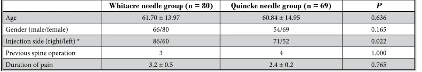
A Quincke needle was used in 69 patients (123 cases) and a Whitacre needle was used in 80 patients (146 cases). There were no significant differences in age, gender, previous spine surgery, and duration of pain except in the injection side (right or left) (Table 1).
The overall incidence of intravascular injection was 10.4% (28/269). We analyzed the overall incidence of in- travascular injection according to the 2 different needle types. The incidence of intravascular injection with the Whitacre needle was 5.4% (8/146), and the incidence of intravascular injection with the Quincke needle was 16.2% (20/123). The incidence of intravascular injection
Table 1. Patient demographic characteristics.
Whitacre needle group (n = 80) Quincke needle group (n = 69) P
Age 61.70 ± 13.97 60.84 ± 14.95 0.636
Gender (male/female) 66/80 54/69 0.165
Injection side (right/left) * 86/60 71/52 0.022
Previous spine operation 3 4 1.000
Duration of pain 3.2 ± 0.5 2.4 ± 0.2 0.765
Values are mean ± SD or number of patients.
*P = 0.022
was 3 times higher in the Quincke needle group com- pared to that in the Whitacre needle group. This differ- ence was statistically significant (P = 0.023). The L4-5 level was the most frequently performed TFESI level. There was no significant difference regarding the incidence of intravascular injection at each level (Table 2).
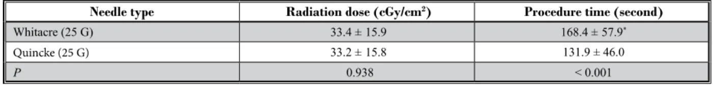
Total procedure time required to complete the TFESI in the Whitacre needle group and the Quincke needle group was 168.4 ± 57.9 (seconds) and 131.9 ± 46.0 (seconds), respectively (P < 0.001). The amount of radiation required to complete the TFESI in the Whita- cre needle group and the Quincke needle group was 33.4 ± 15.9 (cGy/cm2) and 33.2 ± 15.8 (cGy/cm2), respec- tively (P = 0.938) (Table 3).
During the TFESIs, several minor complications oc- curred, including dizziness (4/269), transient headache (2/269), post-injection back soreness (9/269), increased pain sensation (5/269), and transient leg weakness (3/269). No major complications such as paraplegia, quadriparesis, epidural hematoma, or abscess were found.
d
iscussionThe major finding of this study is that the Whitacre needle could significantly reduce the incidence of in- travascular injection compared to the Quincke needle.
Moreover, the incidence of intravascular injection using the Quincke needle was 3 times higher than that using the Whitacre needle.
Shin et al (14) also demonstrated the lower inci- dence rate of intravascular injection using the Whitacre needle compared to that using the Quincke needle.
However, the difference of intravascular incidence be-
tween the 2 needle types in their study was less than 2 times. Several reasons might have contributed to the difference in incidence of intravascular injection be- tween the result of Shin et al (14) and our results. In this study, we used DSA to confirm the intravascular injec- tion in all cases, whereas Shin et al (14) used DSA only when the cases were unclear. In addition, the injected TFESI level (S1) was different from our study and 12 pain physicians participated in the study to perform the TFESIs. Shin et al (14) focused on identifying risk factors contributing to intravascular injection rather than the incidence rate. Therefore, some numerical differences might exist between the 2 studies.
In this study, DSA was used in all cases of TFESI to confirm the intravascular injection. Our previous study demonstrated that the DSA method was superior to real-time fluoroscopy for detecting intravascular in- jection (20). In addition, to minimize the variability in placement of needles and to provide more homog- enous conditions, only one board certified physician performed all cases of TFESI. Therefore, our results about the difference in incidence rate of intravascular injection between the 2 needle types could be more precise and reliable.
Although DSA has the disadvantage of more ra- diation exposure to the patient and medical staff with high cost of up-graded medical equipment, subtracted DSA images could provide more distinguishing images between epidurograms and intravascular injections (20,21).
Differentiating the intravascular injection as ar- terial vs. venous is important because intra-arterial particulate steroid injection is one of the suggested
Table 2. Intravascular injection rate during lumbar transforaminal epidural injection.
Needle type Spinal level
Total (%)
L2-3 L3-4 L4-5 L5-S1
Whitacre (25 G) 1/10 (0.1) 2/20 (0.1) 4/69 (5.7) 1/47 (2.1) 8/146 (5.4) *
Quincke (25 G) 0/5 (0) 1/16 (6.2) 16/68 (23.5) 3/34 (8.8) 20/123 (16.2)
Total 1/15 (6.6) 3/36 (8.3) 20/137 (14.5) 4/81 (4.9)
Data are number of intravascular injections of all transforaminal epidural injections; numbers in parentheses are percentages. The difference between needle type (Whitacre vs. Quincke) was statistically significant. *P = 0.023
Table 3. Radiation dose and total procedure time required for lumbar transforaminal epidural injection.
Needle type Radiation dose (cGy/cm2) Procedure time (second)
Whitacre (25 G) 33.4 ± 15.9 168.4 ± 57.9*
Quincke (25 G) 33.2 ± 15.8 131.9 ± 46.0
P 0.938 < 0.001
Values are mean ± SD. *P < 0.001
mechanisms of serious neurological consequence (11).
However, it is difficult to differentiate the intravascular injections as arterial vs. venous clearly in most of the cases. Although arterial injection can result in more serious complications than that of venous injection, all intravascular injections should be avoided if possible.
Considering the serious morbidity of inadvertent intra-arteral injection of particulate corticosteroids, al- though the reported incidence is infrequent, using the Whitacre needle could be one method to minimize such a disastrous consequence. The Whitacre needle may be more valuable for TFESIs at the sacral and cervical level, at which the incidence of intravascular injection has been reported to be high (14,22,23).
Originally, the Whitacre needle was not intended to minimize the intravascular injection rate during TFESI but to avoid the occurrence of postdural puncture headache (17-19). The Whitacre needle has a tapering pencil-point tip and a side hole rather than the con- ventional sharp bevel and end hole. Similarly, blunt tip needles have almost the same structure but with a more rounded tip (16). Although the Whitacre needle tip is sharper than that of a blunt needle, its intravascu- lar injection incidence is lower than that of the Quincke needle in lumbar TFESI. Shin et al (14) suggested several mechanisms on how the Whitacre needle could mini- mize the intravascular entry. The tapered pencil-point tip of the Whitacre needle may slide by the vessel without injury. Even if a vessel was punctured with the Whitacre needle, the intravascular contrast dye appear- ance might not appear because the needle opening is on the side (14). Although further study is necessary to prove these hypothesis, it seems evident that using a less traumatic needle type can reduce the intravascular injection rate. As a result, using such a needle type can improve overall patient safety.
In spite of such a benefit of the Whitacre needle, it did not gain widespread use in spine intervention due to its inherent disadvantages. The steering abil- ity of the needle gives the technical easiness to the
physician while approaching the final target structure during spine injection. However, the Whitacre needle lacks this steering ability and requires more force to puncture the subcutaneous tissues such as muscles and ligaments (16). This might create technical difficulties with increased procedure time and patient discomfort.
To confirm these technical difficulties of the Whitacre needle, we measured the total procedure time with the amount of radiation exposure during TFESI. In cases of the Whitacre needle, approximately an extra of 30 seconds of time was required compared to that required when using a Quincke needle to complete the TFESI. However, such difference in time at less than one minute is not likely to significantly impact technical dif- ficulties or increase patient discomfort. In addition, the difference in the amount of radiation exposure during TFESI was minimal between the 2 needle groups.
There are several limitations of this study. First, the physician who performed the TFESI was not blinded to the type of needle for detecting intravascular injection.
However, this confirmation bias could be minimized by using 2 physicians who were not performing TFESI.
Second, this study was focused on lumbar TFESIs, however, most TFESIs are performed at the L4-5 and L5-S1 level. Third, we could not conclude that Whitacre needles could reduce the incidence of intra-arterial in- jection which has more clinical significance than that of intra-venous injection.
This study suggests that the establishment of a safe method for performing TFESI is an important issue. Us- ing a Whitacre needle could be one of the methods.
c
onclusionThe Whitacre needle has the benefit of reducing the incidence of intravascular injection with minimal differences in technical difficulties and the amount of radiation exposure during lumbar TFESI. Our findings suggest using Whitacre needles during TFESI is safer with minimal technical difficulties compared to using Quincke needles.
R
efeRences1. Quraishi NA. Transforaminal in- jection of corticosteroids for lum- bar radiculopathy: systematic review and meta-analysis. Eur Spine J 2012;
21:214-219.
2. Gupta R, Singh S, Kaur S, Singh K, Aujla K. Correlation between epidurographic contrast flow patterns and clinical rf-
fectiveness in chronic lumbar discogenic radicular pain treated with epidural ste- roid injections via different approaches.
Korean J Pain 2014; 27:353-359.
3. McGrath JM, Schaefer MP, Malkamaki DM. Incidence and characteristics of complications from epidural steroid in- jections. Pain Med 2011; 12:726-731.
4. Karaman H, Kavak GO, Tufek A, Yldrm ZB. The complications of transforaminal lumbar epidural steroid injections. Spine (Phila Pa 1976) 2011; 36:E819-E824.
5. Lee HK, Choi EJ, Lee PB, Nahm FS. Ana- phylactic shock caused by the epidural- ly-administered hyalurinidase. Korean J Pain 2011; 24:221-225.
6. Houten JK, Errico TJ. Paraplegia after lumbosacral nerve root block: Report of three cases. Spine J 2002; 2:70-75.
7. Bose B. Quadriparesis following cervical epidural steroid injections: Case report and review of the literature. Spine J 2005;
5:558-563.
8. Ludwig MA, Burns SP. Spinal cord in- farction following cervical transforami- nal epidural injection: A case report.
Spine (Phila Pa 1976) 2005; 30:E266-E268.
9. Kennedy DJ, Dreyfuss P, Aprill CN, Bog- duk N. Paraplegia following image- guided transforaminal lumbar spine epidural steroid injection: Two case re- ports. Pain Med 2009; 10:1389-1394.
10. Ay B, Gercek A, Konya D, Ozgen S. Spi- nal abscess after epidural anesthesia:
Need for more vigilance and better patient advice. J Neurosurg Anesthesiol 2004; 16:184-185.
11. Scanlon GC, Moeller-Bertram T, Ro- manowsky SM, Wallace MS. Cervical transforaminal epidural steroid injec- tions: More dangerous than we think?
Spine (Phila Pa 1976) 2007; 32:1249-1256.
12. Dawley JD, Moeller-Bertram T, Wallace MS, Patel PM. Intra-arterial injection in the rat brain: Evaluation of steroids used for transforaminal epidurals. Spine (Phila Pa 1976) 2009; 34:1638-1643.
13. Ozcan U, Sahin S, Gurbet A, Turker G, Ozgur M, Celebi S. Comparison of blunt and sharp needles for transforaminal epidural steroid injections. Agri 2012;
24:85-89.
14. Shin J, Kim YC, Lee SC, Kim JH. A com- parison of Quincke and Whitacre nee- dles with respect to risk of intravascu- lar uptake in S1 transforaminal epidural steroid injections: A randomized trial of 1376 cases. Anesthesia & Analgesia 2013;
117:1241-1247.
15. Smuck M, Yu AJ, Tang CT, Zemper E.
Influence of needle type on the inci- dence of intravascular injection during transforaminal epidural injections: A comparison of short-bevel and long- bevel needles. The Spine Journal 2010;
10:367-371.
16. Smuck M, Leung D. Inadvertent injec- tion of a cervical radicular artery using an atraumatic pencil-point needle. Spine (Phila Pa 1976) 2011; 36:E220-223.
17. Vallejo MC, Mandell GL, Sabo DP, Ra- manathan S. Postdural puncture head- ache: A randomized comparison of five spinal needles in obstetric patients.
Anesth Analg 2000; 91:916-920.
18. Luostarinen L, Heinonen T, Luostarin- en M, Salmivaara A. Diagnostic lumbar puncture. Comparative study between
22-gauge pencil point and sharp bevel needle. J Headache Pain 2005; 6:400-404.
19. Hatfield MK, Handrich SJ, Willis JA, Beres RA, Zaleski GX. Blood patch rates after lumbar puncture with Whitacre versus Quincke 22- and 20-gauge spi- nal needles. AJR Am J Roentgenol 2008;
190:1686-1689.
20. Hong JH, Huh B, Shin HH. Comparison between digital subtraction angiogra- phy and real-time fluoroscopy to detect intravascular injection during lumbar transforaminal epidural injections. Reg Anesth Pain Med 2014; 39:329-332.
21. Lee MH, Yang KS, Kim YH, Jung HD, Lim SJ, Moon DE. Accuracy of live fluo- roscopy to detect intravascular injection during lumbar transforaminal epidural injections. Korean J Pain 2010; 23:18-23.
22. Nahm FS, Lee CJ, Lee SH, Kim TH, Sim WS, Cho HS, Park SY, Kim YC, Lee SC.
Risk of intravascular injection in trans- foraminal epidural injections. Anaesthe- sia 2010; 65:917-921.
23. Kim do W, Han KR, Kim C, Chae YJ.
Intravascular flow patterns in transfo- raminal epidural injections: A compara- tive study of the cervical and lumbar vertebral segments. Anesth Analg 2009;
109:233-239.