• 검색 결과가 없습니다.

Intracranial Hemorrhage in the Corpus Callosum Presenting as Callosal Disconnection Syndrome: FDG-PET and Tractography: A Case Report

N/A
N/A
Protected

Academic year: 2021

Share "Intracranial Hemorrhage in the Corpus Callosum Presenting as Callosal Disconnection Syndrome: FDG-PET and Tractography: A Case Report"

Copied!
5
0
0

로드 중.... (전체 텍스트 보기)

전체 글

(1)

Intracranial Hemorrhage in the Corpus

Callosum Presenting as Callosal Disconnection Syndrome: FDG-PET and Tractography:

A Case Report

In Hwan Kim, MD

1

, Soyoung Lee, MD, PhD

1

, Chang-Young Lee, MD

2

, Dong Gyu Lee, MD

1

Departments of

1

Rehabilitation Medicine and

2

Neurosurgery, Keimyung University School of Medicine, Daegu, Korea

We report the findings of

18

F-fluorodeoxyglocese positron emission tomography (FDG-PET) and diffusion tensor tractography (DTT) in a right-handed patient presenting with callosal disconnection syndrome, including alien hand syndrome, after an anterior communicating artery aneurysmal rupture. The 49-year-old patient had right hemiparesis and unintended movement of the right hand during action of the left hand. A brain magnetic resonance imaging revealed lesions in the upper part of the genu and body in the corpus callosum as well as hemorrhage in the inter-hemispheric fissure. We observed extensive disruption of corpus callosum fibers in the upper genu and trunk by DTT for the evaluation of inter-hemispheric connection. FDG-PET revealed severe hypometabolism in the left cerebral hemisphere, including basal ganglia and thalamus, and hypermetabolism in the right cerebral hemisphere. Based on findings of FDG-PET and DTT, the callosal disconnection syndrome presented in the patient could be the result of loss of transcallosal inhibition in the contralateral hemisphere.

Keywords Corpus callosum, Positron emission tomography, Diffusion tensor imaging

Annals of Rehabilitation Medicine

Ann Rehabil Med 2014;38(6):871-875 pISSN: 2234-0645 • eISSN: 2234-0653 http://dx.doi.org/10.5535/arm.2014.38.6.871

INTRODUCTION

Callosal fibers connecting both hemispheres allow

communication of information as well as integrative and reciprocal inhibitory influences. Injury to corpus callo- sum may disturb an individual’s emotions, motor con- trol, and decision-making. Alien hand syndrome (AHS), first described in 1972, is one of the symptoms of callosal disconnection. The characteristic symptom of AHS is a purposeful action of one hand against the patient’s inten- tions, while the ipsilateral hand could not make intended movements. Several cases of AHS due to deficits in the corpus callosum have been reported [1]. However, the mechanism of AHS after brain injury has not been fully elucidated.

Diffusion tensor tractography (DTT) derived from dif-

Received March 12, 2014; Accepted June 20, 2014 Corresponding author: Dong Gyu Lee

Department of Rehabilitation, Keimyung University School of Medicine, 56 Dalseong-ro, Jung-gu, Daegu 700-712, Korea

Tel: +82-53-250-7980, Fax: +82-53-250-7268, E-mail: [email protected] This is an open-access article distributed under the terms of the Creative Commons Attribution Non-Commercial License (http://creativecommons.

org/licenses/by-nc/3.0) which permits unrestricted noncommercial use, distribution, and reproduction in any medium, provided the original work is properly cited.

Copyright © 2014 by Korean Academy of Rehabilitation Medicine

(2)

fusion tensor imaging (DTI) is a method used to reveal structural connectivity and injury of white matter. Cor- pus callosum is composed of bundles of white matter for communication between the two hemispheres. There- fore, DTT is a useful tool to investigate structural inju- ries to the corpus callosum [2].

18

F-fluorodeoxyglocese positron emission tomography (FDG-PET) is used to ex- amine the functional state of whole brain by measuring glucose metabolism. A change of metabolism detected by FDG-PET may reveal functional activity and pathologi- cal changes after brain injury [3,4]. Herein, we report the findings of DTT and FDG-PET in a patient who presented with callosal disconnection syndrome accompanied by AHS caused by an anterior communicating artery (A- com) aneurysm rupture.

CASE REPORT

A 49-year-old woman suffered from spontaneous sub- dural arachnoid and intracranial hemorrhages in the corpus callosum genu and body, resulting from a rup- tured A-com aneurysm. One month later, after a coiling procedure of the A-com aneurysm, she was transferred to our rehabilitation department. Clinical symptoms of the patient included global aphasia, apraxia, and right hemi- paresis. Features of AHS were observed in her right hand while she demonstrated improved motor strength and performance. Her right hand often reached forward and

grasped objects within sight unintentionally. She experi- enced difficulty voluntarily releasing the grasp similar to a foreign limb in the subacute period. The coordination of the hands was insufficient to be able to do buttons on clothes. The right hand would hold the clothes against the patient’s will, so the left hand had to free right hand’s grip on clothes. After several weeks, the impulsive action disappeared. However, the inter-manual conflict and vol- untary hand release problems still remained. Her func- tional state was severely impaired after one month from onset, at which time FDG-PET was conducted. Recorded performance measures were as follows: 3 for the Modi- fied Barthel Index (MBI) score, 3 for the Berg Balance Scale (BBS), and 16 points for the National Institutes of Health Stroke Scale (NIHSS). One month after rehabili- tation, the functional state of the patient showed much improvement, from 3 to 38 for MBI, 3 to 40 for BBS, and 16 to 6 points for NIHSS. Three months later, inter-man- ual conflict and voluntary hand release had improved enough to undo buttons.

T2-weighted MRI and DTI were acquired from the pa- tient at four weeks after onset. The DTI was acquired using a sensitivity-encoding head coil on a 3T MR scanner (GE Healthcare, Milwaukee, WI, USA). A diffusion-weighted echo-planar imaging sequence was performed with the following parameters for each of the 26 noncollinear dif- fusion-sensitizing gradients: TR/TE/NEX=10000 ms/95.9 ms/2.0; slice thickness=4.0 mm; b=1000 s/mm

2

; matrix

A B C

Fig. 1. Sagittal T2-weighted brain magnetic resonance imaging (A) and axial images (B) showed hyperintensity in the

corpus callosum genu and body as the result of anterior communicating artery aneurysm rupture. (C) Diffusion tensor

tractography of the corpus callosum fibers shows extensive disruption.

(3)

128×128; FOV=250×250 mm. Fiber tracking was per- formed using DTI-Studio software (CMRM; John Hopkins Medical Institute, Baltimore, MD, USA) for reconstruct- ing corpus callosum fibers by a fraction anisotropy <0.2 and angle change >60°. A region of interest was placed on the whole corpus callosum by a sagittal color map.

Eddy current-induced image distortions were removed using affine multi-scale two-dimensional registration in the Oxford Centre for Functional Magnetic Resonance Imaging of the Brain (FMRIB) Software Library (FSL;

www.fmrib.ox.ac.uk/fsl). FDG-PET/CT was performed to assess metabolic activity in the brain. PET scans were ac- quired starting at 50 minutes after intravenous injection of 0.2 mCi/kg

18

F-FDG using an Advance PET scanner (GE Healthcare). Axial resolution of the scanner was 2.14 mm full width at half maximum. The PET images were recon- structed using a Hanning filter (cutoff frequency=10.9 mm) and displayed in a 128×128 matrix with a slice thickness of 4.0 mm. Spatial preprocessing and statistical analysis were performed using the Statistical Parametric Mapping (SPM) method to compare the patient to age- matched control subjects using an unpaired t-test. SPM generates an SPM statistic for the entire brain image which was then transformed to a normal distribution.

Regions were considered significant at an uncorrected level of p<0.001.

DTT of the corpus callosum showed bilateral extensions to both hemispheres in normal subjects. However, the DTT of the patient revealed extensive disruptions at the

upper portion of the genu and whole body area (Fig. 1).

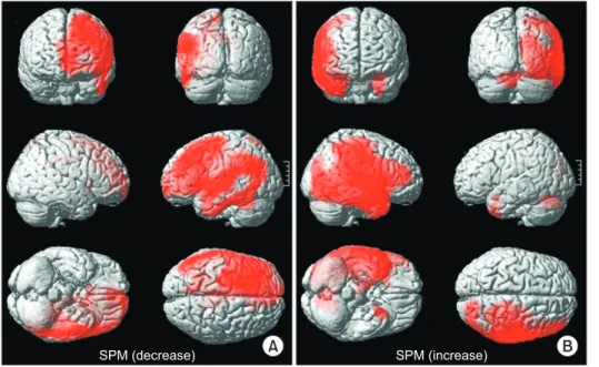
Compared to healthy control subjects (n=18; mean age, 49 years; range, 44–55 years; 8 men), we found abnormal results in the FDG-PET/CT showing reduced intensity in the left hemisphere, including the basal ganglia and thalamus but excluding the occipital lobe (Fig. 2). More- over, metabolism in the right hemisphere was extensively increased. According to the findings of tractography and FDG-PET/CT, disconnection of both hemispheres at the corpus callosum led to metabolic changes in the brain and manifestation of AHS.

DISCUSSION

We report the findings of DTT and FDG-PET/CT for a patient with callosal disconnection syndrome with AHS due to a ruptured A-com aneurysm. MRI scans revealed subdural arachnoid and intracranial hemorrhages in the corpus callosum genu and body. Clinical symptoms included right hemiparesis, right somatosensory deficit, aphasia, gait disturbance with a wide base, apraxia, and AHS. DTT of the corpus callosum showed extensive dis- ruption. FDG-PET/CT showed hypometabolism in the left hemisphere with the exception of the occipital lobe.

Appropriate movement requires coordination of the posterior parietal lobe and supplementary motor area, transcallosal inhibitory influence, and so on [5]. Injury to the corpus callosum could lead to dysfunction of the communication in these brain territories, resulting in

A B

SPM (decrease) SPM (increase)

Fig. 2. Three-dimensional render- ing, showing the spatial distri- bution of significant metabolic changes by Statistical Parametric Mapping (SPM) (p<0.001). (A)

18

F-fluorodeoxyglocese positron emission tomography (FDG-PET) scans reveal diffuse hypometabo- lism in the left hemisphere with the exception of the occipital lobe.

(B) Metabolism of the right hemi-

sphere is generally increased.

(4)

various clinical symptoms associated with callosal dis- connection syndrome. AHS, one of these symptoms, is a state of autonomy causing patient lose the ability to initi- ate intended movement. AHS has two types, frontal and callosal. Pure callosal AHS occurs with lesion restricted to the middle trunk of the corpus callosum. The primary symptom of callosal AHS is inter-manual conflict and difficulties in initiating desired actions. Frontal AHS is caused by lesions in the callosal and frontal cortex with distinguishable clinical symptoms, such as impulsive reaching and grasping behaviors of the contralesional hand. The patient in this study exhibited unwilling un- intentional movement patterns and inter-manual con- flict with difficulty in voluntary hand release during the subacute period. Subsequently, inter-manual conflict remained for three months after onset. Based on these symptoms, it can be concluded that the patient presented frontal and callosal AHS during the subacute period.

Moreover, clinical symptoms of callosal AHS remained for three months.

For setting goals and establishing a treatment plan for stroke rehabilitation, it is very important to determine the location and extent of brain injury. Although MRI is a very sensitive and accurate method for the detection of brain injuries, it is sometimes difficult to determine the degree of injury due to various factors, such as perile- sional edema, volume effect of the stroke, and penumbra lesion. DTT is a reconstruction of tracts of diffusion ten- sor images according to fraction anisotropy in white mat- ter tracts. The corpus callosum is composed of bundles of white matter [6,7]. There have been two cases showing correlation between clinical symptoms and disruption of corpus callosum fibers passing through the corpus callo- sum body using DTT [8]. Our case also presented severe disruption of fibers in the body of corpus callosum.

The patient presented with hypometabolism in the left hemisphere with the exception of the occipital lobe. This was detected by FDG-PET/CT conducted one month after onset. FDG-PET/CT revealed that hemorrhage of the corpus callosum produced degradation and hypo- metabolism of the left hemisphere in the left frontal and parietal lobes. This injury produced right hemiparesis, aphasia, and apraxia of right upper extremities. Remote metabolic effects in PET/CT allowed for the identifica- tion of functional and structural neural connectivity.

Each portion of the corpus callosum connects the cere-

bral cortex between the two halves of the brain, allow- ing communication of information, as well as reciprocal inhibitory influence in both sides of the brain. Therefore, we hypothesized that the loss of transcallosal inhibition influenced by injury to the corpus callosum as well as the mass effect of the hemorrhage was responsible for the hypometabolism of the left hemisphere, which subse- quently led to symptom of AHS in the right hand and dif- ficulty in bimanual activities.

The right hemisphere, including frontal, parietal, and temporal lobes, presented hypermetabolism in FDG- PET/CT when compared to normal control subjects. To the best of our knowledge, this study is the first report concerning contralateral cerebral hypermetabolism of static PET/CT in a stroke patient. Since the brain can- not store energy substrates, the majority of glucose in the brain is consumed for signaling. Hypermetabolism of the resting brain on the

18

F-FDG/PET indicated that cell activity and energy demand of the right hemisphere continuously increased, even at a resting state. Although the mechanism of increased metabolism has not been fully elucidated, we presume that recovery mechanisms (caused by severe injury of left hemisphere) and inter- ruption of transcallosal inhibition (caused by the corpus callosum hemorrhage) lead to hypermetabolism in the right hemisphere. Rehme et al. [9] reported that fMRI findings in stroke patients with great corticospinal tract damage and more marked motor impairments showed enhanced contralesional motor cortex activity as a result of reduced inhibition of the contralesional hemisphere.

Furthermore, some stroke patients use the ipsilateral motor pathway as a recovery mechanism. Repetitive transcranial magnetic stimulation (TMS) at the contral- esional dorsal premotor cortex and primary motor cortex resulted in deterioration of motor performance for some stroke patients. Many other studies reported a relation- ship between functional recovery and the ipsilateral motor pathway of the affected extremities using fMRI, TMS, and DTI [10]. However, little studies concerning hy- permetabolism of resting PET/CT after stroke have been conducted. Therefore, this case report has a limitation in discussing hypermetabolism in PET/CT.

In conclusion, we suggest that callosal disconnection

syndrome is the result of the loss of transcallosal inhibi-

tion and metabolic changes in both hemispheres based

on findings of FDG-PET and DTT. Although studies on

(5)

hypermetabolism in PET/CT is not sufficient, this case showed the possibility that metabolic changes using FDG PET/CT could give useful information to evaluate brain injury and recovery mechanism after stroke. Further study using FDG-PET in stroke patients is needed to elu- cidate brain injury and recovery mechanisms.

CONFLICT OF INTEREST

No potential conflict of interest relevant to this article was reported.

REFERENCES

1. Suwanwela NC, Leelacheavasit N. Isolated corpus ca llosal infarction secondary to pericallosal artery disease presenting as alien hand syndrome. J Neurol Neurosurg Psychiatry 2002;72:533-6.

2. Hong JH, Choi BY, Chang CH, Kim SH, Jung YJ, Byun WM, et al. Injuries of the cingulum and fornix after rupture of an anterior communicating artery aneu- rysm: a diffusion tensor tractography study. Neuro- surgery 2012;70:819-23.

3. Kasanuki K, Iseki E, Fujishiro H, Yamamoto R, Higashi S, Minegishi M, et al. Neuropathological investiga- tion of the hypometabolic regions on positron emis- sion tomography with [18F] fluorodeoxyglucose in patients with dementia with Lewy bodies. J Neurol Sci 2012;314:111-9.

4. Kim YK, Lee DS, Lee SK, Kim SK, Chung CK, Chang KH, et al. Differential features of metabolic abnor- malities between medial and lateral temporal lobe epilepsy: quantitative analysis of (18)F-FDG PET us- ing SPM. J Nucl Med 2003;44:1006-12.

5. Wolpe N, Moore JW, Rae CL, Rittman T, Altena E, Hag- gard P, et al. The medial frontal-prefrontal network for altered awareness and control of action in cortico- basal syndrome. Brain 2014;137(Pt 1):208-20.

6. Maeshima S, Osawa A, Nishio D, Hirano Y, Kigawa H, Takeda H. Diffusion tensor MR imaging of the pyramidal tract can predict the need for orthosis in hemiplegic patients with hemorrhagic stroke. Neurol Sci 2013;34:1765-70.

7. Jang SH, Ahn SH, Sakong J, Byun WM, Choi BY, Chang CH, et al. Comparison of TMS and DTT for predicting motor outcome in intracerebral hemorrhage. J Neurol Sci 2010;290:107-11.

8. Chang MC, Yeo SS, Jang SH. Callosal disconnection syndrome in a patient with corpus callosum hemor- rhage: a diffusion tensor tractography study. Arch Neurol 2012;69:1374-5.

9. Rehme AK, Fink GR, von Cramon DY, Grefkes C. The role of the contralesional motor cortex for motor re- covery in the early days after stroke assessed with lon- gitudinal FMRI. Cereb Cortex 2011;21:756-68.

10. Jang SH. A review of the ipsilateral motor pathway as

a recovery mechanism in patients with stroke. Neuro-

Rehabilitation 2009;24:315-20.

수치

Fig. 1. Sagittal T2-weighted brain magnetic resonance imaging (A) and axial images (B) showed hyperintensity in the  corpus callosum genu and body as the result of anterior communicating artery aneurysm rupture
Fig. 2. Three-dimensional render- render-ing,  showing  the  spatial   distri-bution of significant metabolic  changes by Statistical Parametric  Mapping  (SPM)  (p&lt;0.001)

참조

관련 문서

Especially, it was theoretically predicted that x-axis and y-axis polarization states can be obtained with a right-handed TR film and a left-handed

 Bent posture, and as a fast check of the measurement major pectoral muscle case, the patient lying immediately behind the head comes along,

In Annex I countries, national communication should submit the report including emission inventories, trends and policies of Greenhouse Gases (GHGs) to UNFCCC. GHG

Just as we achieved good control with a joint-based controller that was based on a linearizing and decoupling model of the arm, we can do the same for the Cartesian case.

Serum uric acid levels and risk for vascular disease in patients with metabolic syndrome... Prevalence if the metabolic syndrome in a Turkish

F18-FDG uptake in thyroid from PET for evaluation in cancer patients: high prevalence of malignancy in thyroid PET incidentaloma.. Thyroid incidentalomas:

a prospective analysis of 50 cases. Preoperative staging of non-small-cell lung cancer with positron-emission tomography. Staging of non- small-cell lung cancer

레오나르도는 예수의 얼굴에 사랑과 자애, 슬픔과 고통 등 여러 가지 요소가 담긴 완벽한 얼굴이어야 한다고 생각했으므로 예 수의 얼굴을 그릴 수가 없었다. 오랜