약학희지 제36권 제1호 66^72(1992) Yakhak Hoeji Vol. 36, No. 1
단백질 메틸화효소류 및 S-아데노실-L-에처오닌 연결효소의 활성도에 미처는 사염화탄소-유발 간득성의 영향
남궁 석민 • 유 태무 • 홍성렬* • 이 향 우*
성균관대학교 약학대학, 농과대학 유전공학과*
(Received January 16, 1992)
Effects of Carbon Tetrachloride-induced Hepatotoxicity on the Activities of Protein Methylases and SAM-Synthetase in Rat Liver
Suck M in Namkoong, Tae Moo Yoo, Sung Youl Hong* and Hyang Woo Lee**
Department of Biochemistry, College of Pharmacy and Department of Genetic Enginering*
College of Agriculture, Sung Kyun Kwan University, Suwon 440-746, Korea
Abstract —In order to test relationships between hepatotoxicity and transmethylation, activities of protein methylases and SAM (S-adenosyl-L-methionine)-synthetase were examined in liver tis
sues of rats treated with CCL*. Also the concentrations of SAM and SAH were measured by HPLC in rat liver. The results are as follows. (1). Activities of protein methylases were not significantly changed in 24 hours after CCU treatment. However, in 48 hours, activities of protein methylases were significantly increased in comparison with that of control. (2). Activity of SAM-synthetase was increased steadily in the time course after CCU treatment. (3). S-adenosyl-L-methionine conce
ntration of liver tissues in CCU-treated group was elevated in 24 hours, and then declined thereaf
ter. But the SAH concentration was slightly decreased in the time course after CCU treatment.
These results indicate that SAM was very actively used in transmethylation reactions of CCU damaged rat liver, suggesting the strong relationships between hepatotoxicity and transmethylation phenomena.
Keywords □ Protein methylase, SAM-synthetase, CCU, SAM, SAH, hepatotoxicity
Protein methylation은 posttranslational modifica
tion 반응의 하나로서/^ 지난 30여년간 생화학적 견 지에서의 많은 연구를 통해 methylation반응으로 생긴 단백질의 구조와 기능변화2 생체내 여러현상에 꺼 치는 영향들이 조금씩 밝혀겨 왔다. 이러한 protein methylation 현상은 procaryote에서 eucaryote 까지 다 앙한 생물체에서 관찰되었으며 methyl donor로써 S- adenosyl-L-methionine 을 이용하는 단백 특이성, group 특이성 methyltransferase와 methylation양상 에 따라 protein methylase I, II, III, IV 4가지로
않본 논문에 관한 문의는 이 저자에게로.
분류할 수 있다."^ 이들은 기질 단백의 특정 아미노산 잔기에 대한 높은 특이성을 가지며, 이러한 protein methylation이 관계되는 생화학적 현상에는 현재까지 bacterial chemotaxis,**■타 exocytosis,® ®^ gene regula- tion,으'고*^^ muscle contraction/*^ electron transport/요■표크^ 그리고 carnitine 생합성바^^ 등에 관계된다고 알려 겨왔다. 특히 protein carboxylmethyltransferase (protein methylase II) 는 bacterial chemotaxis와 관 련됨이 발혀진 이래 손상된 protein 의 repair mecha- nism에도 관여하며,배 ^ 최근에는 mammalian ras oncogene product효®^와 brain G-protcin외 y subunit외 C-terminal cysteine residue외 a-carboxyl group이
66
단백질 메틸화효소류 및 S-아데노설-L-메치오닌 연결효소외 활성도에 미처는 사염화란소-유발 간득성의 영향_ ^
methylation되어 있옴이^^이 보고되고 었다.
Methyiation반응에서 methyl donor로 사용되는 S- adenosylmethionine(SAM)은 포유동물외 거외 모든 조직에 분포되어 었으며으*^ protein transmethylation 이외에:£ polyamine biosynthesis, ethylene biosyn
thesis, transsulfuration의 조절,퍼 differentiation모잇에 도 관여하는 것으로 알려 겨 왔다. 또한 SAM-synthe- tase [ATP:L-methionine S-adenosyltransferase, EC 2.5.1.6] 는 L-methionine과 ATP로부터 SAM외 생합 성을 촉매하는 효소로써 분자량파 value에 따라 a, Y 세가지외 동종효소형태로 존재24■25)한다고 밝 혀 졌다.
현재까지 간장에서 transmethylation의 생화학적 작용에 관한 연구보고로는 등이 hepatoma에서 protein methylase I activity가 hepatoma의 성장속 도와 일처함을 보고하였으며 Duerre"가는 흰쥐 간장의 protein methylase II 활성도가 흰쥐의 성장과 일치 하며 이 효소의 세포내 농도는 흰쥐외 연령에 외존 적이라고 보고하였다. 또한 SAM-synthetase의 경우 Tsukada혜 등은 carcinogenesis도중 y 체 동종효소는 증가히는 반면, a 와 P 체 동중 효소는 감소하며, 한 편
인간의 간경화증에서는 이 효소 활성이 50%정도 감소^^
하는데, 이는 a 체 동종 효소량이 크게 감소한 때문 이며 이에 반해 세포내 SAM의 농도는 이 대사산물외 세포내 이용감소로 인해 보상된다는 연구결과가 있다.
Carbon tetrachiorideCCCU)는 실험적 간장해를 일 으키는 약물괴로서 체내외 mixed function (cytoch
rome P-450) oxidase system에 외해 생긴 CCla'free radical이 간 microsome의 막 단백 thiol기와 결함하 여 막의 지질파산화 반웅을 촉 진 하 며 간 에 서 의 단 백함성 억제, 간 glycogen량의 감소, 혈중으로의 GOT, GPT이탈을 일으키고, 조직학적으로는 간세포외 괴사 및 섬유화동을 일으키는 것으로 알려져있다.
이에 본 실험에서는 이와같은 연구보고를 바탕으 로하여 정상 흰쥐의 간장에서 연령별로 SAM파 SAM외 대사물인 SAH(S-adenosylhomocysteine)외 농도분포를 살펴본 다움, CC14를 사용하여 실험적으로 흰쥐에 hepatotoxicity를 유발시킨 후 protein methy- lases 및 SAM-synthetase활성도 번화를 검색하고*
동시에 간조직내의 SAM 과 SAH 의 농도번화를 HPLC로 분석하여 간기능 번화시 protein transmeth-
ylation에 미처는 영향에 관하여 고찰하고자 하였다.
설험 방법
설험동울
체중 200~250g의 건강한 Sprague-Dawley계통외 웅성 흰쥐를 일정기간 cage내에서 사육하여 실험실 환경에 적응시킨 후 사용하였다.
시약
S-Adenosyl-L-[methyl-'^C] methionine (specific activity: 56 mCi/mmole), L-[methyl-^**C] methionine (specific activity; 56 mCi/mmole) (Amersham), Hi- stone type II-As, 2,5-diphenyloxazole (PPO), 1,4-bis [2-(5-phenyloxazolyD ] benzene (POPOP), 1-octa- nesulfonic acid sodium salt (Sigma), P-81 ion ex
change chromatography paper (Whatman), carbon tetrachloride (Duk San), GOT and GPT kit (Asan), 기타외 시약은 시관 특급시약을 사용하였다.
기기
Liquid Scintillation Counter (1211 Rackbeta, LKB, U.S.A.), Spectrophotometer (SP-240, Shimazu, Japan), High Speed Centrifuge (KR-20000T, Ku
bota, Japan), HPLC (501 Solvent Delivery System, 486 Tunable Absorbance Detector, U6K universal Injector, Waters, U.S.A) 를 각각 사용하였다.
흰쥐 간장 조적내 연령별 SAM 과 SAH의 농도 흰쥐 간장내 연령별 농도 분포 실험에서는 new- born, 1 week, 2 weeks, 4 weeks되는 흰쥐 간장을 적출한 후 각각 HPLC sample처리 과정을 거친후 HPLC분석을 하였다,
CCI4 투여로 인한 간득성 유발
대 조군은 corn oil을 5.0 m//kg썩 1희 복강투여 하 였으며 CCU투여군은 1.0 m//kg(in corn oil)썩 대조 군과 동일한 방범으로 투여하여 간득성을 유발시켰다.
약물 투여후 간조직의 처리
CCU투여후 24, 48시간 경과시 흰쥐를 단두시켜 실 혈 시 킨 후 간조직 을 적 출하여 냉 각된 0.25 M suc
rose 용액으로 깨끗이 씻고 수분을 제거하였다. 간조 직 일부를 냉 각된 0.25 M sucrose용액 9 volunie(w/v) 으로하여 teflon glass homogenizer로 균질화하고 Ci- nti 등의 방범^ M) 따라 subcellular fractionation을 한후 whole homogenate와 cytosol fraction을 protein
68 남궁석민 • 유 태 무 • 홍성렬 * 이향우
methylases와 SAM-synthetase활성도 측정을 위한 효소원으로 사용하였다.
SGO T 및 SGPT 활성도 측정
혈청중 SGOT 및 SGPT외 활성도는 Reitman-Fra- nkel외 방법체에 외하여 측정하였으며 GOT, GPT kit를 사용하였다.
Cytochrome P-450 함랑측정
Microsoe fraction중의 cytochrome P-450 함량측 정은 Omura와 Sato의 방범^^을 참조하고 Matsubara 등의 번법^^Ml 준하여 측정하였다.
효소의 활성도 측정
1) Protein methylases 황성도 측정一Paik외 방범효^
에 따라 기절로써 histone type II-As, methyl do- nor로써 [methyl-나C]SAM을 사용하여 protein me- thylase I, II외 효소활성도를 측정하였다.
2) SAM-synthetase 활성도 측정一Geller등외 방법뢰 에 의하여 측정하였다, 50 mM Tris-HCKpH 7.3), 20 mM MgCb, 25 mM KCl, 0.5 mM EDTA, 2.5 mM ATP, 0.02 mM BSA, 0.02 mM [methyl-^"*C] methio
nine, 그러고 효소원을 함유하는 total volume 0 . 1
m/의 incubation mixture를 37t 에서 10분간 incuba- tion 한 후 5 mM methione을 함유하는 2 M HCIO4
용액 20m
/
를 가하고 빙 욕상에 서 냉 각하여 반응을 정 지시컨 후 1,000 X 당에서 10분간 원심분러하여 침 전을 제 거 하였다. 상등액 1 0 0 |j/를 cellulose phosphate paper disc(직경 2.5 cm) 에 점적하고 5mIVl phosphate buffer(pH 7.0)로 일정하게 세척하고 건조 시킨 후 disc를 1 0m/의 scintillation cocktail에 넣어 radioactivity를 측정하여 SAM-synthetase의 specific activity를 산출하였다.
단백질 정랑
BSA를 standard protein으로 하여 Lowry 등의 방법37Ml 의하여 측정하였다.
SAM 및 SAH 농도측정
1) H PLC sample 처러■간조직 일부를 세절한 후 0.4 M HCIO4 용액 2 volume(w/v)으로 하여 teflon glass homogenizer로 균질화한 후 9,000 X g, 4 t 에 서 10분간 원 심분리 하였다. 상동액 일 정 량을 0.45 |im fil
ter unit를 사용하여 filtration 한 후 HPLC 정량에 사용하였다.
2) H PLC 분석 조건一Column은 |j-Bondapak C18
(3.9 X 300 mm, i.d.)를 사용하였으며 이동상으로는
n m o le s /g tis s u e
A ga(w e eks)
- B - SAM - SAH
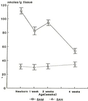
Fig. I —Effect of age on the concentrations of SAM and SAH in rat liver.
5 mM 1-octanesulfonic acid를 함유한 20% methanol (pH 4.0) 을 사용하였고 flow rate는 1.7 pre- ssure는 약 2800 psi 이며 detector는 UV absorbance (254 nm) detector를 사용하였다. aufs는 0.이이 었다.
injection volume은 SAM을 정 량하기 위 하여 50 injection 하였고 SAH를 정 량하기 위하여 20 injec
tion 하였다. SAM 및 SAH의 농도는 SAM파 SAH 에 대한 P근3k area의 관계로 검량선을 작성하여 구하였다. SAM과 SAH peak의 확인은 일정량의 sta- ndard를 sample에 혼합한 후 injection하여 peak area번화를 비교하여 확인하였으며, SAM peak의 경 우에는 [methyl-구*^] SAM을 injection하여 각 peak 위처에서 유출되어 나오는 분획을 scintillation coun- ter로 측정하여 확인하였다.
실험결과
환쥐 간장 조직내 연령별 SA M 및 SAH 농도 변화 Fig, 1에서 보는 바와 같이 SAM 농도는 newborn (111.16) 에서 가장 높았으며, 1주째에 약간 감소(82.
02) 하고 2주째에 다시 증가(94.00) 한 후 4주째에는 상당히 낮은농도 (53.99) 로 떨어졌다. 이에 반해 SAH외 농도는 흰쥐외연령에 상관없이 거의 일정한
J. Pharm. Soc. Korea
단백질 메틸화효소류 및 S-아네노실-L-메처-수닌 연결효소의 활성도에 미처는 사염화탄소-유발 간득성의 영향 69
14 00
1 2 0 0
10 00
8 0 0
6 0 0
4 0 0
2 0 0
SGOT and SGPT (K arm en/m l)
C y to c h ro m e P -4 5 0 (n m o le s/m g p rotein)
p m o te s S A M /m ln /m g p r o te in
24 hrs. 4 8 hrs.
h rs. a fte r tre a tm e n t
H I SQO T<eo«tieU^헬 S G 0 T ( i r * i i * t f ) B 9 S Q P T (co n lr« l)
HD S Q P T ( u * . u « ) H I C y t .p- 4 5 0 i i i C y t . P ^ 4 6 0 ( ir « .u d ) (oenlreO
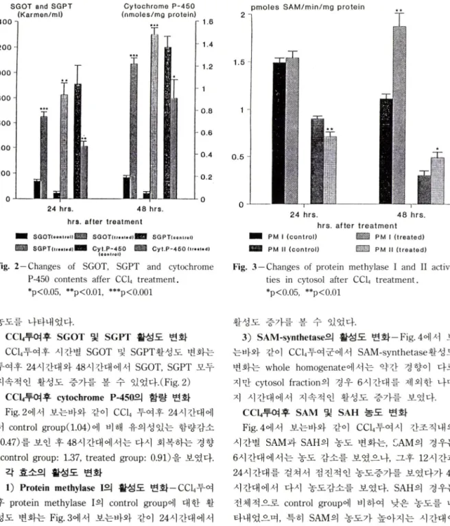
Fig. 2—Changes of SGOT, SGPT and cytochrome P-450 contents affer CCU treatment.
♦p<0.05, *꽤■p<0.01, ***p<0.001
0 .5
2 4 h rs . 4 8 h rs .
h rs . a ft e r tr e a tm e n t P M I ( c o n t r o l ) P M I ( t r e a t e d ) P M II ( c o n t r o l ) i H P M II ( t r e a t e d )
Fig. 3 —Changes of protein methylase I and II activi
ties in cytosol after CCU treatment.
*p<0.05, **p<0.01
농도를 나타내었다.
e c u 투여후 SGOT 및 SGPT 활성도 번화 e cu투여후 시간별 SGOT 및 SGPT활성도 번화는 투여후 24시간대와 48시간대에서 SGOT, SGPT 모두 지속적 인 활성도 증가를 볼 수 있었다.(Fig. 2)
CCI4투여후 cytochrome P-450의 함량 변화 Fig, 2에서 보는바와 같이 CCU 투여후 24시간대에 서 control group(1.04) 에 비해 유의성있는 함량감소 (0.47) 를 보인 후 48시간대에서는 다시 회복하는 경향 (control group: 1.37, treated group: 0.91) 을 보였다.
각 효소의 활성도 변화
1) Protein methylase I의 활성도 변화一CCU투여 후 protein methylase I외 control group에 대한 활 성도 번화는 Fig. 3에서 보는바와 같이 24시간대에서 는 이렇다할 활성도 변화를 보이지 않았으며, 48시 간대의 cytosol fraction에서 control group (l.ll) 에 비해 유외성 있는 활성도 증가(1.87) 를 보였다.
2) Protein methylase II의 활성도 변화一Fig.3에서 보는바와 같이 CCU투여후 protein methylase II외 활성도 번화는 24시간대의 cytosol fraction에서는 활 성도 감소를 보인 반면, 48시간대에서는 유의성있은
활성도 증가를 볼 수 있었다.
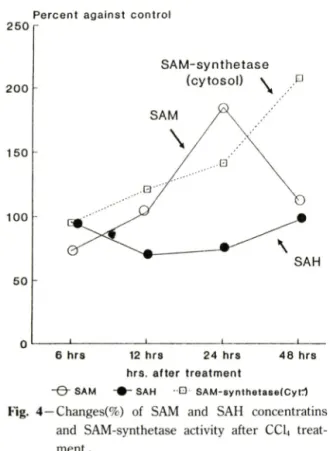
3) SAM-synthetase의 활성도 번화一Fig. 4에 서 보 는바와 같이 CCLt투여군에서 SAM-synthetase활성도 번회*^ whole homogenate에서는 약간 경향이 다르 지만 cytosol fraction의 경우 6시간대를 제외한 나머 지 시간대에서 지속적인 활성도 증가를 보였다.
e c u 투여후 SAM 및 SAH 농도 번화
Fig. 4에 서 보는바와 같이 CC14투여 시 간조직 내 의 시간별 SAM과 SAH외 농도 번화는, 외 경우는
6시간대에서는 농도 감소를 보였으나, 그후 1 2시간과 24시간대를 걸쳐서 점진적인 농도증가를 보였다가 48 시간대에서 다시 농도감소를 보였다. SAH외 경우는 전체적으로 control group에 비하여 낮은 농도를 나 타내었으며, 특히 SAM의 농도가 높아지는 시간대인 12시간과 24시간대에서 보다 낮은 농도 감소 경향을 보였다.
고 찰
Protein methytransferase는 methylation 시 SAM- synthetase 에 의해 만들어진 methyl donor 인 S-ade-
70 남궁석민 • 유 태 무 ■ 홍성렬 * 이 항우
P e rc e n t a g a in s t c o n tr o l
20 0
1 5 0
100
5 0
SAM-synthetase (cytosol)
6 h r s 12 h rs 2 4 h rs 4 8 h rs h rs . a ft e r tr e a tm e n t
" O * S A M SAH " D S A M - s y n t h e t a s e f C y t t l
Fig. 4—Changes(%) of SAM and SAH concentratins and SAM-synthetase activity after CCI4 treat
ment .
nosylmethionine(SAM)을 이용하며, SAM은 methyl 기를 제공한 후 S-adenosylhomocysteine(SAH) 으로 바뀌게 된다.
본 설험에서는 이와같은 protein transmethylation 의 pathway에 입각하여, 정상 흰쥐 간장 조적내 SAM과 SAH의 연령별 조직 분포를 살펴본 다옴, he- patotoxicity를 유발시켜 protein methyltransferase와 SAM-synthetase활성도 번화 및 이들 효소돌에 의해 작용받는 대사산물의 간조직내 농도변화를 관찰함으 로써 간기능 번화시 transmethylation에 미처는 영향 에 관하여 알아보고자 하였다.
Eloranta오고^는 phosphocellulose-column-chromato- graphy방범과 electrophoretic purification을 이용하 여 흰쥐 간장에서 SAM농도는 연령에 별 영향을 받 지않고 SAH농도는 연령에 따라 중가한다고 보고한 바있으나, HPLC를 이용한 본 설험에서는 newborn 에서 SAM농도가 가장 높게 나타난후 연령에 따라 점차 감소하는 경향을 보인데 반해 SAH는 연령에 별 영향을 받지 않는 것으로 나타났다.
e cu treatment test에서 SGOT 및 SGPT활성도는
투여후 시간 경파에 따라 지속적인 증가경향을 나타 내었으며, cytochrome P-450 함량도 24시간대에서 급격한 함량감소룔 나타낸후 차차 회복되는 경향을 보임으로써 1.0m//kg외 CCLi 투여로 착실한 hepato
toxicity 를 유발시킬 수 있었다. protein methylase I과 II외 활성도는 24시간대에서는 대조군과 별차이를 보이지 않는 반면, 48시간대에서는 protein methylase I과 II 모두 전반적으로 유의성있는 활성도 증가를 볼 수 었었다. Paik^®^ 등이 hepatoma에서 protein methylase I 활성도가 hepatoma의 성장시기에 중가 함을 보고하였는데. 본 실험의 결과도 간조직이 심한 손상을 받았을때 protein methylases외 활성도 증가를 보였다. SAM-synthetase의 경우는 CCU투여후 시간이 경과할수록 활성도가 점전적으로 증가하였으며, 이는 Cabrero꾀 등이 사람외 간경화에서 SAM-synthetase 활성도가 현저히 감소했다고 보고했는데 이러한 연 구와 상반되는 결과라고 할 수 있다. SAM과 SAH의 간조직내 농도번화는, SAM농도는 24시간대까지 증 가하다가 48시간대에서는 SAM-synthetase의 활성도 가 가장높게 나온데 반해 오히려 SAM 농도는 정상 상태까지 떨어졌다. 이처럼 48시간대에서 SAM농도와 SAM-synthetase활성도가 일치하지앉는 이유는 48시 간대에 protein methylases활성도 증가로 인해 SAM 이 transmethylation에 많이 사용되었기 때문이라고 추측된다. SAH농도는 전 시간대에서 대조군보다 낮은 농도를 보였으며 특히 SAM농도가 높은 시간대에서 보다 낮은 농도를 보였다. 48시간대에서 transmethy- lation외 활성화로인해 SAM농도가 감소될시 SAM의 대사산물인 SAH의 농도가 뚜렷한 증가를 보이지않은 이유는. SAH는 세포내에서 SAH hydrolase의 작용 에의헤 빠르게 adenosine과 homocysteine으로 가수 분해되어 SAH의 세포내농도가 조절되는데,^^ 이러한 이유인것으로 짐작된다.
이상과 같은 결과를 놓고 볼때, CC14의 작용에 의해 급성 간득성이 유발되었을때 protein transmethyla
tion system이 전반적으로 활성화되는 것을 볼 수 있었으며, 좀더 정확한 작용기 전을 규명하기 위해서는 간득성이 유발된 상태에서 SAM-synthetase isozyme pattern의 연^ , protein methylases^ endogeneous substrate외 확인 및 substrate의 성질파악 등 보다 많은 연구가 수행되어야 할 것이다.
J. Pharm. Soc. Korea
단백질 메틸화효소류 및 S-아데노설-L-메치오닌 연결효소외 활성도에 미처는 사염화란소-유발 간득성의 영향 71
결 론
정상 흰쥐 간장 조직내에서 연령별로 SAM과 SAM외 대사물언 SAH외 농도 분포를 살펴본 다용, 급성 간 득성을 유발시키는 약물인 ecu를 투여한 후 protein methylases활성도와 SAM-synthetase활성도 번화 및 간조직내의 SAM과 SAH농도 번화를 검색 하여 다옴과 같은 결과를 얻었다.
1. 정상 흰쥐 간장 조적내 연령별 SAM농도는 new- born에서 가장 높았고 연령 증가에 따라 점차 감소 했으며, SAH농도는 연령에 별다른 영향을 받지 않 았다.
2. CCU투여후 protein methylases의 활성도는 24시 간대에서는 별 번화가 없었고 48시간대에서 대조군에 비해 유의성있는 활성도 증가를 나타냈다.
3. SAM-synthetase의 활성도는 CCI4투여후 6시간대 에서 48시간대까지 지속적인 활성도 증가를 보였다.
4. 간조직내외 SAM과 SAH농도 번화는, e c u 투여후 SAM농도는 24시간대까지 중가하다가 48시간대에는 감소했으며 SAH의 경우는 전체적으로 대조군에 비 하여 낮은 농도롤 나타내었다.
문 헌
1) Kim, S. and Paik, W.K.: Protein methylation. Bw- chemistry: A Series of Monographs. John Wiley and Sons, New York, p.l(1980).
2) Kim, S. and Paik, W.K.: Protein methylation. Scie
nce 174, 114(1971).
3) Paik, W.K. and Kim, S.: Protein Methylation. CRC Press, Inc., Boca Raton, Florida, p.23(1990).
4) Stock, J. and Stock, A.: What is the role of recep
tor methylation in bacterial chemotaxis ? Trends Biochem. Sci, 12, 371(1987).
5) Boyd, A. and Simon, M.: Bacterial chemotaxis.
Ann. Rev. Physiol. 44, 501(1982).
6) Diliberto, E JJr. and Axelrod, J.: Characterization and substrate specificity of a protein carboxymeth- ylase in the pituitary gland. Proc. Natl Acad. Sci, 71, 1701(1974).
7) Strittmatter, W.J., Gagnon, C. and Axelrod, J.: Beta adrenergic stimulation of protein carboxylmethyla- tion and amylase secretion in rat parotid gland.
J. Pharmacol Exp. Ther., 207, 419(1978).
8) Gagnon, C. and Heisler, S.: Protein carboxyl-me- thylation: Role in exocytosis and chemotaxis. Life Science 25, 993(1979).
9) Borun, T.W., Pearson, D. and Paik, W.K.: Study on histone methylation during HeLa S-3 cell cycle.
/ Biol Chem.. 247, 4288(1972).
10) Lee, H.W., Paik, W.K. and Borun, T.W.: The perio
dic synthesis of S-adenosylmethionine: protein methyltransferase during the HeLa S-3 cell cycle.
J. Biol Chem., 248, 4194(1973).
11) Trayer, I.P., Harris, C.I. and Perry, S.V.: 3-methyl histidine and adult and foetal forms of skeletal muscle myosine. Nature, 217. 452(1968).
12) Polastro, E., Deconinek, M.M., Devogel, M.R., Mai
ler, E., Looze, Y., Schmeck, A.G. and Leonis, J.:
Biological significance of methylation of cytoch
rome from ascomycetes and yeast. FEBS Lett, 8 6, 17(1978).
13) Polastro, E., Looze, Y. and Leonis, J.: Study of the biological significance of cytochrome methyla
tion: I. Thermal, acid and guanidinium hydrochlo
ride denaturations of baker's yeast ferricytochro- mes c. Biochim. Biophys. Acta., 446, 310(1976).
14) Paik, W.K., Nochumson, S. and Kim, s.: Carnitine biosynthesis via protein methylation. Trends in Biochem. Sci, 2, 159(1977).
15) Horne, D.W. and Broquist, H.P.: Role of lysine and e-N-trimethyllysine in carnitine biosynthesis.
J. Biol Chem., 218, 2170(1973).
16) McFadden, P.N. and Clarke, S.: Conversion of isoaspartyl peptides to normal peptides; Implica
tions for the cellular repair of damaged proteins.
Proc. Natl Acad. Sci, 84, 2595(1987).
17) Ingrosso, D., Kagan, R.M. and Clarke, S.: Distinct C-terminal sequences of isozymes I and II of the human erythrocyte L-isoaspartyl/D-aspartyl pro
tein methyltransferase. Biochem. Biophys. Res. Co- mmun., 175, 351(1991).
18) Clarke, S., Vogel, J.P., Deschenes, R.T. and Stock, J.: The mammalian ras oncogene protein is medi- fied by new types of carboxyl methylation reaction.
Proc. Natl. Acad. Sci, 85, 464(1988).
19) Fung, B.K.K., Yamane, H.K., Ota, I.M. and Clark,
72 남궁석민• 유 태 무 • 홍 성 렬 ■이향우
S.: The Y subunit of brain G-proteins is methyl esterified at a C-terminal cysteine. FEBS Lett, 260, 313(1990).
20) Tan, E.W., Perez-Sala, D., Canada, F.J. and Rando, R.R.: Identifying the recognition unit for G protein methylation. / Biol. Chem., 266, 10719(1991).
21) Eloranta, T.O.: Tissue distribution of S-adenosyl- methionine and S-adenosylhomocysteine in the rat; Effect of age, sex and methionine administra
tion on the metabolism of S-adenosylmethionine, S-adenosylhomocysteine and polyamines. Biochem.
I, 166, 521(1977).
22) Giulidori, P., Galli-Kienle, M., Catto, E. and Stra- mentinoli, G.: Transmethylation, transsulfuration and aminopropylation reactions of S-adenosyl-L- methionine in vivo. J. Biol Chem., 259, 4205(1984).
23) Taber, C.W. and Taber, H.: Methionine adenosylt- ransferase (S-adenosylmethionine synthetase) and S-adenosylmethionine decarboxylase. Adv. Enzy- moL, 56, 251(1984).
24) Okada, G., Teraoka, H. and Tsukada, K.: Multiple species of mammalian S-adenosylmethionine syn
thetase: Partial purification and characterization.
Biochemistry 20, 934(1981).
25) Suma, Y., Shimazu, K. and Tsukada, K.: Isozymes of S-adenosylmethionine synthetase from rat liver;
Isolation and charaterization. J. Biochem., 100, 67 (1986).
26) Paik, W.K., Kim, S., Ezirike, J. and Morris, H.P.:
S-adenosylmethionine; Protein methyltransferase in hepatomas. Cancer Res., 35, 1159(1975).
27) Duerre, J A and Fetters, H A : Protein carboxyl methylation-demethylation system in developing rat livers. Biochemistry, 24, 6848(1985).
28) Tsukada, K. and Okada, G.: S-adenosylmethionine synthetase isozyme patterns from rat hepatoma induced by N-2-fluorenylacetamide. Biochem. Bio- phys. Res. Commun., 94, 1078(1980).
29) Cabrero, C., Duce, A.M., Ortiz, P., Alemany, S. and Mato, J.M.: Specific loss of the high-molecular-
weight form of S-adenosyl-L-methionine synthe
tase in human liver cirrhosis. Hepatology 8, 1530 (1988).
30) Recknagel, R.O.: A new direction in the study of carbon tetrachloride hepatotoxicity. Life Sciences, 33, 401(1983).
31) Glende, E.A., Hruszkewycz, A.M. and Recknagel, R.O.: Critical role of lipid peroxidation in carbon tetrachloride-induced loss aminopyrine demethy- lase, cytochrome P-450 and glucose 6-phosphatase.
Biochem. Pharmacol, 25, 2163(1976).
32) Cinti, D.L, Moldeus, P. and Schenkman, J.B.: Ki
netic parameters of drug-metabolizing enzymes in Ca^^-sedimented microsomes from rat liver. Bio
chem. Pharmacol., 21, 3249(1972).
33) Reitman, S. and Frankel, S.: A colorimetric method for the determination of serum glutamic oxalacetic and glutamic pyruvic transaminases. Am. J. Clin.
Pathol., 28, 56(1957).
34) Omura, T. and Sato, R.: The carbon monoxide-bi
nding pigment of liver microsomes: I. Evidence for its hemoprotein nature. J. Biol Chem., 239, 2370(1964).
35) Matsubara, T., Koike, M., Touchi, A.Y. and Sugeno, K.: Quantitative determination of cytochrome P- 450 in rat liver homogenate. Anal. Biochem., 75, 596(1976).
36) Geller, A.M., Kotb, M.Y.S., Jernigan, H.M., Jr. and Kredich, M.K.: Purification and properties of rat lens methionine adenosyltransferase. Exp. Eye.
Res., 43. 997(1986).
37) Lowry, O.H., Rosebrough, N.J., Farr, A.L and Ran
dall R.J.: Protein measurement with the folin phe
nol reagent. J. Biol Chem., 93, 265(1951).
38) Hoffman, D.R., Cornatzer, W.E. and Duerre, J.A.:
Relationship between tissue levels of S-adenosyl
methionine, S-adenosylhomocysteine, and trans
methylation reactions. Can. J. Biochem., 57, 56 (1976).
J. Pharm. Soc. Korea