Otolaryngology & Dermatology 2013;26(1):75-81 pISSN 1738-6640 eISSN 2234-4020
http://www.ood.or.kr http://dx.doi.org/10.6114/jkood.2013.26.1.075
Original Article / 원저
황기 메탄올 추출물이 HaCaT 세포에서 Hyaluronic acid 생성에 미치는 영향
이평재1)·김희택2)·윤경섭3)·박현철3)·하헌용1) 1) 세명대학교 자연약재과학과
2) 세명대학교 한의과대학 안이비인후피부과학교실 3) 사임당화장품 기술연구소
The effect of Astragalus membranaceus methanol extract on hyaluronic acid production in HaCaT cells
Pyeong-Jae Lee
1)·Hee-Taek Kim
2)·Kyung-Sup Yoon
3)·Hyun-Chul Park
3)·Hun-Yong Ha
1)1) Dept. of Natural Medicine Resources, Semyung University
2) Dept. of Oriental Medical Opthalmology &Otolaryngology &Dermatology, Semyung University Oriental Medical Hospital
3) Saimdang Cosmetics Co. Ltd, R&D Center
Abstract
Objectives: Hyaluronic acid, high molecular glycosaminoglycan, exists in extracellular matrix of tissue, especially, in skin and has been known to be deeply involved in skin hydration. In this study, we investigated the effect of methanol extract of Hwang-gi, Astragalus membranaceus root, on hyaluronic acid production in human keratinocyte HaCaT cells.
Methods: We determined hyaluronic acid synthase 2 gene expression and hyaluronic acid production in HaCaT cells by using RT-PCR and ELISA, respectively.
Results: Hwang-gi extract didn't show the toxicity to HaCaT cells within the treated concentration and increased the hyaluronic acid synthase 2 gene expression and hyaluronic acid production.
ⓒ 2013 the Society of Korean Medical Ophthalmology & Otolaryngology & Dermatology
This is an Open Access journal distributed under the terms of the Creative Commons Attribution Non-Commercial License (http://creativecommons.org/license/by-nc/3.0/) which permits unrestricted non-commercial use, distribution, and reproduction in any medium, provided the original work is properly cited.
교신저자 : 하헌용, 충북 제천시 세명로 65 세명대학교 자연약재과학과 (Tel : 043-649-1416, E-mail : [email protected])
∙
접수 2013/1/8∙
수정 2013/1/31∙
채택 2013/2/7Conclusions: Hyaluronic acid production increased by Hwang-gi could be, partially, contribute to the moisturing effect in skin by it.
Key words : Astragalus membranaceus; Hyaluronic acid; Hyaluronic aid synthase; HaCaT
I. 서 론
피부에서 일정 수분을 유지한다는 것은 물분자들이 증발한다는 점을 고려하면 수분을 잃지 않기 위한 장 벽을 구성한다는 의미가 포함된다고 생각한다. 이러 한 장벽이 원활히 작동하기 위해 여러 종류의 지질, 당, 단백질 등이 관여하고 상호작용한다. 이중 Hyaluronic acid (이하 HA)는 세포외 연결 조직에 존재하는 고분자의 glycosaminoglycan(Fig. 1)으로서 수분 증발을 막는 장벽의 역할을 수행한다고 알려져 있고1), 나이가 들어감에 따라 HA의 함량이 줄어든다 고 보고되었다2). 외부의 환경적 요인 중 특히 자외선 의 장기 노출은 피부의 일정한 HA 유지에 부정적 영 향을 준다3). 이러한 내재적 혹은 외재적 요인으로 인 한 HA의 함량 감소는 피부의 탄력 감소, 거친 피부, 주름의 한 요인이 된다. 따라서 피부에서 HA의 함량 유지는 피부 보습은 물론 미용상 젊은 피부를 유지하 는데 있어 매우 중요하다 할 수 있다. HA는 지속적 인 Hyaluronic acid Synthase (HAS)에 의한 합성과 Hyaluronidase (HYAL)에 의한 분해에 의해 함량이 조절된다4). 피부에서 HA의 함량을 유지 혹은 증대 시키는 방법 중에 하나는 HA의 합성을 증가시킴과 동시에 HA의 분해를 억제하는 것이라 생각한다. 따 라서 HAS의 유전자 발현의 증가와 HYAL 활성 감소 를 시키는 능력이 있는 약재 혹은 이 두 가지 기능이 있는 약재의 혼합은 피부의 보습력을 강화시키는 방 법이 될 수 있을 것으로 생각한다. 실제로 많은 천연 물과 물질들이 HAS의 유전자 발현 증가를 통해 HA
의 생산량을 증가 시킨다고 보고되고 있으며 대표적 으로 Black rice, 인삼에서 분리한 compound K, kaempferol, quercetin, retinoic acid 등이 있다5-8). HYAL 억제 약재 및 천연물로는 감초의 glycyrrhizin, isoliquiritigenin, Keiskea japonica, 등이 있다9-11). 황기는 보익약으로서 補氣升陽의 효능이 있으며 특히 衛氣가 건실하지 못해서 흘리는 식은땀에 효과가 있 다12). 피부와 관련하여 황기는 피부수분함량 측정 시 험에서 보습효과가 확인되었다13). 앞서 설명하였듯이 HA의 함량 증가는 보습효과에 한 기전으로 황기가 어떻게 보습효과를 보이는지 알기위하여 human keratinocyte인 HaCaT에서 HA 생산과 HYAL 저해 에 미치는 영향을 알아보았다.
Fig. 1. Hyaluronic acid structure
II. 재료 및 방법
1. 재료 및 추출
감초와 황기는 퓨어마인드제약 (경북, 영천)에서 구 입하였으며 시료는 세명대 자연약재과학과 천연물실 험실에 보관 중 사용하였다. 약재는 100 g을 측정하 여 80% (v/v) 메탄올 용매에 담가 냉침하였다. 여과 하여 Bottle에 포집하였고 이 과정을 3회 반복하였다.
감압농축기로 농축한 이후에 동결건조 하였다.
2. Hyaluronidase enzyme assay
Hyaluronidase enzyme assay는 Morgan-Elson method에 따랐다. Bovine hyaluronidase 50 ㎕ (8
㎎/㎖, 0.1 M acetate buffer, pH3.6)과 50 ㎕의 시료 를 섞은 후에 37℃에서 20분간 방치하였다. CaCl2
200 ㎕ (12.5 mM)를 넣어준 후 다시 37℃에서 20분 간 방치하였다. Sodium hyaluronate 250 ㎕ (12 ㎎/
5 ㎖의 농도로 acetate buffer에 녹였다.)를 넣어주고 다시 37℃에서 40분간 방치한 후 NaOH 100 ㎕ (0.4 M)와 potassium tetraborate 100 ㎕ (0.4 M)를 넣어 주었다. 끓는 물에 3분간 놓아둔 후 실온에서 식힌 후에 DMAB 용액 (1.5 ㎖)를 넣어주고 다시 37℃에서 20분 간 방치한 후 585 nm에서 흡광도를 측정하였다. HYAL 저해제로 알려진 glycyrrhizin과 isoliquiritigenin을 함유 한 감초 및 항알러지 약물인 disdium cromoglycate (DSCG)와 비교하였다. 저해율은 [(ODc - ODs) / ODc] x 100 (%)로 계산하였다. (ODc: control의 흡 광도, ODs: 시료의 흡광도)
3. 세포배양
HaCaT 세포는 Dulbecco's Modified Eagle Medium (DMEM, Gibco)에 10% Fetal bovine serum (FBS, Gibco)과 1% penicillin-streptomycin (PS, Gibco)이 함유된 배지에서 37℃, 5% CO2 조건 에서 배양하였다.
4. MTT assay
세포를 2 x 10⁵/㎖ 농도로 96 well plate에 seeding
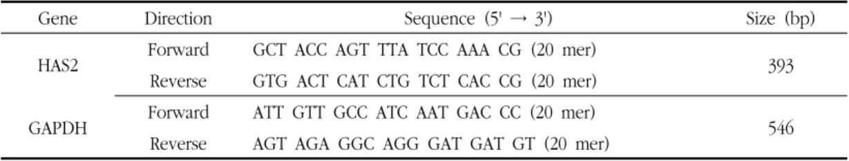
Table 1. Primers Sequence
Gene Direction Sequence (5' → 3') Size (bp)
HAS2 Forward GCT ACC AGT TTA TCC AAA CG (20 mer) Reverse GTG ACT CAT CTG TCT CAC CG (20 mer) 393
GAPDH Forward ATT GTT GCC ATC AAT GAC CC (20 mer) Reverse AGT AGA GGC AGG GAT GAT GT (20 mer) 546
하였다. 24시간 후에 serum-free DMEM으로 교체해 준 후에 황기추출물을 처리해주었다. 24시간동안 배 양해준 후에 media를 걷어내고 20 ㎕의 3-(4, 5-dimethyl-2-thiazolyl)-2,5-diphenyl-2H-tetrazolium bromide (MTT, 5 ㎎/㎖)을 넣어주고 CO2 배양기에 서 2시간 배양하였다. 100 ㎕의 DMSO로 결정을 용 해시킨 후에 570 nm에서 흡광도를 측정하였다. 생존 율은 control과 비교하여 %로 표시하였다. 세포에 처 리한 샘플에 포함된 DMSO는 최종농도 0.1%에 맞추 었다.
5. RT-PCR
세포를 2 x 10⁵/㎖ 농도로 6 well plate에 seeding 하였다. 24시간 후에 serum-free DMEM으로 교체해 준 후에 황기추출물을 처리해주었다. 최종 DMSO는 농도는 0.1%로 맞추어 주었다. 24시간동안 배양해준 후에 easyBlue (intron)로 RNA를 추출하였다. RNA 농도와 순도 (OD260 / OD280)를 측정한 후에 2 ㎍ RNA로 power cDNA synthesis kit (intron)을 이용 하여 cDNA 합성을 하였다. PCR은 premix PCR kit (Solgent)을 이용하였다. PCR product는 1.5%
agarose gel에 전기영동 하여 밴드를 확인하였다.
All-trans-retinoic acid (ATRA, sigma) 1 µM을 양 성대조군으로 사용하였다. Primer는 기존 논문에 보 고된 sequence로 제작하였다(Table 1)5).
PCR조건은 다음과 같다. 94℃ 15 min, 32~35 cycles: 94℃ 30s 50℃ 30s 72℃ 60s 72℃ 10 min 최 종 합성.
6. ELISA
세포를 2 x 10⁵/㎖ 농도로 6 well plate에 seeding 하였다. 24시간 후에 serum-free DMEM으로 2번 washing 해준 후에 serum-free DMEM 교체하고 샘 플을 처리해주었다. DMSO는 0.1%로 맞추어 주었다.
24시간 후에 350 ㎕의 media를 걷어내었고 다시 24 시간 후에 동량 걷어내었다. 15,000 x g에서 5분간 원심분리하고 상층액을 걷어내어 ELISA 할 때 까지 -20℃에서 보관하였다. ELISA는 HA-ELISA kit (echelon)를 이용하였으며 제조사에서 제공한 방법에 의해 진행하였다. ATRA 1 µM을 양성대조군으로 사 용하였다.
7. 통계
통계는 Student's t-test를 이용하였으며 유의성 기 준을 p value가 0.05 미만일 경우로 설정하였다.
III. 결과 및 고찰
1. MTT assay
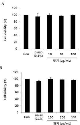
80% 메탄올 황기 추출물을 10, 50, 100 ㎍/㎖로 처 리하여 먼저 MTT assay를 하였고 다음에 100 ~ 300
㎍/㎖ 농도 범위에서 생존율을 확인하였다. 10 ~ 300
㎍/㎖에서 독성을 보이지는 않았다(Fig. 2 A와 B). 총 추출물임을 감안하여 최대농도를 200 ㎍/㎖까지 정하 였다.
2. Hyaluronidase enzyme assay
HA 함량 증가는 HA 생성의 증가와 분해의 감소 두 가지를 생각해 볼 수 있다. HA 감소는 HYAL의 작용으로 알려져 있다. 본 실험에 사용한 HYAL은 사람유래는 아니나 피부에서 HYAL 저해와 관련하여 간접적으로 확인할 수 있을 것으로 생각한다. 황기의 HYAL 활성저해에 관해서는 보고된 바가 있다14). 황
A
B
Fig. 2. MTT assay, A: concentration of treated Astragalus membranaceus (10 - 100 ㎍/㎖), B: concentration of treated Astragalus membranaceus (100 - 300
㎍/㎖), Values are represented as percentage relative to control. Results are presented as mean±S.D.
Fig. 3. Hyaluronidase enzyme assay. Values are represented as percentage of hyaluronidase inhibition rate. Results are presented as mean±S.D.
기 처리 농도를 200 ㎍/㎖로 했을 때 저해율은 8.73%
이었고 감초와 DSCG는 각각 35.78%와 31.17%의 저 해율을 보였다(Fig. 3). 메탄올 황기 추출물을 125 ㎍/㎖
로 처리했을 때 7.63% 저해율을 보인 최근 논문과 비슷 한 경향이었다15). 다소간 HYAL의 활성을 저해 하는 것 으로 보이나 감초에 비해 저해율은 저조했다.
3. 황기의 HAS2 유전자 발현 증가
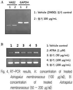
DMSO (0.1%) 처리군과 80% 메탄올 황기추출물 100 ㎍/㎖ 처리군의 HAS2 유전자발현 정도를 비교하 여 황기 처리군에서 HAS2 유전자의 발현이 증가함을 확인하였다. 80% 메탄올 황기 추출물을 50, 100, 200
㎍/㎖ 처리했을 때의 HAS2 유전자의 발현 양상을 RT-PCR로 확인하였다. 양성대조군인 ATRA 1 µM 처리했을 때 HAS2의 유전자 발현이 매우 현저하게 늘어남을 확인 할 수 있었으며 처리 농도 범위 내에 서 HAS2의 유전자 발현량이 늘어남을 확인 할 수 있 었다(Fig. 4). HAS2 유전자 발현량의 증가는 HA합성 을 증가 시킬 수 있음을 보여준다.
A
B
Fig. 4. RT-PCR results, A: concentration of treated Astragalus membranaceus (100 ㎍/㎖), B:
concentration of treated Astragalus membranaceus (50 - 200 ㎍/㎖)
4. HA-ELISA assay
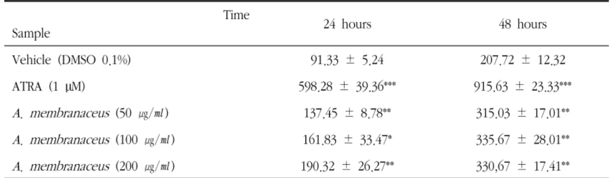
황기 추출물 50, 100, 200 ㎍/㎖ 처리 했을 때 HA 의 생성량을 확인하기 위해 ELISA kit를 사용하였다.
48시간 처리가 24시간 처리보다 모든 샘플에서 HA 생성량이 늘었음을 확인 하였으며 양성대조군으로 사 용한 ATRA (1 µM) 처리했을 때 HA 생성량이 24시 간과 48시간에서 각각 598.28±39.36 ng/㎖ 과 915.63±23.33 ng/㎖로 현저히 늘어남을 확인하였다.
황기 처리군 (50 ~ 200 ㎍/㎖)의 경우 DMSO 0.1%
처리한 vehicle 군과 비교했을 때 24시간에서는 각각 50.55%, 77.2 %, 108.33% 증가하였고 48시간에서는 각각 51.67%, 61.59%, 59.19% 증가하였다. 48시간 처리에서 황기 처리농도 100과 200 ㎍/㎖에서는 차이 가 나타나지 않았다(Table 2). Hsu와 Chiang은 황기 의 Bacillus subtilis 발효가 skin cell에서 HA의 생성 량을 늘린다고 보고하였다16). 동일한 실험에서 물 추 출물은 HA 생성을 늘리지 못했다. 황기는 ononin, calycosin, formononetin등의 isoflavonoids를 함유하 고 있다17,18). 위 이 논문에서 저자는 황기에 포함되어 있는 isoflavone인 ononin, calycosin, formononetin 의 함량이 발효에 의해 줄어들며 이는 isoflavone들이 aglycone의 형태나 혹은 다른 관련 물질로 대사함으 로서 HA의 함량을 증가시키는데 관여함을 제시하였 다. Genistein과 daidzein이 HA의 함량을 늘린다는 보고19)가 있는 것으로 보아 isoflavone들이 HA 생성 에 관여할 수도 있을 것으로 생각한다. Isoflavone 천 연물을 함유하는 한약재들이 HA함량증가를 통한 보 습효과를 보일 가능성이 있으며 보습 관련 화장품의 원료로서 이용가치가 있을 것으로 생각한다. 본 실험 에서 황기의 메탄올 추출물은 양성대조군 ATRA과 비교하여 HA를 급격히 증가 시키지는 못했으나 HAS2 유전자 발현을 유도하고 소폭 HA의 함량을 증가시켰다. 이는 황기가 갖는 보습효과에 부분적으 로 HA 증가가 관여할 수도 있음을 보여준다. 인삼에 서 분리한 compound K의 경우 1 µM의 24시간 처
Time
Sample 24 hours 48 hours
Vehicle (DMSO 0.1%) 91.33 ± 5.24 207.72 ± 12.32
ATRA (1 µM) 598.28 ± 39.36*** 915.63 ± 23.33***
A. membranaceus (50 ㎍/㎖) 137.45 ± 8.78** 315.03 ± 17.01**
A. membranaceus (100 ㎍/㎖) 161.83 ± 33.47* 335.67 ± 28.01**
A. membranaceus (200 ㎍/㎖) 190.32 ± 26.27** 330,67 ± 17.41**
Results are presented as mean±S.D. ***: p < 0.001, **: p < 0.01, *: p < 0.05
Table 2. Hyaluronic Acid Production (ng/㎖)
리에서 약 200%넘는 효과를 보였다6). 많은 한약재 추출물과 함유 성분에서 HA 증가 효과탐색이 가능할 것으로 사료된다.
IV. 결 론
1. 황기추출물은 비교적 독성이 약했으며 50, 100, 200 ㎍/㎖을 처리 농도로 정하였다.
2. 황기추출물 200 ㎍/㎖에서 Hyaluronidase에 대해 8.73%의 저해율을 보였으며 감초와 DSCG는 각각 35.78%와 31.17%의 저해율을 보였다.
3. HaCaT 세포에서 황기추출물 처리는 HAS2 유전 자의 발현량을 증가 시켰다.
4. 황기 추출물 50, 100, 200 ㎍/㎖을 처리했을 때 DMSO 0.1% 처리한 vehicle 군과 비교하여 24시 간에서는 각각 50.55%, 77.2 %, 108.33% 증가하 였고 48시간에서는 각각 51.67%, 61.59%, 59.19% 증가하였다.
감사의 글
본 연구는 보건복지부 한의약산업육성제품화지원사 업의 지원에 의하여 이루어진 것임 (F110018)
참 고 문 헌
1. Verdier-Sévrain S, Bonté F. Skin hydration: a review on its molecular mechanisms. J Cosmet Dermatol. 2007;6(2):75-82.
2. Ghersetich I, Lotti T, Campanile G, Grappone C, Dini G. Hyaluronic acid in cutaneous intrinsic aging. Int J Dermatol. 1994;33 (2):119-22.
3. Dai G, Freudenberger T, Zipper P, Melchior A, Grether-Beck S, Rabausch B, et al.
Chronic ultraviolet B irradiation causes loss of hyaluronic acid from mouse dermis because of down-regulation of hyaluronic acid synthases. Am J Pathol. 2007;171(5):1451-61.
4. Stern R, Maibach HI. Hyaluronan in skin:
aspects of aging and its pharmacologic modulation. Clin Dermatol. 2008;26(2):
106-22.
5. Sim GS, Lee DH, Kim JH, An SK, Choe TB, Kwon TJ, et al. Black rice (Oryza sativa L.
var. japonica) hydrolyzed peptides induce expression of hyaluronan synthase 2 gene in HaCaT keratinocytes. J Microbiol Biotechnol.
2007;17(2):271-79.
6. Kim S, Kang BY, Cho SY, Sung DS, Chang HK, Yeom MH, et al. Compound K induces expression of hyaluronan synthase 2 gene in transformed human keratinocytes and increases hyaluronan in hairless mouse skin. Biochem Biophys Res Commun. 2004;316 (2):348-55.
7. Kim SH, Nam GW, Kang BY, Lee HK, Moon SJ, Chang IS. The Effect of Kaempferol, Quercetin on Hyaluronan-Synthesis Stimulation in Human Keratinocyte (HaCaT). J Soc Cosmet Scientists Korea. 2005;31(1):97-102.
8. Saavalainen K, Pasonen-Seppänen S, Dunlop TW, Tammi R, Tammi MI, Carlberg C. The human hyaluronan synthase 2 gene is a primary retinoic acid and epidermal growth factor responding gene. J Biol Chem.
2005;280(15):14636-44.
9. Furuya T, Yamagata S, Shimoyama Y, Fujihara M, Morishima N, Ohtsuki K.
Biochemical characterization of glycyrrhizin as an effective inhibitor for hyaluronidases from bovine testis. Biol Pharm Bull. 1997;20(9):
973-77.
10. Kakegawa H, Matsumoto H, Satoh T.
Inhibitory effects of some natural products on the activation of hyaluronidase and their anti-allergic actions. Chem Pharm Bull.
1992;40(6):1439-42.
11. Murata T, Miyase T, Yoshizaki F.
Hyaluronidase inhibitors from Keiskea japonica. Chem. Pharm. Bul. 2012;60(1):
121-28.
12. Ahn DK. Illustrated Book Of Korean Medicinal Herbs. 3rd ed. Seoul:Kyo-Hak Publishing Co. Ltd. 2000.
13. Jung TK, Kim MJ, Lim KR, Yoon KS.
Moisturing and Anti-oxidation Effect of Astragalus membranaceus. J Soc Cosmet Scientists Korea. 2006;32(3):193-200.
14. Lee YH, Choi SI, Lee JW, Jung SM, Park SM, Heo TR. Isolation of Hyaluronidase Inhibitory Component from the Roots of Astraglus membranaceus Bunge (Astragali Radix). Food Sci Biotechnol. 2005;14(2):263-67.
15. Cha BC. Tyrosinase, Hyaluronidase Inhibitory Effect and Antioxidant Activity of Medicinal Plants. Kor J Pharmacogn. 2011;42(1):89-97.
16. Hsu MF, Chiang BH. Stimulating effects of Bacillus subtilis natto-fermented Radix astragali on hyaluronic acid production in human skin cells. J Ethnopharmacol. 2009;
125(3):474-81.
17. Wu T, Annie Bligh SW, Gu LH, Wang ZT, Liu HP, Cheng XM, et al. Simultaneous determination of six isoflavonoids in commercial Radix Astragali by HPLC-UV.
Fitoterapia. 2005;76(2):157-65.
18. Qi LW, Yu QT, Li P, Li SL, Wang YX, Sheng LH, et al. Quality evaluation of Radix Astragali through a simultaneous determination of six major active isoflavonoids and four main saponins by high-performance liquid chromatography coupled with diode array and evaporative light scattering detectors. J Chromatogr A.
2006;1134(1-2):162-9.
19. Miyazaki K, Hanamizu T, Iizuka R, Chiba K.
Genistein and daidzein stimulate hyaluronic acid production in transformed human keratinocyte culture and hairless mouse skin.
Skin Pharmacol Appl Skin Physiol.
2002;15(3):175-83.