∙ Received: December 30, 2010. Accepted: March 7, 2011.
∙ Corresponding author: Min Soo Park
Department of Nuclear Medicine, Severance Hospital, Yonsei University Health System, 250 Seongsanno, Seodaemun‐gu, Seoul, 120‐752, Korea
Tel: +82‐2‐2228‐6064, Fax: +82‐2‐312‐0578 E‐mail: [email protected]
Original Article 월경 주기에 따른 18 F‐FDG PET/CT에서의 유방 섭취 변화에 관한 고찰
연세의료원 세브란스병원 핵의학과
박민수⋅박훈희⋅김정열⋅임한상⋅김재삼⋅이창호
A Study of the Change of Breast Uptake in Menstrual Cycle on
18
F‐FDG PET/CT
Min Soo Park, Hoon Hee Park, Jung Yul Kim, Han Sang Lim, Jae Sam Kim and Chang Ho Lee Department of Nuclear Medicine, Severance Hospital, Yonsei University Health System
Purpose: 18F‐FDG PET/CT has been known a useful modality to diagnose high‐glucose‐using cells such as cancer cells by glucose metabolism of FDG. Mainly, FDG takes on cancer and inflammatory cells; However, There have been FDG uptakes on normal tissues by individual physiological characteristics, occasionally.
Especially, in fertile females, unusual FDG uptake of breast changes as the menstrual cycle, and disturb diagnosis. Therefore, the study aimed to evaluate the change of breast FDG uptake in menstrual cycle on 18F‐
FDG PET/CT. Materials and Methods: 160 females (34±3.5 years old) who do not undergo a gynecologic anamnesis and have regular menstrual cycle over the previous 6 months were examined, from March 2009 to February 2010. They were divided 4 groups (each 40 patients) as flow phase, proliferative phase, ovulatory phase and secretory phase using Pregnancy Calculator 0.14. and history taking. Discovery Ste (GE Healthcare, Milwaukee, Mi, USA) was used as PET/CT. We analyzed SUVs on accumulated region on breast, and 3 nuclear medicine specialists did the Blind test. Results: SUVs on the Breast were flow phase (1.64±0.25), proliferative phase (0.93±0.28), ovulatory phase (1.66±0.26) and secretory phase (1.77±0.28). It showed high uptake value in secretory, flow phase and ovulatory phase (p<0.05). In gross analysis, the accumulation of breast was divided into 3 grades as comparing with lung and liver. The breast’s uptake was equal to lung (Grade Ⅰ); between lung and liver (Grade II); equal to or greater than liver (Grade III). The results showed high uptake value in secretory, flow phase and ovulatory phase (p<0.05). Conclusion: In fertile females, FDG uptake of breast changed as menstrual cycle, and it available to diagnose breast disease. Therefore, we consider reducing false‐negative finding of breast disease, by doing examination on appropriate period through history taking about individual menstrual cycle. (Korean J Nucl Med Technol 2011;15(1):39-44)
Key Words : 18F‐FDG PET/CT, Menstrual cycle, Breast
서 론
18F‐FDG PET/CT는 포도당대사로 인한 암세포의 비정상 적인 섭취를 통해 암을 진단하는 유용한 검사이다.1,2) 일반적
으로 FDG는 암세포와 염증성 질환에 높은 섭취를 보이나, 환자의 생리적 특성에 따라 정상세포에도 비정상적인 섭취 를 나타내기도 한다.3-7) 특히 여성의 월경 주기에 따른 자궁 벽 두께의 구조적 변화는 골반 초음파 결과에 영향을 미치게 되며, 여성 호르몬의 변화로 인해 자궁내막의FDG의 비정상 적인 섭취를 보여 PET/CT검사에도 영향을 줄 수 있다.8) 이 러한 가임기 여성의 월경 주기는 보통 28일을 기준으로 일어 나며, 이는 자궁과 난포의 변화에 따라 월경기(menstrual flow phase), 증식기(proliferative phase), 배란기(ovulatory phase), 분비기 (secretory phase)로 나뉘고 이에 따라 자궁은
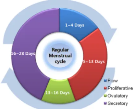
Fig. 1. 160 females (34±3.5 years old) who do not undergo a gynecologic anamnesis and have regular menstrual cycle (28 days) over the previous 6 months were examined. 1‐4 days was classified as flow phase, 5‐13 days was classified as proliferative phase, 13‐16 days as ovulatory; and 16‐28 days as secretory phase.
Fig. 2. Before PET/CT procedure, we were confirmed each menstrual cycle through history taking, and then, Whole Body PET/CT was progressed.
일정 주기로 변화한다. 각 월경 주기 동안 난소는 난포기 (follicular phase)와 황체기(luteal phase)의 두 단계를 거치며, 이 과정에서 여성의 대표 호르몬인 에스트로겐(estrogen)과 프로게스테론(progesterone)이 분비된다. 월경 이후 여포에서 분비되는 에스트로겐의 영향으로 자궁내막은 두터워지고, 자궁선과 혈관도 함께 발달하며, 황체에서 분비되는 프로게 스테론은 자궁 점막을 증식시킨다. 이러한 호르몬의 변화는 자궁 뿐 아니라 유방의 혈류를 자극하며 유방 결합 조직의 탄력성을 증가시키고 더불어 지방 침착을 증가시킨다. 이는 유관 및 유선을 발달시키므로 유방촬영(Mammography)과 PET/CT 결과에 미칠 영향을 배제해서는 안 된다.9)
국내의 암 발생률 중 여성의 경우 유방암은 갑상선 암 다 음으로 높은 빈도를 차지하고 있으며, 최근 유방암에 의한 유용한 병기 결정과 치료 예후 평가를위해 PET‐CT 검사의 중요성이 강조되고 있다.10‐13) 하지만 여성의 월경 주기에 따 른 유방의 비정상적인 섭취는 미세한 유방 병변의 정확한 진 단을 저해하는 요인이 될 수 있다.14‐16) 따라서 본 연구는 월 경 주기에 따른 유방의 FDG 섭취 변화를 비교 하여 PET/CT 검사의 최적시기를 평가함으로써 유방의 미세 병변 에 대한 진단능을 향상시키고자 한다.
대상 및 방법
1. 환자 정보
2009년 3월부터 2010년 2월까지 본원에 내원한 부인과 병 력이 없고 6개월 이상 규칙적인 월경 주기를 가진 160명의 여성환자(평균 연령은 34±3.5세)를 대상으로 하였다(Fig. 1).
환자 문진과 Pregnancy Calculator (Ver. 0.14)를 이용하여 월 경기, 증식기, 배란기, 분비기로 나누어 각각 40명씩 정보를 수집하였다.
2. 장비 및 검사방법
사용 된 PET/CT는 GE사의Discovery STe (GE Health- care, Milwaukee, Mi, USA) Scanner이며, 크리스탈(crystal)은 BGO 를 사용하였다. 고유분해능(intrinsic resolution)은 6.0 mm의 반치폭(full width at half maximum, FWHM)을 사용 하였고, DFOV (display field of view)는 70.0 mm, 1 bed 당 overlap은 9 mm, CT는 2 mm 의 slice thickness로 8 slice로 구성되어 있으며, 재구성 방법은 iterative method를 사용하 여 subset 28회, iterative 2회 시행하였다. 환자 검사 전처치로 는 최소한 8시간 이상 금식을 하도록 하였으며, 검사 전날과 당일에는 무리한 운동을 금하였다. 500~100 ml이상 충분한 수분을 섭취하도록 하였으며, 검사 전 혈당은 6.69 mmol/l (120 mg/dl) 이하였다. 18F‐FDG를 투여는 약 15분 정도 안정 을 시키고 약 5.6 MBq/kg (0.15 mCi) 정맥 투여하였고, 근육 의 섭취를 방지하기 위해 움직임을 제재하였으며, 60‐90분 후 전신 검사를 진행하였다. 전신 검사는 환자가 바로 누운 자 세에서 뇌의 기저부부터 대퇴근위부까지 검사 범위를 설정하 였고, 조영제를 사용하지 않은 140Kvp, 30mAs 조건의 NCCT (non contrast computed tomography)를 진행한 후 1 bed 당 3 분 동안 방출영상(emission scan)을 진행 하였다(Fig. 2). 방출 영상이 끝난 후 CECT (contrast enhanced computed tomo- graphy)를 진행하였으며, 사용 된 조영제는 OMNIPAQ UE (GE Healthcare Co., Ireland, 1 mL 당 요오드 300 mg 포함)를 사용하였다. 이 때 환자의 몸무게 Kg 당 2 cc 용량으로 초당 2 mL의 속도로 주입하였고, 자동 주입기는 dual shot injector 인 Optivantage (Mallinchrodt, LIEBEL‐FLARSHEIM Co., United States of America)를 사용하였다.
Fig. 3. We did semi‐quantitative analysis using each SUV measured of breast, liver, and lung for identifying the change of SUVs as menstrual cycle.
Fig. 4. Three nuclear medicine specialists did the Blind test. (A), (B), (C) were PET whole body images, and (A‐1, B‐1, C‐1) were Fusion image of each Grade. The higher grade was, the more breast FDG uptake increased unusually.
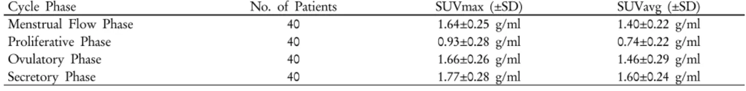
Cycle Phase No. of Patients SUVmax (±SD) SUVavg (±SD)
Menstrual Flow Phase 40 1.64±0.25 g/ml 1.40±0.22 g/ml
Proliferative Phase 40 0.93±0.28 g/ml 0.74±0.22 g/ml
Ovulatory Phase 40 1.66±0.26 g/ml 1.46±0.29 g/ml
Secretory Phase 40 1.77±0.28 g/ml 1.60±0.24 g/ml
Table 1. In secretory phase, SUVmax was the highest value as 1.77±0.28 g/ml, proliferative phase showed the lowest SUVmax as 0.93±0.28 g/ml.
3. 영상분석
Pregnancy Calculator (Ver. 0.14)와 검사 전 문진을 통하여 여성의 월경 주기를 구분하였으며, 주기 별 간과 폐, 유방의 SUV의 변화를 비교 분석하였다(Fig. 3). 또한 핵의학 전문의 3명을 통해 Blind Test로 육안적 평가를 시행하였으며, 각 월 경 주기 별 폐, 간, 유방의 FDG 섭취 정도를 통해 유방의 FDG 섭취가 폐와 유사할 경우 Grade Ⅰ, 폐와 간 중간 정도 의 FDG 섭취를 보일 경우에는 Grade Ⅱ, 간과 같거나 높을 경우에는 Grade Ⅲ으로 구분하였다(Fig. 4).
결 과
유방에서의 SUV를 측정한 결과 월경기(SUVmax 1.64±
0.25 g/ml), 증식기(SUVmax 0.93±0.28 g/ml), 배란기(SUVmax 1.66±0.26 g/ml), 분비기(SUVmax 1.77±0.28 g/ml)로 나타났 으며(Table 1), 분비기에서 가장 높고 다음으로 배란기와 월 경기 순으로 높은 SUV를 나타냈다(p<0.05).
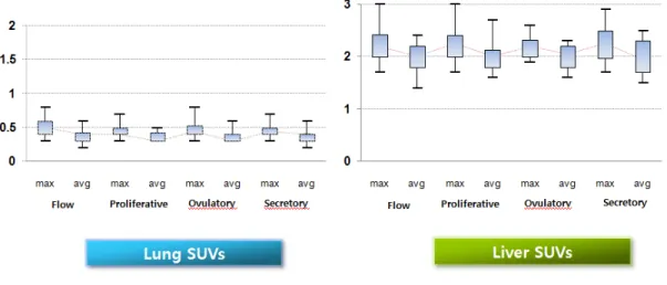
또한 여성 월경 주기 별 유방의 SUV의 변화를 Box Plot으 로 도식한 결과 분비기와 월경기, 배란기에서 증가되어 나타 났고, 증식기에서 SUV가 감소되어 있음을 확인 할 수 있었 다(Fig. 5). 하지만 폐와 간에서는 월경 주기에 따라 SUV의 변화를 볼 수 없었다(Fig. 6).
Fig. 6. The uptake values of lung and liver had almost no variation over menstrual cycle. This suggested that breast’s SUVs were only influenced by menstrual cycle.
FDG Uptake in Breast Flow Proliferative Ovulatory Secretory Total No.
Grade Ⅰ 2 32 0 0 34
Grade Ⅱ 13 8 16 9 46
Grade Ⅲ 25 0 24 31 80
Total No. 40 40 40 40 160
Table 2. The FDG uptake in the breast regions shows Grade I in 34 women (21.2%), grade II in 46 (28.8%) and grade III in 80 (50.0%).In proliferative phase, FDG uptake of breast was the lowest, but, secretory phase showed the highest FDG uptake value.
Fig. 5. Breast uptake values increased in order of secretory, flow, and ovulatory phase, similarly. Proliferative phase showed comparative low SUVs, only.
또한 핵의학 전문의 3명이 Blind Test로 섭취 정도를 평가 한 육안적 분석에서는 Grade Ⅰ으로 구분 된 환자는 총 160 명에서 34명(21.2%)이었으며, 이 중 월경기의 환자는 2명, 증 식기는 32명, 배란기 0명, 분비기 역시 0명이었고, 주로 증식 기 때 검사를 진행한 환자가 grade Ⅰ에 해당하였다. grade
Ⅱ는 46명(28.8%)이었으며, 월경기 13명, 배란기 16명으로 나타났다. FDG섭취가 가장 높은 grade Ⅲ는 총 환자 160명
고 찰
월경 주기에 따른 호르몬의 변화는 PET/CT 전신 검사에 서 유방의 FDG섭취를 증가시켰다. FDG의 섭취 변화는 분 비기에서 가장 컸으며, 증식기에서 가장 영향이 작았다. 본 연구를 통하여 월경 주기 별 유방의SUV 변화와 PET/CT 전 신 검사에서의 육안적 유방 섭취 변화를 관찰 할 수 있었으 며, 진료과와의 협조를 통해 증식기 때 PET/CT 검사를 유도 하여 검사를 진행 한다면 유방의 미세 병변까지 진단 할 수 있는 정확한 검사가 진행될 것으로 생각된다. 다만 본 연구 에서는 규칙적인 월경 주기를 가진 환자를 대상으로 하였으 므로 불규칙적인 월경 주기를 가진 환자의 경우 연구 대상에 서 제외 되었다. 따라서 불규칙 월경 주기의 환자까지 적용 가능 성에 대해서는 추 후 더욱 많은 연구와 논의가 필요할 것이다.
Fig. 7. PET whole body images of secretory phase (D) and flow phase (A) were the highest FDG uptake of breast, and each fusion images (D‐1, A‐1) showed increases remarkably. (B) and (B‐1) were proliferative phase, and breast uptake was almost no increase. (C) and (C‐1) were ovulatory phase that breast FDG uptake was increased more than proliferative.
결 론
가임기 여성은 월경 주기에 따라 유방의 FDG 섭취 정도 가 변화되며, 특히 미세한 유방 병변을 진단하는데 월경 주 기의 정보를 유용하게 활용할 수 있다. 본 연구를 통해 각 주 기 별 유방의 FDG 섭취를 비교 하였으며, 가임기 여성의 경 우 분비기 때 유방의 섭취가 가장 높았고, 증식기 때에는 유 방의 FDG섭취가 가장 낮았다. 따라서 검사 전 문진을 통하 여 가임기 여성의 정확한 월경 주기를 확인하고 적정한 시기 를 유도하여 검사를 시행함으로써 미세한 유방 질환에 대한 위음성 결과를 감소시킬 수 있을 것으로 사료된다.
요 약
18F‐FDG PET/CT는 당대사로 인한 암세포의 비정상적인 섭취를 통해 암을 진단하는 유용한 검사이다. 일반적으로 FDG는 암세포와 염증성 질환에 높은 섭취를 보이나, 환자 의 생리적 특성에 따라 정상세포에도 비정상적인 섭취를 나 타내기도 한다. 특히 여성 월경 주기에 따른 유방의 비정상 적인 섭취는 미세한 유방 병변의 정확한 진단을 저해하는 요 인이 될 수 있다. 따라서 본 연구는 월경 주기에 따른 유방의 FDG 섭취 변화를 비교 평가하고자 한다. 2009년 3월부터 2010년 2월까지 부인과 병력이 없고 6개월 이상 규칙적인 월 경 주기를 가진 160명의 여성(34±3.5세)을 대상으로 하였다.
환자 문진과 Pregnancy Calculator (Ver. 0.14)를 이용하여 월경 기, 증식기, 배란기, 분비기로 나누어 각각 40명씩 정보를 수집 하였으며, PET/CT 장비는 Discovery STe(GE Healthcare, Milwaukee, Mi, USA)를 이용하였다. 분석 방법으로는 월경 주 기 별 유방의 SUV 변화를 비교 분석하였으며, 핵의학 전문의 3명을 통해 Blind Test로 육안적 평가를 시행하였다. 유방의 SUV는 월경기(1.64±0.25), 증식기(0.93±0.28), 배란기 (1.66±0.26), 분비기(1.77±0.28)로 나타났으며 분비기가 가장 높 고 월경기와 배란기에서도 높은 섭취를 나타냈다(p<0.05). 또 한 육안적 분석에서는 간과 폐의 집적에 대하여 유방의 집적 정도를 단계별로 나누어 폐와 유사한 섭취를 보인 경우(grade I), 폐와 간 중간 정도의 섭취를 보인 경우(grade II), 간과 같 거나 높은 섭취를 보인 경우(grade III)로 구분하였으며, 육안 적 평가에서도 분비기와 월경기에서 높은 섭취를 보였다 (p<0.05). 가임기 여성은 월경 주기에 따라 유방의 18F‐FDG 섭취 정도가 변화되며, 특히 미세한 유방 병변을 진단하는데 월경 주기의 정보를 유용하게 활용할 수 있다. 따라서 검사 전 문진을 통하여 가임기 여성의 정확한 월경 주기를 확인하 고 적정한 시기를 유도하여 검사를 시행함으로써 유방 질환 에 대한 위음성 결과를 감소시킬 수 있을 것으로 사료된다.
REFERENCES
1. Engel H, Steinert H, Buck A, Berthold T, Rahel A, von Schulthess GK. Whole‐body PET: physiological and artifactual fluorodeoxy-
Nucl Med 1996;26:308-314.
4. Shreve PD, Anzai Y, Wahl R. Pitfalls in oncologic diagnosis with FDG PET imaging: physiologic and benign variants. Radiographics 1999;19:61-77.
5. Cook GJ, Wegner EA, Fogelman I. Normal variants, artefacts and interpretative pitfalls in PET imaging with 18‐fluoro‐2‐ deoxyglucose and carbon‐11 methionine. Eur J Nucl Med Mol Imaging 1999;26:1363-1378.
6. Blodgett TM, Fukui MB, Snyderman CH, et al. Combined PET‐CT in the head and neck: part 1—physiologic, altered physiologic, and artifactual FDG uptake. Radiographics 2005;25: 897-912.
7. Prabhakar HB, Sahani DV, Fischman AJ, Mueller PR, Blake MA.
Bowel hot spots at PET‐CT. Radiographics 2007;27:145-159.
8. Mijin Yun, Arthur Cho, Jae Hoon Lee, Yun‐Jung Choi, Jong Doo Lee, and Chun K. Kim. Physiologic 18F‐FDG Uptake in the Fallopian Tubes at Mid Cycle on PET/CT. J Nucl Med 2010;51: 682-685.
9. Treloar, A. E.; Boyton, R. E.; Behn, B. G.; and Brown, B. W.
(1967). "Variations of the Human Menstrual Cycle through Reproduc- tive Life." International Journal of Fertility 9:77-126.
of FDG‐PET/CT in suspected recurrence of breast cancer. Cancer 2006;107:2545-2551.
13. Beresford M, Lyburn I, Sanghera B, Makris A, Wong WL. Serial integrated 18F fluorodeoxythymidine PET/CT monitoring neoadju- vant chemotherapeutic response in invasive ductal carcinoma. Breast J 2007;13:424-425.
14. Rosenberg RD, Hunt WC, Williamson MR, et al. Effects of age, breast density, ethnicity, and estrogen replacement therapy on screening mammographic sensitivity and cancer stage at diagnosis:
review of 183,134 screening mammograms in Albuquerque, New Mexico. Radiology 1998; 209:511.
15. Kerlikowske K, Grady D, Barclay J, et al. Effect of age, breast density, and family history on the sensitivity of first screening mammography. JAMA 1996; 276:33-38.
16. Kavanagh AM, Cawson J, Byrnes GB, et al. Hormone replacement therapy, percent mammographic density, and sensitivity of mammo- graphy. Cancer Epidemiol Biomarkers Prev 2005;14:1060-1064.