Ⅰ. INTRODUCTION
18
F-FDG PET/CT is a useful test for detecting cancer by assessing the abnormal uptake of FDG by cancer cells due to the glucose metabolism
1-3). Generally, the
high uptake of FDG by cancer cells and inflammatory diseases was observed
4). Nonetheless, normal cells may show abnormal uptake depending on the physiological characteristic of patients. In particular, the structural changes that occur in the uterine wall according to the menstrual cycle influence the pelvic ultrasonography
Journal of Radiological Science and Technology, 38(1), 31-38 eISSN 2384-1168 ISSN 2288-3509 http://dx.doi.org/10.17946/JRST.2015.38.1.05
<원저>
A Study of the Changes of Breast Uptake in Menstrual Cycle on 18 F-FDG PET/CT
-월경 주기에 따른
18F-FDG PET/CT에서 유방 섭취 변화에 관한 고찰-
Dept. of Radiological Technology, Shingu College Dept. of Nuclear Medicine, Severance Hospital, YUHS
1)Graduate School of Public Health, Yonsei University
2)Yeojin Tak ․ Min Soo Park
1)․ Juyoung Lee
2)․ Hoon-Hee Park
― Abstract ―
18
F-FDG PET/CT has been known a useful modality to diagnose high-glucose-using cells such as cancer cells by glucose metabolism of FDG. Mainly, FDG takes on cancer and inflammatory cells; however, there have been FDG uptakes on normal tissues by individual physiological characteristics, occasionally. Especially, in fertile fe- males, unusual FDG uptake of breast changes as the menstrual cycle, and disturb diagnosis. Therefore, the study aimed to evaluate the change of breast FDG uptake in menstrual cycle on
18F-FDG PET/CT. 160 females (34±3.5 years old) who do not undergo a gynecologic anamnesis and have regular menstrual cycle over the previous 6 months were examined. They were divided 4 groups (each 40 patients) as flow phase, proliferative phase, ov- ulatory phase and secretory phase using Pregnancy Calculator 0.14. and history taking. Discovery Ste (GE Healthcare, Milwaukee, Mi, USA) was used as PET/CT. We analyzed SUVs on accumulated region on breast, and 3 nuclear medicine specialists did the Blind test. SUVs on the Breast were flow phase (1.64±0.25), proliferative phase (0.93±0.28), ovulatory phase (1.66±0.26) and secretory phase (1.77±0.28). It showed high uptake value in secretory, flow phase and ovulatory phase (p<0.05). In gross analysis, the accumulation of breast was divided in- to 3 grades as comparing with lung and liver. The breast’s uptake was equal to lung (Grade Ⅰ); between lung and liver (Grade II); equal to or greater than liver (Grade III). The results showed high uptake value in secre- tory, flow phase and ovulatory phase (p<0.05). In fertile females, FDG uptake of breast changed as menstrual cycle, and it available to diagnose breast disease. Therefore, we consider reducing false-negative finding of breast disease, by doing examination on appropriate period through history taking about individual menstrual cycle.
Key Words :
18F-FDG PET/CT, Menstrual cycle, Breast
Corresponding author: Hoon-Hee Park (452-743) Department of Radiological Technology, Shingu College 377
results
5,6). Moreover, the endometrium shows abnormal FDG uptake and influences the PET/CT test due to the change in female hormones. In fertile women, the menstrual cycle is generally 28 days
7). The menstrual cycle is divided to the Menstrual Flow Phase, Proliferative Phase, Ovulatory Phase, and Secretory Phase according to the changes in the uterus and follicle
8). The uterus undergoes changes as a regular cycle. During each menstrual cycle, the ovary undergoes changes in two stages, Follicular Phase and Luteal Phase. During this process, the representative female hormones estrogen and progesterone are secreted
9-11). After menstruation, the endometrium becomes thick due to the influence of estrogen released from the follicle, and the uterine glands and blood vessels developed simultaneously
12-14). The progesterone released from the corpus luteum stimulates the proliferation of endometrium and blood flow in the breast resulting in an increase in the elasticity of breast connective tissues and fat deposition, which induces the development of the lactiferous duct and mammary glands
15,16). Therefore, its influence on the result of mammography and PET/CT should not be ruled out.
In Korean women, breast cancer is the second highest cancer next to thyroid cancer, and the importance of PET-CT for determining the stage of breast cancer and assessing the prognosis after the treatments has been emphasized. On the other hand, the abnormal FDG uptake by the breast of women according to the menstrual cycle may be a factor that impairs an accurate diagnosis of breast micro-lesions.
Therefore, this study assessed the optimal time for a PET-CT test by comparing the uptake of FDG by the breast according to the menstrual cycle to improve the ability to diagnose micro-lesions in the breast.
Ⅱ. MATERIALS & METHODS
A. Patients information
The subjects were 160 female patients (mean age, 34
± 3.5 years) without a disease history of gynecological
disease, and with a regular menstrual cycle for longer than 6 months(Figure 1). The subjects were divided into the following phases by history taking and the application of Pregnancy Calculator Ver.0.14: the menstrual flow phase, proliferative phase, ovulatory phase and secretory phase. Information of 40 patients in each phase was collected.
Fig. 1 160 females (34±3.5 years old) who do not undergo a gynecologic anamnesis and have regular menstrual cycle (28 days) over the previous 6 months were examined. 1 ~ 4 days was classified as flow phase, 5 ~ 13 days was classified as proliferative phase, 13 ~ 16 days as ovulatory; and 16 ~ 28 days as secretory phase.
B. Equipment and test methods
The Discovery STE scanner (Milwaukee, Wi, GE Healthcare, Co., USA) was used for PET/CT. BGO was used as the crystal. A 6. 0 mm full width at half maximum (FWHM) was used as the intrinsic resolution.
The display field of view (DFOV) was 70.0 mm, and the Overlap per 1 bed was 9 mm. CT consisted of 8 slices with a 2 mm slice thickness. As the reconstruction method, “subset” was performed 28 times and “iterative”
was performed 2 times using the iterative method. As
the pretreatment test, the patients were fasted for a
minimum of 8 hours, and excessive exercise was
prohibited on the day before the test and on the day of
the test. The patients took sufficient liquid, more than
100 ~ 500 ml. The blood glucose levels prior to the test
were <6.69 mmol/l (120 mg/dl). For the administration
of
18F-FDG, after the patients were allowed to rest for approximately 15 minutes, approximately 5.6 MBq/kg (0.15 mCi) was injected intravenously. Movements were restrained to prevent uptake by the muscles, and a full-body scan was performed after 60 ~ 90 minutes.
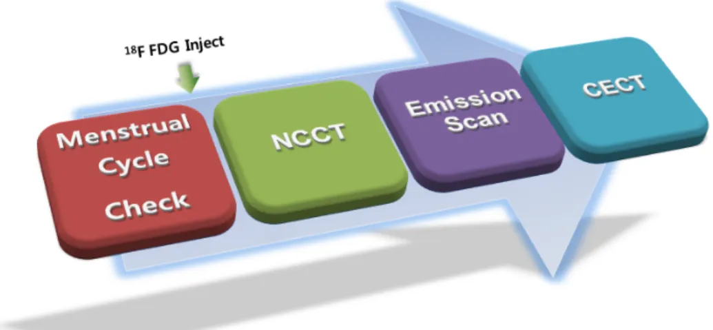
In a full-body scan, the test range was from the base of the brain to the proximal femoral area in the supine position. Non contrast computed tomography (NCCT) without contrast was performed under the condition of 140 kVp and 30 mAs. Subsequently, an emission scan for 3 minutes per bed was performed (Figure 2). After the emission scan, contrast enhanced computed tomography (CECT) was performed. OMNIPAQ UE (300 mg iodine/ml, GE Healthcare Co., Ireland) was used as the contrast. At that time, it was injected at a dose of 2 cc per kg of the patient’s body weight and a speed of 2 ml per second. A dual shot injector optivantage (Mallinchrodt, LIEBEL FLARSHEIM Co., USA) was used as the automatic injector.
C. Image analysis
Using Pregnancy Calculator Ver. 0.14 and history taking prior to the test, the women were classified according to their menstrual cycle, and in each phase, the changes in SUV in the liver, lung and breast were compared and analyzed (Figure 3).
In addition, a macroscopic evaluation was performed by 3 radiologists as a Blind Tests. The level of FDG uptake by the lung, liver and breast was measured in each menstrual phase. Cases in whom the FDG uptake by the breast was comparable to the lung were classified as Grade I. Cases in whom the FDG uptake of the breast was between the lung and liver were Grade II. Cases in whom the FDG uptake of the breast was comparable to the liver or higher than the liver were Grade III (Figure 4).
Fig. 3 We did semi-quantitative analysis using each SUV measured of breast, liver, and lung for identifying the change of SUVs as menstrual cycle.
Fig. 2 Before PET/CT procedure, we were confirmed each menstrual cycle through history
taking, and then, Whole Body PET/CT was progressed.
Ⅲ. RESULTS
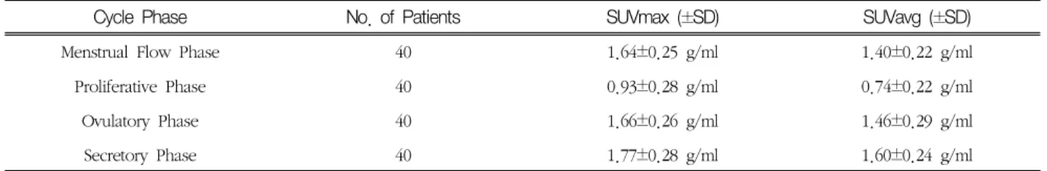
The SUVmax of the menstrual, proliferative, ovulatory and secretory phases was 1.64 ± 0.25 g/ml, 0.93 ± 0.28 g/ml, 1.66 ± 0.26 g/ml and 1.77 ± 0.28 g/ml, respectively(Table 1). The SUV was highest in the secretory phase followed in order by the menstrual flow phase and the ovulatory phase (p < 0.05).
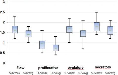
In addition, the change in SUV in each menstrual phase was drawn as a Box Plot, which revealed an increased in the secretory phase, menstrual flow phase and ovulatory phase. The SUV was lower in the proliferative phase(Fig. 5). On the other hand, the SUV in the lung and the liver showed no change according to the menstrual cycle(Fig. 6).
Three radiologists evaluated the uptake of the 160 patients by macroscopic analysis as a blind test. The results revealed, Grade I in 34 patients (21.2 %).
Among them, 2 and 32 cases were in the menstrual flow and proliferative phases, respectively. No cases were observed in the ovulatory and secretory phases.
Most of the patients who underwent the test during the proliferative phase corresponded to Grade I. Forty six (28.8 %) patients were in Grade II; 13 and 16 cases were in the menstrual flow and ovulatory phase, respectively. Eighty cases (50 %) were Grade III, of which the FDG uptake was highest. Thirty one patients were in the secretory phase, and their ratio was highest. In particular, all images of the secretory phase corresponded to Grades II or III, and the FDG uptake by the breast was noticeably higher than the proliferative phase(Figure 7).
Fig. 4 Three nuclear medicine specialists did the Blind test. (A), (B), (C) were PET Whole Body images, and (A-1, B-1, C-1) were Fusion image of each Grade. The higher Grade was, the more Breast FDG uptake increased unusually.
Cycle Phase No. of Patients SUVmax (±SD) SUVavg (±SD)
Menstrual Flow Phase 40 1.64±0.25 g/ml 1.40±0.22 g/ml
Proliferative Phase 40 0.93±0.28 g/ml 0.74±0.22 g/ml
Ovulatory Phase 40 1.66±0.26 g/ml 1.46±0.29 g/ml
Secretory Phase 40 1.77±0.28 g/ml 1.60±0.24 g/ml
Table 1. Analysis of SUV of Breast area.
Fig. 6 The uptake values of lung and liver had almost no variation over menstrual cycle. This suggested that breast’s SUVs were only influenced by menstrual cycle.
FDG Uptake in Breast Flow Proliferative Ovulatory Secretory Total No.
Grade Ⅰ 2 32 0 0 34
Grade Ⅱ 138 16 9 46
Grade Ⅲ 25 0 24 3 1 80
Total No. 40 40 40 40 160
Table 2. Results of blind test.
Fig. 5 Breast uptake values increased in order of secretory, flow, and ovulatory phase,
similarly. Proliferative phase showed comparative low SUVs, only.
Ⅳ. DISCUSSION
In a PET/CT full-body scan, hormonal changes according to the menstrual cycle increase the FDG uptake by the breast. The change in FDG uptake was largest in the secretory phase, and its effect was lowest in the proliferative phase. Based on this study, the SUV changes in the breast according to the menstrual cycle and the macroscopic changes in uptake by the breast could be detected by a full-body PET/CT scan. If a PET/CT test is performed during the proliferative phase in collaboration with the diagnosis department, it can provide an accurate test that could detect even micro lesions in the breast.
Nonetheless, in the present study, the subjects were patients with a regular menstrual cycle. Patients with an irregular menstrual cycle were excluded.
Accordingly, many studies will be needed before this can be applied to patients with an irregular menstrual cycle.
Ⅴ. CONCLUSION
The level of the FDG uptake by the breast in fertile women varies according to the menstrual cycle. In particular, information on the menstrual cycle can be applied widely for a diagnosis of breast micro-lesions.
Through this study, the uptake of FDG by the breast in each phase was compared. In fertile women, the FDG uptake by the breast was highest during the secretory phase and lowest in the proliferative phase.
Therefore, it is believed that false negative results of micro breast lesions may be reduced by assessing the accurate menstrual cycle through history taking before the test and by performing the test at the appropriate phase.
References
1. Engel H, Steinert H, Buck A, Berthold T, Rahel A, von Schulthess GK: Whole-body PET: physiological and artifactual fluorodeoxyglucose accumulations, J Nucl Med , 37, 441–6, 1996
Fig. 7 PET Whole Body images of Secretory phase (D) and flow phase (A) were the highest FDG uptake of breast,
and each fusion images (D-1, A-1) showed increases remarkably. (B) and (B-1) were proliferative phase, and breast
uptake was almost no increase. (C) and (C-1) were ovulatory phase that breast FDG uptake was increased more than
proliferative.
2. Strauss LG: Fluorine-18 deoxyglucose and false- positive results: a major problem in the diagnostics of oncological patients, Eur J Nucl Med, 23, 1409–
15, 1996
3. Cook GJ, Fogelman I, Maisey MN: Normal physio- logical and benign pathological variants of 18-fluoro-2-deoxyglucose positron-emission to- mography scanning: potential for error in inter- pretation, Semin Nucl Med, 26, 308–314, 1996 4. Shreve PD, Anzai Y, Wahl R: Pitfalls in oncologic
diagnosis with FDG PET imaging: physiologic and benign variants, Radiographics, 19, 61–77, 1999 5. Cook GJ, Wegner EA, Fogelman I: Normal variants,
artefacts and interpretative pitfalls in PET imaging with 18-fluoro-2-deoxyglucose and carbon-11 methionine, Eur J Nucl Med Mol Imaging, 26, 1363–
1378, 1999
6. Blodgett TM, Fukui MB, Snyderman CH, et al:
Combined PET-CT in the head and neck: part 1—
physiologic, altered physiologic, and artifactual FDG uptake, Radiographics, 25, 897–912, 2005 7. Prabhakar HB, Sahani DV, Fischman AJ, Mueller
PR: Blake MA, Bowel hot spots at PET-CT, Radiographics, 27, 145–159, 2007
8 . Mijin Yun, Arthur Cho, Jae Hoon Lee, Yun-Jung Choi, Jong Doo Lee, and Chun K. Kim: Physiologic 18F-FDGUptakeintheFallopianTubes at Mid Cycle on PET/CT, J Nucl Med, 51, 682–685, 2010 9. Treloar, A. E.; Boyton, R. E.; Behn, B. G.; and
Brown, B. W: Variations of the Human Menstrual Cycle through Reproductive Life, International Journal of Fertility, 9, 77–126, 1967
10. Zangheri B, Messa C, Picchio M, Gianolli L, Landoni C, Fazio F: PET/CT and breast cancer, Eur J Nucl Med Mol Imaging, 31(suppl 1), 135–142, 2004
11. Tatsumi M, Cohade C, Mourtzikos KA, Fishman EK, Wahl RL: Initial experience with FDG-PET/CT in the evaluation of breast cancer, Eur J Nucl Med Mol Imaging, 33, 254–262, 2006
12. Radan L, Ben-Haim S, Bar-Shalom R, Guralnik L, Israel O: The role of FDG-PET/CT in suspected re- currence of breast cancer, Cancer, 107, 2545–2551, 2006
13. Beresford M, Lyburn I, Sanghera B, Makris A, Wong WL: Serial integrated 18F fluorodeox- ythymidine PET/CT monitoring neoadjuvant che- motherapeutic response in invasive ductal carci- noma, Breast J, 13, 424–425, 2007
14. Rosenberg RD, Hunt WC, Williamson MR, et al:
Effects of age, breast density, ethnicity, and es- trogen replacement therapy on screening mammo- graphic sensitivity and cancer stage at diagnosis:
review of 183,134 screening mammograms in Albuquerque, New Mexico, Radiology, 209, 511, 1998
15. Kerlikowske K, Grady D, Barclay J, et al: Effect of age, breast density, and family history on the sensitivity of first screening mammography, JAMA, 276, 33–38, 1996
16. Kavanagh AM, Cawson J, Byrnes GB, et al:
Hormone replacement therapy, percent mammo-
graphic density, and sensitivity of mammography,
Cancer Epidemiol Biomarkers Prev, 14, 1060–1064,
2005
∙국문초록
월경 주기에 따른
18F-FDG PET/CT에서 유방 섭취 변화에 관한 고찰
탁여진 ․ 박민수
1)․ 이주영
2)․ 박훈희
신구대학교 방사선과 연세대학교 세브란스병원 핵의학과1)연세대학교 보건대학원2)
18