∙ Received: June 15, 2009. Accepted: July 13, 2009.
∙ Corresponding author: Hoon Hee Park
Department of Nuclear Medicine, Severance Hospital, Yonsei University Health System, 250 Seongsanno, Seodaemun-gu, Seoul, 120-752, Korea
Tel: 82-2-2228-6064, Fax: 82-2-2227-7062 E-mail: [email protected]
Original Article 미만성 거대 B 세포 림프종 환자에서 Filgrastim 사용이 PET/CT 영상에 미치는 영향에 대한 고찰
연세의료원 세브란스병원 핵의학과
남궁혁⋅박훈희⋅반영각⋅강신창⋅김상규⋅임한상⋅이창호
A Study of Influence of Filgrastim on PET/CT In Diffuse Large B cell Lymphoma
Hyuk NamKoong, Hoon Hee Park, Yung Gak Ban, Sin Chang Kang, Sang Kyoo Kim, Han Sang Lim, Chang Ho Lee
Department of Nuclear Medicine, Severance Hospital, Yonsei University Health System, Seoul, Korea
Purpose: It has been known that PET/CT is very valuable in follow-up study of diffuse large B cell lymphoma (DLBCL). Generally, in DLBCL, radiotherapy and chemotherapy has been progressed, because the lesion hasn’t been limited to one site. And, it has lead to the decrease of leukocyte like neutropenia, due to myelosuppression of chemotherapy. So, in that case, administration of Filgrastim (Granulocyte colony-stimulating factor; G-CSF) is universal. However, in short time after administration, PET/CT has limitation to offer accurate images, through the uptake of 18F-FDG is increased in the region that is activated bone marrow by hematopoietic growth.
Therefore, the aim of this study is that PET/CT in a certain period of time after administration of Filgrastim is able to show normal degree of 18F-FDG uptake. Materials and Methods: 10 patients under follow-up study of diffuse large B cell lymphoma were examined in this study from January, 2007 to January, 2009 (Male: 4 persons; Female: 6 persons; The mean age: 53.8 years old; The mean weight: 57.3 Kg). Using PET/CT (Discovery STe; GE Healthcare, Milwaukee, WI, USA), whole body images were acquired in 1 hour after
18F-FDG injection. For image analysis, each ROI (120 mm2)was drawn on C6 (the sixth C-spine),L4 (the forth L-spine), liver, spleen, and lung, then SUV (Standard Uptake Value)s were measured. We compared with each uptake between in 1-day and 5~7 days after administration of Filgrastim at same patient, so confirmed significance about these by SPSS version 12. Results: In case of C6, L4, spleen, every SUV of 1 day later was remarkably higher than that of 5~7 days later, but liver and lung were similar. Also, the images acquired after 5~7 days distinct remarkably and show normal degree of 18F-FDG uptake, because uptake of bone was almost disappeared. Conclusions: In this study, each SUV was prominent difference as a period of time after Filgrastim’s administration. And Filgrastim makes concentrate uptake of 18F-FDG in bone, but, after 5~7 days, bone’s uptake was greatly decreased. Therefore, we are able to infer a certain period of time that shows normal degree of uptake, by numerical value proven. Also, we consider that this study contribute to advanced study about the other agent like Pegfilgrastim, Lenograstim besides Filgrastim, afterwards. (Korean J Nucl Med Technol 2009;13(3):17-23)
Key Words: Filgrastim (G-CSF), PET/CT, Diffuse Large B Cell Lymphoma
서 론
림프종이란, 주로 림프 조직에 발병하나 림프 외 조직에 발병하는 경우도 흔하게 있다. 림프절이나 실질장기를 침범 하고 질병의 파급이 일정한 연결계통이 없이 일어나며, 흔히 다발성으로 나타난다. 림프종에는 호지킨스씨 림프종과 비호
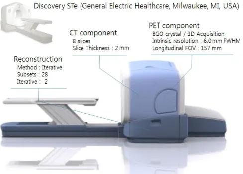
Fig. 1. Discovery-Ste was used. In PET component, it acquired as a 3-dimensional mode. Intrinsic Resolution was 6.0 mm FWHM and DFOV was 70.0 mm and overlapping per bed was 9 slices. CT component used 8 slices using 2 mm thickness. It took iterative mode as a reconstruction method having 28 subsets and 2 iterations.
지킨스씨 림프종이 있는데, 미만성 거대 B 세포 림프종은 비 호지킨스씨 림프종에 속하며 우리나라에서 나타나는 비호지 킨스씨 림프종 중 에서 40~50% 정도 차지하고 있다.
현재 PET/CT 검사는 암의 조기 검진 및 암 진단 환자의 수술 전 또는 수술 후 추적 검사에서 없어서는 안 되는 검사 로 인정되고 있다. 많은 암 환자들 중 특히 미만성 거대 B 세 포 림프종 환자의 추적 검사에서 PET/CT 검사는 환자의 기 타 다른 검사들과 견주어 볼 때 매우 중요하다는 것은 이미 알려져 있다. 미만성 거대 B 세포 림프종 환자의 치료는 대부 분 환자의 장기 한 곳에 국한된 경우가 거의 없다. 그래서, 수 술이 적용되는 경우가 극히 드물기 때문에 방사선 치료나 항암 화학 요법 치료로 시행한다. 항암 화학 치료 요법에 쓰이는 약제로는 플루다라빈, 클라드리빈과 같은 약제를 단독으로 쓰기도 하고, 복합 항암 화학 요법의 표준인 CHOP [Cyclophosphamide (Cytoxan), Hydroxydaunorubicin (Adriamycin), Oncovin (vincristine), Prednisone] 요법에 Rituximab(같은 B세포 항원에 대한 단클론 항체를 이용한 치 료)을 병합하여 쓰기도 한다.
이러한 항암 화학 요법 치료는 골수의 활동을 억제하는 치 료이기 때문에 환자에게서 호중구감소증과 같은 백혈구의 감 소가 나타날 수 있다.1-2) 이러한 증상을 개선하기 위하여 과립 구 자극 요소(Granulocyte colony-stimulating factor)인 Filgrastim 성분의 골수 촉진제를 사용한다. Filgrastim은 대장 균을 숙주로 하여 만들어진 과립구 자극 요소이고, 조혈 호르 몬의 생성, 성숙, 활성에 관련된 조혈 성장 인자(임파구와 골
수에서 생성되며 성숙 혈구 세포의 콜로니를 형성하는 조혈 모세포를 자극하는 인자)이다. 또한, 호중구의 증식, 성장 및 이동을 가속 시키는 역할을 하고 있다.3-5)
요약하자면, 항암 치료 후 떨어진 백혈구(특히, 호중구) 수 치를 올리기 위해 쓰는 약제인 것이다. 과립구 자극 요소 약 제가 투여된 환자에게서 단기간에 PET/CT 검사를 진행하게 되면, PET/CT 검사에 쓰이는 18F-FDG의 섭취는 조혈 세포 가 모여 있는 골수의 활성화로 그 곳에 섭취가 가중되어 정확 한 영상의 정보를 얻을 수 없다.
본 연구는 이러한 사실을 영상에서의 차이점뿐만 아니라 표준화 섭취 계수(Standard Uptake Value)를 통해 수치화 시 키고 통계분석 하여 고찰함으로써 보다 정확한 정보를 얻기 위함을 목적으로 하고 있다. 또한 연구된 환자를 대상으로 하 여 과립구 자극 요소 약제의 투여 후 어느 정도 시간이 지난 후에 PET/CT 검사를 시행하면 18F-FDG의 정상 섭취가 가 능한지도 유추해 보기로 한다.
실험재료 및 방법
1. 환자 정보
2007년 1월~2009년 1월까지 미만성 거대 B 세포 림프종을 진단받고 추적 검사 중인 환자 10명을 대상으로 하였다. 남 자 4명, 여자 6명이고, 평균나이는 53.8세, 평균 체중은 57.3 kg이다.
Scout &
+NCECT
1 min 1 min
Preparation
5 min 5 min
PET scan
18 min 18 min
+CECT
4 min 4 min
Scout &
+NCECT
1 min 1 min
Scout &
+NCECT Scout &
+NCECT
1 min 1 min
Preparation
5 min 5 min
PET scan
18 min 18 min
+CECT
4 min 4 min
Preparation
5 min 5 min
Preparation Preparation
5 min 5 min
PET scan
18 min 18 min
PET scan PET scan
18 min 18 min
+CECT
4 min 4 min
+CECT +CECT
4 min 4 min
Fig. 2. The procedure of the examination consists of 4 stages. First, it takes 5 minutes for preparation. Second, Scout scan and NCECT (Non-Contrast Enhanced CT) Scan needs 1 minute. Third, PET Scan (Emission Scan) takes 18 minutes. Forth, CECT (Contrast Enhanced CT) Scan needs for 4 minutes.
★ ROI (Region Of Interest) size: 12mm
26
thCervical spine : C
6Lung Spleen
Liver
4
thLumbar spine : L
4★ ROI (Region Of Interest) size: 12mm
26
thCervical spine : C
6Lung Spleen
Liver
4
thLumbar spine : L
4★ ROI (Region Of Interest) size: 12mm
26
thCervical spine : C
66
thCervical spine : C
6Lung Lung Spleen Spleen
Liver Liver
4
thLumbar spine : L
44
thLumbar spine : L
4Fig. 3. On each patient, three of ROI (Region of Interest)s were drew PET image. They are drew 6th cervical spine, 4th Lumbar spine, Liver, Spleen and Lung. Each ROI’s diameter was 12 mm2 on a circle shape.
2. 장비
PET/CT Discovery-Ste (General Electric Healthcare, Milwaukee, MI, United States of America) Scanner를 사용하 여 3D (3-Dimension) 방법으로 검사하였으며 크리스탈 (Crystal)은 BGO를 사용하였다. 고유 분해능(Intrinsic Resolution)은 6.0 mm의 반치폭(FWHM)을 사용하였고, DFOV (Display Field Of View)는 70.0 mm, 1 Bed 당 Overlap은 9 slices이고, 재구성 방법으로는 반복 재구성 방법으로 Subset 28회, Iteration 2회 사용하였다. CT는 2 mm의 Slice Thickness로 8 slices로 구성되어 있다(Fig. 1).
3. 검사 방법
검사를 위해 환자에게 18F-FDG를 킬로그램 당 7.4 MBq를 정맥 주사 후 1시간 가량 안정시키고, 환자가 준비되고 검사 준비가 완료되면 먼저 Scout 촬영을 하고 조영제를 사용하지
않는 CT 촬영(140 KV, 30 mA) 후 1 Bed당 2분 30초로 평균 총 7 Bed PET 검사를 진행하였고, 이어서 조영제를 사용하 여 CT 촬영(140 KV, 200 mA)을 진행하였다(Fig. 2). 조영제 로는 OMNIPAQUE (Amersham Health Co., Ireland, 1 mL 당 요오드 300 mg 포함)을 사용하였다.
4. 영상 분석
영상 분석은 환자에게서 획득한 두 가지의 PET 영상을 Advantage. Workstation Version 4.3프로그램에서 PET Volume Viewer Method로 C6 (6 번 경추), L4 (4 번 요추), 간, 비장, 폐에 동일한 관심 영역(Region Of Interests: ROIs)을 설정 하 였다. 관심 영역은 지름 12 mm2 원형으로 하였고 해당 영역에 서 표준화 섭취 계수(SUV)를 측정 후 비교 하였다(Fig. 3). 모 든 관심 영역은 CT, PET, Fusion 영상의 관상면, 시상면, 횡 단면을 참조하여 관상면을 기준으로 그렸다. 같은 환자를 골 수촉진제 주사 1일 후 검사 영상과 5~7일 후 검사 영상을 각
Region SUV After Filgrastim injected, 1-day (Mean±SD)
After Filgrastim injected, 1-day (Mean±SD)
Decrease of SUV (p value)
C6 max 4.8±0.45 2.1±0.35 -56.2% (0.05)
avg 3.8±0.51 1.8±0.31 -52.6% (0.05)
L4 max 6.4±0.36 2.0±0.24 -68.7% (0.05)
avg 5.5±0.47 1.7±0.26 -69.1% (0.05)
Spleen max 4.3±0.36 2.3±0.14 -46.5% (0.05)
avg 3.6±0.45 2.0±0.17 -44.4% (0.05)
Liver max 2.2±0.25 2.2±0.12 -0.06% (0.67)
avg 1.9±0.17 2.0±0.10 +0.5% (0.14)
Lung max 0.8±0.14 0.7±0.14 -1.3% (0.63)
avg 0.6±0.14 0.6±0.12 -0.03% (0.52)
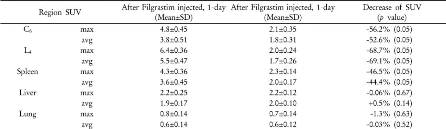
Table 1. Mean and Maximum Standardized Uptake Values (SUV) for Different Regions Averaged Over All Patients 각 분석하였다.
5. 통계 분석
통계 분석은 SPSS version 12. (SPSS Inc., USA)를 사용하 였으며, 두 가지 PET 영상의 정량 분석의 비교에서는 비모수 적 방법 인 윌콕슨 부호순위 검정(Wilcoxon signed-rank test) 을 사용하였다. p<0.05인 경우에 유의한 차이가 있는 것으로 인정하였으며 제시된 모든 값은 평균±표준편차로 표시하고, Filgrastim 주사 후 1일 영상에 대한 5~7일 영상에서의 표준 화 섭취 계수(SUV) 감소율을 표시 하였다.
결 과
검사를 시행한 10명의 환자에게서 C6 (6 번 경추), L4 (4 번 요추), 간, 비장, 폐의 최대 표준화 섭취 계수(maxSUV)와 평 균 표준화 섭취 계수(avgSUV)를 측정하여 100 개의 영역을 분석하였다. Filgrastim 주사 후 1 일 환자에서 C6(6 번 경추) 의 최대 표준화 섭취 계수 평균값과 표준편차는 4.8±0.45 이 었고, Filgrastim 주사 후 5~7일 환자의 최대 표준화 섭취 계 수 평균값과 표준편차는 2.1±0.35로 감소율 56.2%를 보였으 며 유의 확률이 0.05 이므로 통계적으로 유의한 것으로 나타 났다. Filgrastim 주사 후 1일 환자에서 C6 (6 번 경추)의 평 균 표준화 섭취 계수 평균값과 표준편차는 3.8±0.51이었고, Filgrastim 주사 후 5~7 일 환자의 평균 표준화 섭취 계수 평 균값과 표준편차는 1.8±0.31로 감소율 52.6%를 보였으며 마 찬가지로 유의 확률이 0.05이므로 통계적으로 유의한 것으로 나타났다.
Filgrastim 주사 후 1일 환자에서 L4 (4번 요추)의 최대 표준 화 섭취 계수 평균값과 표준편차는 6.4±0.36이었고, Filgrastim 주사 후 5~7일 환자의 최대 표준화 섭취 계수 평균값과 표준
편차는 2.0±0.24로 감소율 68.7%를 보였으며 유의 확률이 0.05 이므로 통계적으로 유의한 것으로 나타났다. Filgrastim 주사 후 1일 환자에서 L4 (4번 요추)의 평균 표준화 섭취 계수 평균값과 표준편차는 5.5±0.47이었고, Filgrastim 주사 후 5~7 일 환자의 평균 표준화 섭취 계수 평균값과 표준편차는 1.7±0.26로 감소율 69.1%를 보였으며 마찬가지로 유의 확률 이 0.05이므로 통계적으로 유의한 것으로 나타났다.
Filgrastim 주사 후 1일 환자에서 비장의 최대 표준화 섭취 계수 평균값과 표준편차는 4.3±0.36이었고, Filgrastim 주사 후 5~7일 환자의 최대 표준화 섭취 계수 평균값과 표준편차 는 2.3±0.14로 감소율 46.5%를 보였으며 유의 확률이 0.05이 므로 통계적으로 유의한 것으로 나타났다. Filgrastim 주사 후 1일 환자에서 비장의 평균 표준화 섭취 계수 평균값과 표준 편차는 3.6±0.45 이었고, Filgrastim 주사 후 5~7일 환자의 평 균 표준화 섭취 계수 평균값과 표준편차는 2.0±0.17로 감소율 44.4%를 보였으며 마찬가지로 유의 확률이 0.05이므로 통계 적으로 유의한 것으로 나타났다.
Filgrastim 주사 후 1일 환자에서 간의 최대 표준화 섭취 계 수 평균값과 표준편차는 2.2±0.25이었고, Filgrastim 주사 후 5~7일 환자의 최대 표준화 섭취 계수 평균값과 표준편차는 2.2±0.12로 감소율 0.06%를 보였으며 유의 확률이 0.67이므 로 통계적으로 유의하지 않은 것으로 나타났다. Filgrastim 주 사 후 1일 환자에서 간의 평균 표준화 섭취 계수 평균값과 표 준편차는 1.9±0.17이었고, Filgrastim 주사 후 5~7일 환자의 평균 표준화 섭취 계수 평균값과 표준편차는 2.0±0.10로 오히 려 증가율이 0.5%로 증가 했으며 유의 확률이 0.14이므로 통 계적으로 유의하지 않은 것으로 나타났다.
Filgrastim 주사 후 1일 환자에서 폐의 최대 표준화 섭취 계 수 평균값과 표준편차 0.8±0.14이었고, Filgrastim 주사 후 5~7일 환자의 최대 표준화 섭취 계수 평균값과 표준편차는 0.7±0.14로 감소율 1.3%를 보였으며 유의 확률이 0.63이므로
Fig. 4. Comparison after Filgrastim injected, 1day and after Filgrastim injected, 5~7days of Maximum Standardized Uptake Values (SUV).
Fig. 5. Comparison after Filgrastim injected, 1day and after Filgrastim injected, 5~7days of Average Standardized Uptake Values (SUV).
통계적으로 유의하지 않은 것으로 나타났다. Filgrastim 주사 후 1일 환자에서 폐의 평균 표준화 섭취 계수 평균값과 표준 편차는 0.6±0.14이었고, Filgrastim 주사 후 5~7일 환자의 평 균 표준화 섭취 계수 평균값과 표준편차는 0.6±0.12로 감소율 0.03%보였으며 유의 확률이 0.52이므로 통계적으로 유의하 지 않은 것으로 나타났다(Table 1).
위의 결과에서 보여 주듯이 C6 (6 번 경추)와 L4 (4 번 요추)
그리고, 비장은 Filgrastim 주사 후 1일 영상과, 5~7일 영상에 서 표준화 섭취 계수의 변동이 크게 보임을 알 수 있고, 간과 폐에서는 큰 변화가 없음을 알 수 있다(Fig. 4, 5). 그러므로, C6 (6 번 경추)와 L4 (4번 요추), 비장은 Filgrastim의 성분의 약제에 영향을 받는 조혈 세포를 가지고 있는 장기이고, 간과 폐는 Filgrastim의 성분의 약제에 큰 영향을 받지 않기 때문에 조혈 세포를 가지고 있지 않는 장기임을 유추 할 수 있다. 또
A B
A B
A
AB
BFig. 6. A image is 1-day image after Filgrasim injected to patient. B image is 7-days after Filgrasim injected to same patient.
한, 영상에서는 Filgrastim 주사 후 1일 환자의 영상에서 다른 장기에 비해 뼈에 18F-FDG 섭취가 집중되어 있는 것을 볼 수 있고, Filgrastim 주사 후 5~7일 같은 환자의 영상에서는 뼈에
18F-FDG 섭취가 현저히 줄어들어 정상 섭취됨을 볼 수 있다 (Fig. 6).
결론 및 고찰
Filgrastim 주사 후 1일째 촬영을 하는 것은 특별한 경우를 제외하고는 거의 없다. 그렇기 때문에 2년여에 걸쳐서 환자 대상군을 모아 왔지만 같은 환자를 두 번 검사하는 경우를 찾 기가 어려워 10명의 환자만을 대상으로 하였다. 해외에서 발 표된 논문에서 Filgrastim이라는 약제를 쓴 후 5일 정도 후에
18F-FDG의 정상 섭취가 가능하다고 발표되어 있다.6) 이 연 구는 발표된 내용을 직접 확인해 보고자 하는데 의미를 두고 진행 하였으며, 본 연구에서도 5일 지나고 18F-FDG 정상 섭 취를 보인 환자가 10명 중 4명이고, 나머지 6명은 7일 후
18F-FDG 정상 섭취를 보인 것이 확인 되었다. 또한, 표준화 섭취 계수라는 것을 이용하여 영상을 분석하고 수치화 하고 자 하는데 목적을 두고 진행 하였다.
위의 연구에서도 보여진 것과 같이, Filgrastim 약제를 투 여한 환자의 검사영상과 5~7일 정도 시간이 지나고 검사한 환자의 영상에서 관심 영역에 포함된 각 장기의 표준화 섭취 계수가 현저한 차이를 보였으며, Filgrastim 약제를 투여한 환
자의 영상에 뼈의 18F-FDG 섭취가 집중된 것을 볼 수가 있다.
서두에도 언급했듯이 이 연구는 실험을 통해 새로운 사실 을 알고자 한 것이 아니고, 기존의 사실에 대한 것을 보다 더 구체적으로 수치화 시켜 통계 분석을 통해 고찰하고자 했고, 직접 연구를 통해 Filgrastim 약제 투여 후에 어느 정도 기간 이 지나고 PET/CT 검사를 진행 하는 것이 좋을 것이라는 것 을 유추하는데 목적을 두었다. 추후에 Filgrastim 이외에 골수 촉진제로 많이 쓰이는 Pegfilgrastim, Lenograstim 등의 성분 으로 구성된 기타약제와의
유사성과 차이점에 대한 유사 연구에도 기여할 수 있으리 라 사료된다.
요 약
많은 암 환자들 중 특히 미만성 거대 B 세포 림프종 환자 의 추적 검사에서 PET/CT 검사는 기타 다른 검사들과 견주 어 볼 때 매우 중요하다는 것은 이미 알려져 있다. 이들 환자 의 치료는 대부분 환자의 장기 한 곳에 국한된 경우가 없으므 로, 방사선 치료나 항암 화학 요법 치료로 시행한다. 이러한 항암 화학 요법 치료는 골수의 활동을 억제하는 치료이기 때 문에 환자에게서 호중구감소증과 같은 백혈구의 감소가 나타 날 수 있다. 이러한 증상을 개선하기 위하여 과립구 자극요소 (Granulocyte colony-stimulating factor)인 Filgrastim 성분의 골수촉진제를 사용하는 경우가 일반적이며, 이 약제가 투여
된 환자에게서 단기간에 PET/CT 검사를 진행하게 되면, 검 사에 사용되는 18F-FDG의 섭취가 조혈 세포가 모여 있는 골 수의 활성화로 그 곳에 섭취가 가중되어 정확한 영상의 정보 를 얻는데 한계가 있다. 이 연구의 목적은 Filgrastim의 약제 투여 1일 후의 영상과 어느 정도 시간이 지난 후 영상의 SUV 를 각각 비교하여 수치화하고, 대략 어느 정도 시간이 지난 후에 정확한 영상의 정보를 줄 수 있는 18F-FDG의 섭취가 일 어나는지를 유추해 보고자 한다.
환자대상은 2007년 1월~2009년 1월까지 미만성 거대 B 세 포 림프종을 진단받고 추적 검사 중인 환자 10명을 대상으로 하였다. 남자 4명, 여자 6명이고, 평균나이는 53.8세 평균 체 중은 57.3 kg이다. PET/CT (Discovery STe; GE Healthcare, Milwaukee, WI, USA) 검사를 위해 환자에게 18F-FDG 정맥 주사 후 1시간 가량 안정시키고, PET/CT 영상을 획득하였 다. 영상 분석은 ROI (12 mm2)를 C6 (6 번 경추), L4 (4 번 요추), 간, 비장, 폐에 각각 그린 후 표준화 섭취 계수(SUV)를 측정하였다. 같은 환자를 골수촉진제 주사 1일 후 영상과 5~7 일 후 영상을 각각 분석하였으며, 통계 프로그램으로는 SPSS version 12.
Wilcoxon signed rank test를 사용하여 두 집단 간의 유의 성을 확인하였다.
검사를 시행한 10명의 환자의 SUV를 분석한 결과, 골수촉 진제를 투여하고 1일 후의 SUV가 5~7일 후에 비해 C6 (6 번 경추), L4 (4 번 요추), 비장에서 현저하게 높게 나타났고, 간 과 폐에서는 비슷하게 나타났다. SUV의 차이와 더불어, 5~7 일 후의 영상에서는 뼈의 섭취가 거의 없어진 정상 섭취 영상 을 얻을 수 있었다.
위의 연구에서도 보여진 것과 같이 과립구 자극요소 약제 투여 후 1일째 환자의 영상과 5~7일 후의 영상에서 SUV의 현저한 차이가 있었으며, 약제를 투여한 환자의 영상에 뼈의
18F-FDG 섭취가 집중되었으며, 평균 5~7일 후에 검사에서는 뼈의 섭취가 현저하게 줄어든 정상적이 영상을 얻을 수 있었 다. 본 연구를 통하여 SUV를 측정하고, 통계분석 함으로서
기존의 사실을 수치화 하였으며, 정상 섭취가 가능한 기간을 유추 할 수 있었다. 추후에 과립구 자극요소인 Filgrastim 이 외에 Pegfilgrastim, Lenograstim등 기타약제와의 유사성과 차 이점에 대한 유사 연구에도 기여할 수 있으리라 사료된다.
REFERENCES
1. Crawford J, Ozer H, Stoller R, et al. Reduction by granulocyte colony-stimulating factor of fever and neutropenia induced by chemotherapy in patients with small-cell lung cancer. N Engl J Med 1991;325:164-170.
2. Green MD, Koelbl H, Baselga J, et al. A randomized double- blind multicenter phase Ш study of fixed-dose single-administration pregfilgrastim versus daily filgrastim in patients receiving myelosuppressive chemotherapy. Ann Oncol 2003;14:29-35.
3. Groopman JE, Molina JM, Scadden DT, Hematopoietic growth factors: biology and clinical applications. N Engl J Med 1989;
321:1449-1459.
4. Lieschke GJ, Burgess AW. Granulocyte colony-stimulating factor and granulocyte-macrophage colony-stimulating factor (1). N Engl J Med 1992;327:28-35.
5. Lieschke GJ, Burgess AW. Granulocyte colony-stimulating factor and granulocyte-macrophage colony-stimulating factor (2). N Engl J Med 1992;327: 99-106.
6. Dmitri M, Edward MB. Interaction of colony-stimulating factor and Fluorodeoxyglucose 18F Positron Emission Tomography. Ann Pharmacother 2002;36:1796-1799.
7. Heather AJ, Takayoshi I, James ME, et al. Effects of Pefilgrastim on normal bildistribution of 18F-FDG: Preclinical and Clinical studies. J Nucl Med 2006;47:950-956.
8. Robert KD, Lisa KD, Erin KS, et al. Dynamic and Static approaches to quantifying 18F-FDG uptake for measuring cancer response to therapy, including the effect of Granulocyte colony- stimulating factor. J Nucl Med 2007;48:920-925.
9. Kaye LJ, John PF, Brian RM et al. Abnormal PET scan secondary to the use of hematopoietic growth factors. Heamatologica 2005;
90(8):e88.
10. Yoshifumi S, Kenneth RZ, Paul VK, et al. Splenic Fluorodeoxyglucose uptake increased by granulocyte colony-stimulating factor therapy:
PET imaging results. J Nucl Med 1999;40:1456-1462.