1. INTRODUCTION
Katsura tree (Cercidiphyllum japonicum Sieb.
Et Zucc), is the only species belonging to Cercidiphyllum genus, which is well repre- sented in the fossil record, with occurrences in the late Cretaceous and Tertiary of North America and Europe. However, it is now con- fined to East Asian countries (Manchester et al., 2009). The tree is a long-lived, deciduous, wind-pollinated tree with dimorphic leaves and up to 30 to 45 m tall with a symmetrical can- opy and new growth is reddish turning a light pale green. Fall color is a spectacular yellow,
with some red. Thus, it is valued as an orna- mental or a shade tree for landscape (Zhang et al., 2009). The natural populations of the tree inhabit distribute sites (600 to 2000 m) of tem- perate deciduous forests scattered across East China and Japan (Isagi et al., 2005).
A hydrolyzable tannin or pyrogallol-type tannin is a type of tannin that, on heating with hydrochloric or sulfuric acids, yields gallic or ellagic acids, and they are mixtures of poly- galloyl glucoses and/or poly-galloyl quinic acid derivatives containing in between 3 up to 12 gallic acid residues per molecule (Haslam, 1989). Ellagitannins are a diverse class of hy-
Original Article
A Polyoxygenated Ellagitannin from Cercidiphyllum japonicum Bark 1
Min-Sung Lee
2⋅Hee-Jeong Min
2⋅Jin-Kyu Kim
3⋅Young-Soo Bae
2,†ABSTRACT
Katsura tree (Cercidiphyllum japonicum Sieb. Et Zucc) bark was collected, air-dried and extracted with 70%
aqueous acetone, then concentrated and sequentially fractionated using n-hexane, methylene chloride (CH
2Cl
2), ethylacetate (EtOAc), and H
2O. The H
2O fraction was chromatographed on a Sephadex LH-20 column with various aqueous MeOH eluting solvents to isolate an ellagitannin. The isolate was elucidated as macabarterin, a polyoxygenated ellagitannin by NMR analysis, including HSQC, HMBC, Q-TOF MS, and with the compar- ison of authentic literature data. The compound was an ellagitannin which was isolated, for the first time, from the extracts of Katsura tree bark, and has never been reported before in domestic tree or plant sources.
Keywords : Cercidiphyllum japonicum bark, H
2O fraction, macabarterin, ellagitannin, column chromatography
1
Date Received June 1, 2016, Date Accepted July 7, 2016
2
Department of Forest Biomaterials Engineering, College of Forest and Environment Sciences, Kangwon National University, Chuncheon 24341, Korea
3
Bio-Center, Gyeonggi Institute of Science & Technology Promotion, Suwon 16229, Korea
†
Corresponding author: Young-Soo Bae (e-mail: [email protected])
drolyzable tannins, a type of polyphenol primar- ily formed from the oxidative linkage of galloyl groups in 1,2,3,4,6-pentagalloyl glucose (Kwon and Bae, 2009; Sepulveda et al., 2011;
Steinmetz, 2010). Ellagitannins contain various numbers of hexahydroxydiphenoyl (HHDP) units, as well as galloyl units and/or sangui- sorboyl units bounded to sugar moiety. In order to determine the quantity of every individual unit, the hydrolysis of the extracts with tri- fluoroacetic acid in methanol/water system is performed. Hexahydroxydiphenic acid, created after hydrolysis, spontaneously lactonized to el- lagic acid, and sanguisorbic acid to sanguisorbic acid dilactone, while gallic acid remains intact (Yoshida et al., 2009).
Recently there have been many studies to evaluate biological activities of various natural resources and to develop pharmaceutical or functional food or cosmetic products. However, there are little studies on katsura tree extracts for functional uses in domestic or abroad (Tada and Sakurai, 1991; Takasugi and Katui, 1986;
Towatari et al., 2002). This study was carried out to investigate the extracts of katsura tree bark, and to elucidate the structure of a poly- oxygenated ellagitannin isolated from the extracts.
2. MATERIALS and METHODS 2.1. Plant material
Fresh Cercidiphyllum japonicum bark was collected at Samcheok, Gangwon-do in June
2014, air dried for two weeks and then ground to fine particles to be extracted.
2.2. Sample preparation
The ground bark (1.6 kg) was immersed in 70% aqueous acetone at room temperature for 3 days. After three times extraction and filtration, the filtrates were combined together and evapo- rated on a vacuum evaporator under reduced pressure at 40 ℃. The residue was successively fractionated on a separatory funnel and freeze dried to give n-hexane (1.77 g), CH
2Cl
2(1.48 g), EtOAc (31.1 g), and H
2O (56.3 g) soluble fractions.
2.3. ESI-Q-TOF MS
The following ESI-Q-Tof (AB SCIEX Triple Tof 5600+, Miami, UK) conditions, quadrupole and orthogonal acceleration time-of-flight tan- dem mass spectrometer, were used: ionspray voltage, 5.5 kV; declustering potential (DP), 80 V; the turbo spray temperature, 500 ℃; neb- ulizer gas (Gas 1) of 50 psi; heater gas (Gas 2), 50 psi; curtain gas, 25 psi. Nitrogen was kept as nebulizer and auxiliary gas. The TOF MS scan was operated with the mass range of m/z 300-1800. Recalibration was carried by EasyMass Accuracy
®device before analysis.
2.4. Structure analysis
1
H and
13C NMR spectra, including 2D-NMR
such as HSQC (Heteronuclear Single Quantum
Coherence), HMBC (Heteronuclear Multiple
Bond Correlation), were recorded on a Bruker (USA) Avance DPX 700 MHz spectrometers using TMS (Tetramethylsilane) as an internal standard and chemical shift was given in δ (ppm). Thin layer chromatography (TLC) was done on DC-Plastikfolien Cellulose F (Merck) plates and developed with TBAW (t-BuOH-HOAc-H
2O (3 : 1 : 1, v/v/v)) and 6%
aqueous HOAc. The spot was detected by illu- minating ultraviolet light (UV, 254 and 365 nm).
2.5. Column chromatography
A portion of H
2O fraction (6.0 g) was chro- matographed on a Sephadex LH-20 column, eluting with MeOH-H
2O (1 : 3, v/v) to afford 8 fractions. Fraction 8 was retreated with MeOH-H
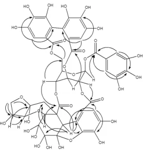
2O (3 : 1, v/v) to isolate the poly- oxygenated ellagitannin (Fig. 1).
Yellowish amorphous powder
R
f: 0.02 (TBAW) and 0.40 (6% HOAc).
Q-TOF-MS : Found m/z 1103 [M+H]
+.
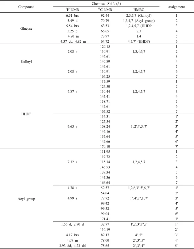
1
H (700 MHz) and
13C (700 MHz) NMR : See Table 1.
3. RESULTS and DISCUSSION
The compound was isolated from the H
2O fraction of the extracts of katsura tree (Cerdidiphyllum japonicum Sieb, Et Zucc) bark by column chromatography using Sephadex LH-20, and the structure was elucidated by NMR analysis and comparison with the liter- ature data.
In the
1H NMR spectrum, D-(+)-glucose of
the compound showed a double doublet signal at δ4.37 and δ4.82 for two H-6 protons and H-5 gave a triplet signal at δ4.80. H-2 and H-4 indicated a doublet signal at δ5.49 and δ5.25, respectively. H-3 also showed a singlet signal at δ5.54. H-1 gave a doublet signal at δ6.51 with 8.37 Hz of the coupling constant suggesting the β-anomeric glucose. These proton signals were very close to the previous literature data (Ngoumfo et al., 2008). The one galloyl symmetrical protons, which is attached to C-1 of D-(+)-glucose, indicated a singlet sig- nal at δ7.08 for H-1 and H-6 (Kwon, 2010;
Xianbin et al., 2009). Also two HHDP (hexahy-
droxydiphenoyl) galloyl protons, which are
bound to C-3 and C-6 of D-(+)-glucose, in-
dicated two singlets at δ6.87 and δ6.63 for
H-3 and H-3', respectively (Ngoumfo et al.,
2008; Xianbin et al., 2009). Above proton sig-
Fig. 1. Structure of the isolated compound.
Compound Chemical Shift ( δ)
assignment
1